The document summarizes the major arteries of the systemic and pulmonary circulations. It describes the branching patterns of the aorta and its major divisions including the ascending aorta, arch of aorta, descending aorta, abdominal aorta, and their tributaries. It also discusses the pulmonary arteries and veins. Venous drainage of the head, neck, upper limbs, thorax, and abdominal regions are outlined. Common sites used for intravenous injections are noted.
The document describes the anatomy and structures of the heart. It discusses:
- The heart is surrounded by the pericardium, a double-walled sac that has an outer fibrous layer and inner serous layers separated by fluid.
- The heart has four chambers - two upper atria that receive blood and two lower ventricles that pump blood out. Blood flows through valves between the chambers.
- The heart wall has three layers - the outer epicardium, middle muscular myocardium, and inner endothelial endocardium.
- The heart is supplied by the right and left coronary arteries and drains into the coronary sinus vein.
- Nerves from the autonomic nervous system
The thoracic aorta begins where the aortic arch ends at the fourth thoracic vertebrae and extends down to the diaphragm. It supplies blood to the thoracic cavity and has several important branches including the bronchial arteries which supply the lungs, esophageal arteries which supply the esophagus, and posterior intercostal arteries which supply the spaces between the ribs. The thoracic aorta also gives off mediastinal and pericardial branches before passing through the diaphragm and becoming the abdominal aorta.
The pleura consists of two layers - the parietal pleura which lines the thoracic wall and diaphragm, and the visceral pleura which covers the lungs. Between these two layers is a potential space called the pleural cavity, which contains pleural fluid that lubricates the pleural surfaces and allows the lungs to slide smoothly against the chest wall during respiration. The pleura has several recesses that allow for full expansion of the lungs during deep inspiration.
The document summarizes the anatomy of the pleura and lungs. It describes the pleura as a membrane that surrounds the lungs and lines the chest cavity. It has a parietal layer on the chest wall and visceral layer covering the lungs. The two layers form a pleural cavity containing pleural fluid. Each lung is cone-shaped and divided into lobes separated by fissures. The lungs receive deoxygenated blood from the pulmonary arteries and return oxygenated blood to the heart via pulmonary veins. Lymph drains from the lungs through plexuses and nodes in the hilum. The lungs are innervated by the pulmonary plexus and have segments supplied by segmental bronchi, arteries and veins.
The arch of the aorta begins behind the upper border of the second right sternochondral joint and arches over the root of the left lung before ending at the lower border of the fourth thoracic vertebra. It has anterior relations including nerves and veins and posterior relations such as the trachea and esophagus. Its main branches are the brachiocephalic trunk, left common carotid artery, and left subclavian artery. Developmental anomalies of the arch of the aorta include right sided aortic arch, double aortic arch, abnormal origin of the right subclavian artery, and coarctation of the aorta.
Right Atrium of human heart
This PPT help to understand the external and internal structures of right atrium.
sulcus terminalis on external surface of rt atrium,
crista terminalis on internal side of rt. atrium,
interior is divided into rough anterior part and smooth posterior part ( sinus venarum)
superior and inferior venae cavae drains deoxygenated blood into rt. atrim
there is Eustachian valve to guard the opening of IVC and Thebesian valve to guard the opening of coronary sinus
septal wall presents fossa ovalis with its border limbus fossa ovalis
The document provides an overview of heart anatomy. It describes the heart's location in the thorax and its dimensions. The four chambers - right and left atria and ventricles - are introduced. Each chamber's function is summarized as receiving or pumping blood. The three layers of the heart - epicardium, myocardium, and endocardium - are defined. The cardiac skeleton is described as a fibrous structure that partitions the chambers and anchors the heart valves.
The document summarizes the anatomy and flow patterns of the major veins in the human body. It describes the three major systemic veins that return deoxygenated blood to the heart - the coronary sinus, inferior vena cava (IVC), and superior vena cava (SVC). It then provides details on the venous drainage of specific regions including the head and neck, upper limbs, thorax, abdomen, and pelvis. Key veins discussed include the internal and external jugular veins, azygos vein system, hepatic portal vein, common iliac veins, and renal veins.
The heart is a hollow, pyramidal-shaped organ located in the thoracic cavity. It is composed of 4 chambers - the right and left atria which receive blood, and the right and left ventricles which pump blood out of the heart. The heart has external features including an apex, base, surfaces and borders that divide the surfaces. The apex points downward and leftward. The base is posterior. The surfaces are sternocostal, diaphragmatic, right and left pulmonary. Borders include right, left, superior and inferior borders. The heart is about the size of a clenched fist.
The abdominal aorta originates at the T12 vertebrae and runs downward in front of the lumbar vertebrae, terminating at the L4 vertebrae by dividing into the common iliac arteries. It gives off several unpaired branches including the celiac trunk, superior mesenteric artery, and inferior mesenteric artery. It also provides paired arteries such as the inferior phrenic, suprarenal, renal, gonadal, lumbar, and common iliac arteries that supply the abdominal organs and structures.
The median cubital vein is a branch of the cephalic vein located within the cubital fossa of the arm. It receives branches from deep forearm veins before joining with the basilic vein. As the largest, most superficial vein in the cubital fossa, the median cubital vein is preferred for venipuncture due to being easier to access, less likely to move or bruise compared to other arm veins.
The diaphragm is a dome-shaped musculotendinous sheet that separates the thoracic and abdominal cavities. It has peripheral muscular parts and a central tendinous portion. It arises from the lumbar vertebrae, ribs, sternum and xiphoid process. The diaphragm inserts into its central tendon. It contains openings for structures like the aorta, inferior vena cava and esophagus. The diaphragm is innervated by the phrenic nerves and functions in respiration and exertion. Clinical implications include hernias, hiccups and paralysis.
The heart is a hollow, muscular organ located slightly left of center in the chest. It is surrounded by three layers of tissue - the outer fibrous pericardium, middle muscular myocardium, and inner endothelial endocardium. The heart is divided into four chambers - right atrium, right ventricle, left atrium, and left ventricle - with valves that ensure one-way blood flow. Deoxygenated blood enters the right atrium from the body and is pumped to the lungs via the right ventricle. Oxygenated blood returns to the left atrium from the lungs and is pumped back out to the body by the left ventricle.
he spleen is a fist-sized organ found in the upper left side of your abdomen, next to your stomach and behind your left ribs. It's an important part of your immune system but you can survive without it. This is because the liver can take over many of the spleen's functions
The document describes the structure and features of the heart chambers. It states that the heart is composed of 4 chambers - the right atrium, right ventricle, left atrium, and left ventricle. Blood enters the atria and is then pumped into the ventricles. From the left ventricle, blood passes into the aorta for systemic circulation, and from the right it enters the pulmonary circulation via the pulmonary arteries. Each chamber has distinct internal and external features and relations to other cardiac structures. The septa divide the atrial and ventricular chambers.
The document discusses the pleurae, which are thin membranes that cover the lungs and line the thoracic cavity. It describes the two layers - the parietal pleura covering the thoracic wall and the visceral pleura covering the lungs. Between these layers is the pleural cavity, which contains a small amount of fluid and allows the lungs to expand and contract during breathing. The document outlines the structure and supply of the pleurae and conditions like pneumothorax that can occur when air enters the pleural space.
The lungs are located in the thoracic cavity and are separated by the mediastinum. Each lung has an apex, base, borders, and surfaces. The root of the lung contains structures like the bronchi, pulmonary arteries and veins. The lungs are divided into lobes by fissures. Bronchopulmonary segments are supplied by their own artery. Alveoli have type I and II pneumocytes, endothelium, and macrophages. Surfactant is produced by type II cells and reduces surface tension in the alveoli.
The heart is pyramidal in shape with an apex pointing leftward and downward. It has four chambers - two atria which receive blood and two ventricles which pump blood. The right atrium receives deoxygenated blood from the body and pumps it to the right ventricle to be sent to the lungs. The left atrium receives oxygenated blood from the lungs and pumps it to the left ventricle to be sent to the body. Blood flows through the heart via the tricuspid, bicuspid/mitral, pulmonary and aortic valves. The heart's conduction system coordinates contractions and is composed of the sinoatrial node, atrioventricular node and bundle of His.
The heart contains four chambers - two upper chambers called atria and two lower chambers called ventricles. The atria receive blood from veins into the heart while the thicker-walled ventricles pump blood out of the heart and into arteries. There are three types of circulation - systemic circulation pumps oxygenated blood from the left ventricle to the body, pulmonary circulation pumps deoxygenated blood from the right ventricle to the lungs, and hepatic portal circulation transports blood from the intestines to the liver. Valves between the chambers regulate blood flow through the heart.
The document summarizes the bones of the upper limb, including the clavicle, scapula, humerus, radius, and ulna. It describes the location and markings of each bone, as well as their articulations with other bones and attachments of muscles. Key points include that the clavicle and scapula form the shoulder girdle, connecting the humerus to the axial skeleton. The humerus extends from the shoulder to elbow joints. The radius and ulna articulate at the elbow and wrist, allowing rotation of the forearm.
right ventricle internal and external features-
interior is divided into inflowing and outflowing parts (infundibulum)
inflowing part is rough due to trabeculae corneae, - ridges, bridges, pillars. Chordae tendineae- are attached to pillars and cusps of tricuspid valve.
outflowing part is smooth, semilunar valve guards opening of pulmonary valve
The pericardium has two layers - the fibrous pericardium and serous pericardium. The fibrous pericardium is a dense connective tissue that protects the heart. The serous pericardium contains two layers that lubricate the heart and prevent friction. It also contains the pericardial cavity filled with fluid. Too much fluid in the cavity can cause compression of the heart.
1. The document describes the anatomy of the thoracic wall including bones, joints, muscles, blood vessels, and nerves.
2. It discusses the 12 pairs of ribs, their classification as true, false, or floating ribs, and their articulations with the sternum.
3. The intercostal spaces contain intercostal muscles like the external and internal intercostals that act in respiration, as well as vessels and nerves.
The clavicle is an S-shaped bone that transmits weight from the upper limb to the sternum. It has two ends - a lateral end that articulates with the acromion of the scapula to form the AC joint, and a medial end that articulates with the manubrium sterni to form the SC joint. The shaft of the clavicle is divided into a lateral one-third and a medial two-thirds, with various surfaces, borders, and tubercles that provide attachments for muscles and ligaments like the deltoid, trapezius, pectoralis major, and coracoclavicular ligaments. The clavicle is the only long bone that lies horizontally
The document summarizes the major arteries and veins of the systemic and pulmonary circulations. It describes the path of blood flow from the aorta and its major branches that supply the head, neck, upper limbs, thorax, abdomen, pelvis and lower limbs. It also details the pulmonary circulation from the pulmonary trunk to the lungs and back to the heart via the pulmonary veins.
The carotid arteries are the primary vessels supplying blood to the brain and face. The right common carotid artery (RCCA) originates in the neck from the brachiocephalic artery while the left common carotid artery (LCCA) arises in the thorax from the arch of the aorta.
The document summarizes the major arteries of the head and neck, including their origins, courses, and branches. It begins with an overview of the aorta and its branches that supply the head and neck (brachiocephalic artery, common carotid artery, subclavian artery). It then describes the external and internal carotid arteries in detail, outlining their branches which include the facial, lingual, occipital, and maxillary arteries. Throughout, clinical correlations and examples of surgical/traumatic implications are provided.
The head and neck receive their primary blood supply from the carotid and vertebral arteries. The common carotid arteries divide into external and internal carotids. The external carotid supplies neck and face structures, branching into the superior thyroid, lingual, facial, occipital, and maxillary arteries. The internal carotid supplies the brain, branching into the anterior and middle cerebral arteries. Venous drainage is performed by the internal and external jugular veins, which drain into the subclavian vein.
The major blood vessels include the arteries, which carry oxygen-rich blood away from the heart, and veins, which carry deoxygenated blood back to the heart. The circulatory system is divided into the pulmonary and systemic circuits. The pulmonary circuit carries deoxygenated blood to the lungs and returns oxygenated blood to the heart. The systemic circuit pumps oxygenated blood to the entire body through progressively smaller arteries and returns deoxygenated blood to the heart via veins. The major arteries include the aorta, carotid, brachial and iliac arteries. The major veins include the jugular, subclavian, brachial and iliac veins.
arteries of human body including head and neck, upper extremities, lower extremities
different between the veins and arteries. common carotid arteries, internal carotid arteries, external carotid arteries, subclavian arteries
circle of Willis
The heart is a hollow muscular organ located in the middle mediastinum. It is approximately the size of a fist and weighs 250-300 grams. The heart has four chambers - two upper atria and two lower ventricles. It is surrounded by a double-walled sac called the pericardium. The heart pumps blood through two circuits - the pulmonary circulation and the systemic circulation - using a series of valves to ensure one-way blood flow.
Vascular structure head and neck Almas khan , Khorfakkhan hospitalalmasmkm
This document discusses the vascular structure of the head and neck. It begins by explaining the components of the circulatory system, including the cardiovascular and lymphatic systems. It then describes the three main circuits of circulation: coronary, pulmonary, and systemic. The document focuses on the head and neck vasculature, detailing arteries like the aorta, carotid, vertebral and various branches. It also discusses venous drainage and the circle of Willis. Common clinical indications involving the head and neck vessels are presented. In conclusion, the circulatory system of the head and neck is reviewed to aid in angiography identification and understanding normal and abnormal vascular distributions.
The cardiovascular system summary is as follows:
1. The heart is located in the chest behind the sternum and is about the size of a fist. It has four chambers - two atria for receiving blood and two ventricles for pumping blood out.
2. The pericardium is a sac that surrounds and protects the heart. It has two layers - an outer fibrous layer and an inner serous layer that secretes fluid.
3. Blood enters the right atrium from the body and is pumped into the pulmonary artery to the lungs. Oxygenated blood returns to the left atrium and is pumped by the left ventricle through the aorta to the body.
MEDISTINUM and The HEART.pptx1111111111111111111marrahmohamed33
The document discusses the mediastinum and structures contained within it, including the heart. It provides details on the different compartments of the mediastinum (superior, anterior, middle, posterior), and structures found in each. It describes the pericardium and heart in detail. The pericardium surrounds and protects the heart. It has fibrous and serous layers. Conditions like pericarditis and pericardial effusion are discussed. The heart is located in the middle mediastinum and has an apex pointing left and downward and a base facing posteriorly.
The heart has four chambers. The two superior receiving chambers are the atria (= entry halls or chambers), and the two inferior pumping chambers are the ventricles (= little bellies).
On the anterior surface of each atrium is a wrinkled pouchlike structure called an auricle
Be the first to comment
Anatomy And Physiology of Human Heart
1. ANATOMY OF THE HEART By: Dr Mohammed Faez
2. The Heart The heart is a chambered muscular organ that pumps blood received from the veins into the arteries, thereby maintaining the flow of blood through the entire circulatory system.
3. The Heart • The heart is surrounded by membrane called Pericardium.
4. The Pericardium • The pericardium is a fibroserous sac that encloses the heart and the roots of the great vessels. • The pericardium lies within the middle mediastinum.
5. The Pericardium
6. The Pericardium • Its function is to restrict excessive movements of the heart as a whole and to serve as a lubricated container in which the different parts of the heart can contract.
1) The document describes the major arteries of the systemic circulation, including branches off the aorta such as the coronary, carotid, and brachial arteries.
2) It also details important veins that drain blood from the head, neck, arms and abdomen, including the internal and external jugular veins, brachial veins, and hepatic portal vein.
3) The hepatic portal system is summarized as carrying nutrient-rich blood from the abdominal viscera to the liver through the hepatic portal vein and its tributaries, before the blood continues to the inferior vena cava and heart.
The document summarizes the major arteries and veins supplying the head and neck region. It begins by listing the objectives which are to describe the major arteries including the common carotid arteries, external carotid artery, internal carotid artery, and subclavian artery. It then provides details on the branches and relations of these arteries. Next, it discusses the major veins including the external jugular vein, internal jugular vein, and their tributaries. It concludes by describing the formation of the brachiocephalic veins and superior vena cava which drain the head and neck veins.
The document describes the structure and function of the heart. It discusses the location of the heart in the mediastinum and its external and internal anatomy. The four chambers of the heart (right and left atria and ventricles) are described along with the valves that regulate blood flow. The circulations of blood through the pulmonary system and systemic circulation are also summarized. Key details about the layers of the heart wall, coronary circulation and blood flow through arteries, capillaries and veins are provided.
Anatomy & physiology of cardiovascular systemvinayanerurkar
This document provides an overview of the anatomy and physiology of the cardiovascular system. It describes the location and structure of the heart, including its chambers and layers. It explains the circulation of blood through the heart, into the pulmonary circulation to oxygenate blood and into the systemic circulation to distribute oxygenated blood to the body. It details the coronary circulation which provides blood supply to the heart muscle.
The document provides an overview of the anatomy of the heart and its associated vessels. It describes the internal and external structures of the heart including the chambers, valves, arteries and veins. Key points include:
- The heart is located in the middle mediastinum and is surrounded by the pericardium. It has four chambers - right and left atria on top which receive blood and right and left ventricles on bottom which pump blood out.
- Blood flows from the vena cava and pulmonary veins into the atria, through valves into the ventricles, and out through the pulmonary artery and aorta into the lungs and body.
- Coronary arteries supply blood to the heart muscle and coronary
Cardiovascular anatomy and imaging TechniquesMilan Silwal
The heart receives blood through four major vessels - the superior and inferior vena cava and the right and left pulmonary veins. It pumps blood out through two major vessels - the pulmonary trunk and aorta. The heart has four chambers - two upper atria and two lower ventricles. It is surrounded by layers including the pericardium. The heart lies in the middle mediastinum and has four surfaces and four borders defined by its chambers. Valves between the chambers include the tricuspid, pulmonary, mitral and aortic valves. The heart has a complex electrical conduction system to coordinate contractions. Arteries supplying the heart include the coronary arteries while veins draining it include the cardiac veins.
Similar to systemic and pulmonary circulation.pdf (20)
Retinopathy of prematurity.pdf ROP newbornPooja Rani
Retinopathy of prematurity (ROP) is a retinal disorder that affects premature infants, caused by abnormal blood vessel growth in the retina. It develops due to retinal immaturity and hyperoxia from high oxygen therapy in preterm infants less than 37 weeks gestational age. Early stages involve mild abnormal vessel growth, while later stages involve retinal detachment. Treatment depends on the stage, and may include laser therapy, scleral buckling surgery, or prevention through careful oxygen management for preterm infants.
The Braden Scale and Norton Scale are tools used to assess risk for pressure ulcer development. The Braden Scale consists of 6 subscales that are scored from 1-4 or 1-3, with lower total scores indicating higher risk. The Norton Scale consists of 5 subscales scored from 1-4, with total scores below 14 indicating higher risk. Both scales provide structured assessments of factors like sensory perception, mobility, nutrition, and moisture that are used to determine a patient's pressure ulcer risk level.
Neonatal Intensive Care Unit. NICU. neonatePooja Rani
The document discusses the organization and management of neonatal intensive care units (NICU) in India. It outlines a 3-tier system of neonatal care based on weight and gestational age. Level I care is for healthy newborns, Level II care is for preterm or low birth weight babies, and Level III care is for very preterm and low birth weight babies requiring advanced equipment. It provides details on NICU unit size, location, design, equipment, staffing, and infection control practices to optimize care for sick newborns.
Kangaroo Mother Care . neonate. newborncarePooja Rani
Kangaroo Mother Care (KMC) is a technique for caring for low birth weight babies that provides skin-to-skin contact between the mother and baby to promote thermal control, breastfeeding, infection prevention, and bonding. Key components of KMC include prolonged, continuous skin-to-skin contact; exclusive breastfeeding; and early discharge from the hospital with regular follow-up care. KMC has benefits like increased breastfeeding rates, better temperature control for the baby, earlier discharge from the hospital, and lower morbidity for the infant.
methods of data collection in nursing researchPooja Rani
This document discusses various tools and methods used for data collection in research studies. It describes primary and secondary sources of data, as well as common data collection methods like interviews, questionnaires, attitude scales, and observations. Interviews can be structured, semi-structured, or unstructured, while questionnaires contain open-ended or closed-ended questions. Attitude scales assess perspectives using tools like Likert scales or semantic differentials. Observations are structured, unstructured, participant, or non-participant. Proper planning is required to select the most appropriate data collection methods and tools for different research objectives.
Nose anatomy & physiology sensory systemPooja Rani
The nose consists of the external nose and nasal cavity divided by a septum. The external nose has two nostrils separated by the nasal septum. The framework of the external nose is made up of nasal bones, frontal processes of maxillae, and nasal part of frontal bone. The nose contains 10-100 million olfactory receptors for smell within the olfactory epithelium in the nasal cavity. Olfactory receptors are neurons that transmit signals from inhaled chemicals to the olfactory bulb.
fertilization and implantation-converted.pdfPooja Rani
Fertilization occurs when a sperm fuses with an egg in the fallopian tube, forming a zygote. Thousands of sperm are ejaculated into the vagina during sex and travel to meet the egg released from the ovary. Within 24 hours, a single sperm will fertilize the egg. The zygote then begins dividing rapidly as it travels down the fallopian tube and into the uterus over the next few days. By 8 weeks, it is called an embryo and implants in the uterine wall, beginning pregnancy.
moving and lifting of patient-converted.pdfPooja Rani
This document provides guidelines for moving and lifting patients, including indications, contraindications, instructions, and nursing responsibilities. It describes how to transfer patients from bed to wheelchair or stretcher safely with assistance. Key points include planning the movement, supporting the head/shoulders/hips/legs, using lifting devices like gait belts, and positioning patients with pillows when rolling or logrolling them. Nursing assessments and preparations are outlined to ensure transfers are performed correctly.
The document discusses the structure and function of the tongue. It describes how taste is sensed on the tongue and transmitted through cranial nerves to the brainstem and cortex. Taste pathways are discussed for the front, middle, and back of the tongue. Factors influencing taste sensation and abnormalities of taste are also summarized, along with pathological terms related to the tongue and changes in tongue color.
The cardiac cycle describes the repeating sequence of events in the heart from one heartbeat to the next. It involves systole, when the heart contracts to pump blood, and diastole, when the heart relaxes and refills with blood. The normal cardiac cycle lasts 0.8 seconds and includes atrial systole, ventricular systole, and relaxation. An electrocardiogram (ECG) records and measures the heart's electrical activity through electrodes placed on the skin, detecting the P wave, QRS complex, ST segment, and T wave to evaluate the heart's rhythm and electrical conduction.
This document discusses the nutritional needs of patients from a nursing perspective. It defines nutrients and their importance for energy, tissue maintenance, and bodily functions. Nutritional needs are affected by various factors like age, activity level, medical conditions, and more. A nutritional assessment evaluates intake, lifestyle, medical history, and includes anthropometric measurements, biochemical tests, clinical exams, and dietary analyses. Nurses play an important role in meeting patients' nutritional needs by considering preferences, assisting with meals, discussing diet importance, and more.
There are three main types of joints in the body: fibrous or fixed joints which do not allow movement; cartilaginous or slightly movable joints which allow limited movement; and synovial or freely movable joints which allow the greatest range of motion. Synovial joints contain synovial fluid that lubricates and nourishes the joint space. The six types of synovial joints each allow different ranges and axes of movement and include ball-and-socket, hinge, pivot, gliding, saddle, and condyloid joints.
The lymphatic system removes excess fluid from tissues, absorbs fat and transports white blood cells and antigens. It comprises a network of lymphatic vessels that carry lymph fluid towards the heart. Lymph is filtered through lymph nodes which contain lymphocytes and phagocytes that help fight infection and disease. The major components are lymph, lymph vessels, lymphoid tissues and lymphocytes.
The document summarizes the ABO and Rh blood group systems. It explains that the ABO system categorizes blood into four main groups - A, B, AB, and O - based on the presence or absence of antigens A and B on red blood cells. The Rh system further distinguishes blood as Rh positive or negative based on the presence of the D antigen. The document outlines Karl Landsteiner's discovery of the ABO system in 1901 and the discovery of the Rh system in 1940. It notes that the ABO system is important for blood transfusions and describes the potential for Rh incompatibility between mother and fetus.
Hemostasis is the process by which bleeding is stopped. It involves four key mechanisms:
1) Vasoconstriction of blood vessels to reduce blood flow from the injury site.
2) Formation of a platelet plug through adhesion, activation, and aggregation of platelets at the injury.
3) Coagulation of blood to form a fibrin clot from fibrinogen.
4) Later lysis of the clot through fibrinolysis to restore blood flow.
The eye is a spherical organ that allows for vision. It has three layers - an outer fibrous coat, middle vascular coat, and inner nervous coat containing photoreceptor cells. Light enters through the cornea and is focused by the lens onto the retina. Photoreceptor cells in the retina convert light into neural signals via the optic nerve. Accessory structures like the eyelids and lacrimal system help protect and lubricate the eye. Common refractive errors that impact vision like myopia, hyperopia, and astigmatism can often be corrected using lenses.
The kidneys are located in the back of the abdomen and are bean-shaped organs that filter waste from the blood to produce urine. The kidneys regulate blood volume, pressure, pH, and electrolyte levels. They remove waste and toxins from the body. The kidneys receive blood through the renal artery and filter it through nephrons to remove waste, which is collected by minor and major calyces and exits through the ureters as urine.
The nephron is the basic structural and functional unit of the kidney that filters blood and forms urine. Each nephron consists of 10 parts including the glomerulus, Bowman's capsule, proximal convoluted tubule, loop of Henle, distal convoluted tubule, and collecting duct. The glomerulus filters blood in the renal corpuscle which is composed of the glomerulus and Bowman's capsule. Filtrate then passes through the renal tubule where reabsorption and secretion occur to regulate water, salt, and waste removal before final collection in the ureters. Each kidney contains about 1 million nephrons to perform these vital filtration and regulatory functions.
This document discusses anti-tubercular drugs, classifying them into first and second line drugs. It describes the mechanism of action, dosage, contraindications, adverse effects and drug interactions of major first line drugs - isoniazid, rifampicin, pyrazinamide, ethambutol and streptomycin. It also summarizes the same details for several second line drugs used when tuberculosis is resistant to first line treatment or these cannot be tolerated, including para-amino salicylate, ethionamide, cycloserine, thiacetazone, fluoroquinolones, clarithromycin and aminoglycosides like kanamycin, amikacin and capreomycin.
Dr. Tan's Balance Method.pdf (From Academy of Oriental Medicine at Austin)GeorgeKieling1
Home
Organization
Academy of Oriental Medicine at Austin
About AOMA: The Academy of Oriental Medicine at Austin offers a masters-level graduate program in acupuncture and Oriental medicine, preparing its students for careers as skilled, professional practitioners. AOMA is known for its internationally recognized faculty, award-winning student clinical internship program, and herbal medicine program. Since its founding in 1993, AOMA has grown rapidly in size and reputation, drawing students from around the nation and faculty from around the world. AOMA also conducts more than 20,000 patient visits annually in its student and professional clinics. AOMA collaborates with Western healthcare institutions including the Seton Family of Hospitals, and gives back to the community through partnerships with nonprofit organizations and by providing free and reduced price treatments to people who cannot afford them. The Academy of Oriental Medicine at Austin is located at 2700 West Anderson Lane. AOMA also serves patients and retail customers at its south Austin location, 4701 West Gate Blvd. For more information see www.aoma.edu or call 512-492-303434.
Understanding Atherosclerosis Causes, Symptoms, Complications, and Preventionrealmbeats0
Definition: Atherosclerosis is a condition characterized by the buildup of plaques, which are made up of fat, cholesterol, calcium, and other substances, in the walls of arteries. Over time, these plaques harden and narrow the arteries, restricting blood flow.
Importance: This condition is a major contributor to cardiovascular diseases, including coronary artery disease, carotid artery disease, and peripheral artery disease. Understanding atherosclerosis is crucial for preventing these serious health issues.
Overview: We will cover the aims and objectives of this presentation, delve into the signs and symptoms of atherosclerosis, discuss its complications, and explore preventive measures and lifestyle changes that can mitigate risk.
Aim: To provide a detailed understanding of atherosclerosis, encompassing its pathophysiology, risk factors, clinical manifestations, and strategies for prevention and management.
Purpose: The primary purpose of this presentation is to raise awareness about atherosclerosis, highlight its impact on public health, and educate individuals on how they can reduce their risk through lifestyle changes and medical interventions.
Educational Goals:
Explain the pathophysiology of atherosclerosis, including the processes of plaque formation and arterial hardening.
Identify the risk factors associated with atherosclerosis, such as high cholesterol, hypertension, smoking, diabetes, and sedentary lifestyle.
Discuss the clinical signs and symptoms that may indicate the presence of atherosclerosis.
Highlight the potential complications arising from untreated atherosclerosis, including heart attack, stroke, and peripheral artery disease.
Provide practical advice on preventive measures, including dietary recommendations, exercise guidelines, and the importance of regular medical check-ups.
A congenital heart defect is a problem with the structure of the heart that a child is born with.
Some congenital heart defects in children are simple and don't need treatment. Others are more complex. The child may need several surgeries done over a period of several years.
Breast cancer :Receptor (ER/PR/HER2 NEU) Discordance.pptxDr. Sumit KUMAR
Receptor Discordance in Breast Carcinoma During the Course of Life
Definition:
Receptor discordance refers to changes in the status of hormone receptors (estrogen receptor ERα, progesterone receptor PgR, and HER2) in breast cancer tumors over time or between primary and metastatic sites.
Causes:
Tumor Evolution:
Genetic and epigenetic changes during tumor progression can lead to alterations in receptor status.
Treatment Effects:
Therapies, especially endocrine and targeted therapies, can selectively pressure tumor cells, causing shifts in receptor expression.
Heterogeneity:
Inherent heterogeneity within the tumor can result in subpopulations of cells with different receptor statuses.
Impact on Treatment:
Therapeutic Resistance:
Loss of ERα or PgR can lead to resistance to endocrine therapies.
HER2 discordance affects the efficacy of HER2-targeted treatments.
Treatment Adjustment:
Regular reassessment of receptor status may be necessary to adjust treatment strategies appropriately.
Clinical Implications:
Prognosis:
Receptor discordance is often associated with a poorer prognosis.
Biopsies:
Obtaining biopsies from metastatic sites is crucial for accurate receptor status assessment and effective treatment planning.
Monitoring:
Continuous monitoring of receptor status throughout the disease course can guide personalized therapy adjustments.
Understanding and managing receptor discordance is essential for optimizing treatment outcomes and improving the prognosis for breast cancer patients.
This presentation gives information on the pharmacology of Prostaglandins, Thromboxanes and Leukotrienes i.e. Eicosanoids. Eicosanoids are signaling molecules derived from polyunsaturated fatty acids like arachidonic acid. They are involved in complex control over inflammation, immunity, and the central nervous system. Eicosanoids are synthesized through the enzymatic oxidation of fatty acids by cyclooxygenase and lipoxygenase enzymes. They have short half-lives and act locally through autocrine and paracrine signaling.
Giloy in Ayurveda - Classical Categorization and SynonymsPlanet Ayurveda
Giloy, also known as Guduchi or Amrita in classical Ayurvedic texts, is a revered herb renowned for its myriad health benefits. It is categorized as a Rasayana, meaning it has rejuvenating properties that enhance vitality and longevity. Giloy is celebrated for its ability to boost the immune system, detoxify the body, and promote overall wellness. Its anti-inflammatory, antipyretic, and antioxidant properties make it a staple in managing conditions like fever, diabetes, and stress. The versatility and efficacy of Giloy in supporting health naturally highlight its importance in Ayurveda. At Planet Ayurveda, we provide a comprehensive range of health services and 100% herbal supplements that harness the power of natural ingredients like Giloy. Our products are globally available and affordable, ensuring that everyone can benefit from the ancient wisdom of Ayurveda. If you or your loved ones are dealing with health issues, contact Planet Ayurveda at 01725214040 to book an online video consultation with our professional doctors. Let us help you achieve optimal health and wellness naturally.
TEST BANK For Brunner and Suddarth's Textbook of Medical-Surgical Nursing, 14...Donc Test
TEST BANK For Brunner and Suddarth's Textbook of Medical-Surgical Nursing, 14th Edition (Hinkle, 2017) Verified Chapter's 1 - 73 Complete.pdf
The Children are very vulnerable to get affected with respiratory disease.
In our country, the respiratory Disease conditions are consider as major cause for mortality and Morbidity in Child.
Selective alpha1 blockers are Prazosin, Terazosin, Doxazosin, Tamsulosin and Silodosin majorly used to treat BPH, also hypertension, PTSD, Raynaud's phenomenon, CHF
congenital GI disorders are very dangerous to child. it is also a leading cause for death of the child.
this congenital GI disorders includes cleft lip, cleft palate, hirchsprung's disease etc.
Can Traditional Chinese Medicine Treat Blocked Fallopian Tubes.pptxFFragrant
There are many traditional Chinese medicine therapies to treat blocked fallopian tubes. And herbal medicine Fuyan Pill is one of the more effective choices.
Storyboard on Acne-Innovative Learning-M. pharm. (2nd sem.) CosmeticsMuskanShingari
Acne is a common skin condition that occurs when hair follicles become clogged with oil and dead skin cells. It typically manifests as pimples, blackheads, or whiteheads, often on the face, chest, shoulders, or back. Acne can range from mild to severe and may cause emotional distress and scarring in some cases.
**Causes:**
1. **Excess Oil Production:** Hormonal changes during adolescence or certain times in adulthood can increase sebum (oil) production, leading to clogged pores.
2. **Clogged Pores:** When dead skin cells and oil block hair follicles, bacteria (usually Propionibacterium acnes) can thrive, causing inflammation and acne lesions.
3. **Hormonal Factors:** Fluctuations in hormone levels, such as during puberty, menstrual cycles, pregnancy, or certain medical conditions, can contribute to acne.
4. **Genetics:** A family history of acne can increase the likelihood of developing the condition.
**Types of Acne:**
- **Whiteheads:** Closed plugged pores.
- **Blackheads:** Open plugged pores with a dark surface.
- **Papules:** Small red, tender bumps.
- **Pustules:** Pimples with pus at their tips.
- **Nodules:** Large, solid, painful lumps beneath the surface.
- **Cysts:** Painful, pus-filled lumps beneath the surface that can cause scarring.
**Treatment:**
Treatment depends on the severity and type of acne but may include:
- **Topical Treatments:** Such as benzoyl peroxide, salicylic acid, or retinoids to reduce bacteria and unclog pores.
- **Oral Medications:** Antibiotics or oral contraceptives for hormonal acne.
- **Procedures:** Such as chemical peels, extraction of comedones, or light therapy for more severe cases.
**Prevention and Management:**
- **Cleanse:** Regularly wash skin with a gentle cleanser.
- **Moisturize:** Use non-comedogenic moisturizers to keep skin hydrated without clogging pores.
- **Avoid Irritants:** Such as harsh cosmetics or excessive scrubbing.
- **Sun Protection:** Use sunscreen to prevent exacerbation of acne scars and inflammation.
Acne treatment can take time, and consistency in skincare routines and treatments is crucial. Consulting a dermatologist can help tailor a treatment plan that suits individual needs and reduces the risk of scarring or long-term skin damage.
2. Systemic Arteries:
• It resembles a tree trunk that continue to branch
and re-branch forming arterioles and capillaries.
• Aorta is the major artery that serves as main trunk.
It consist of ascending aorta, arch of aorta and
descending aorta.
• The descending aorta when it pass through thoracic
cavity and abdominal cavity it is called as thoracic
aorta and abdominal aorta respectively.
• The branches of arch of the aorta is different in
right and left.
4. THE AORTA
• The Ascending
aorta:
• Short section of
aorta, 5 cms –
located behind
the sternum.
• Branches are the
R and L Coronary
Arteries.
• They arise at the
level of the aortic
valve
5. The Arch of Aorta:
• Lies behind the Manubrium of the sternum.
Runs upwards, backwards and to the left in
front of the trachea.
• 3 Main Branches of the Aortic Arch deliver
blood to head and neck:
1. Brachio-cephalic artery
2. Left common carotid artery
3. Left subclavian artery
7. • The Brachiocephalic Artery 5 cms long - at
the level of the sterno-clavicular joint it
divides into Branches to form:
– Right subclavian artery
– Right common carotid artery
• The Subclavian Arteries branches within
thoracic cavity:
– Internal thoracic artery
– Vertebral artery
– Thyro-cervical trunk
9. All the blood supply to head and neck arise
from arch of aorta.
The major branches of arch of aorta are;
1. Brachiocephalic artery (innominate)
a. Right Common Carotid (right internal carotid
and right external carotid arteries)
b. Right Subclavian (right vertebral, right axillary
and right brachial.)
2. Left Common carotid artery (left internal carotid
and left external carotid)
3. Left Subclavian artery (left vertebral, left axillary
and left brachial.)
10. The Vertebral Arteries
▪ Also supply brain with
blood
• Left and right vertebral
arteries:
– arise from subclavian
arteries
– enter cranium through
foramen magnum
– fuse to form basilar
artery
11.
12. The Common Carotid Arteries
• Carry blood to head and neck (Left and right
common carotid artery)
• At the upper border of the Thyroid gland -
common carotid divides into:
– External carotid artery- Supplies Neck,
lower jaw, face.
- Internal carotid artery- Enters skull and
divides into: ophthalmic artery and
cerebral artery.
13.
14. External Carotid Artery
It supplies superficial tissues of the neck and
face. Branches are
• Superior thyroid artery
• Ascending pharyngeal artery
• Lingual artery
• Facial artery
• Occipital artery
• Posterior auricular
• Maxillary artery
• Superficial temporal artery
15. Internal carotid artery
• Major contributor of the Circle of
Willis.
• It enters skull and divides into:
ophthalmic artery and cerebral
artery
17. The Circulus Arteriosus
The Circle of Willis is the
joining area of several
arteries at the bottom
(inferior) side of the brain.
At the Circle of Willis, the
internal carotid arteries
branch into smaller
arteries that supply
oxygenated blood to over
80% of the cerebrum.
18.
19. Carotid Body/ Carotid Sinus
• The carotid body is a
chemoreceptor located in the
bifurcation of the common
carotid artery and senses for
pCO2, and pO2.
• The carotid sinus is a
baroreceptor that senses
changes in systemic blood
pressure and is located in the
carotid bulb of the internal
carotid artery.
20. Veins of the Head and neck
• Venous drainage from the face is entirely
superficial and it drains to external jugular veins
which further joins to subclavian vein.
• Venous drainage from the head and neck
terminate in the internal jugular vein which join
the subclavian vein to form the brachio-cephalic
vein.
• Two brachio-cephalic veins unite to form superior
vena cava.
22. Internal Jugular Vein
➢ The internal jugular vein receives blood
from the head & neck.
➢ It descends through the neck in the carotid
sheath & unites with the subclavian vein to
form the brachiocephalic vein.
➢ The main branches of internal jugular veins
are Pharyngeal vein, lingual veins, Facial
veins and veins of larynx and thyroid.
24. External Jugular Vein
➢It drains mainly scalp and face.
➢It is formed by the union of posterior
division of the retro-mandibular vein with
the posterior auricular vein.
➢It descends obliquely superficial to the
sterno-cleidomastoid (muscle lateral to
neck) to the root of the neck and then it
ends in the subclavian vein.
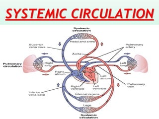
29. The Pulmonary Circuit
• Deoxygenated blood arrives at heart from
systemic circuit:
– passes through right atrium and ventricle
– enters pulmonary trunk
• At the lungs:
– CO2 is removed
– O2 is added
• Oxygenated blood:
– returns to the heart via pulmonary veins and
distributed to systemic circuit
30. Pulmonary Vessels
• Pulmonary arteries: Carry deoxygenated blood
• The Pulmonary trunk branches to left and right
pulmonary arteries.
• The Pulmonary arteries branch into pulmonary
arterioles and further into capillary networks
that surround alveoli.
• Pulmonary veins carry oxygenated blood to the
heart. Capillary networks around alveoli join to
form venules. Venules join to form 4 pulmonary
veins. Pulmonary veins empty into left atrium.
33. • The Subclavian Arteries Branches in thoracic
cavity:
– internal thoracic artery or mammary artery.
– vertebral artery
– thyrocervical trunk (cervical artery & thyroid
artery)
• Leaving the thoracic cavity:
– become axillary artery in arm and brachial
artery distally
– Further divides into radial artery and ulnar
artery and then Superficial Palmar arch is
formed
34.
35.
36. Arteries of the upper limb
• Axillary artery- it is a continuation of subclavian
artery from lateral margin of first rib.
• Brachial artery- it is continuation of the axillary
artery and supplies arm.
• Radial artery- it lies along the radial side of
forearm. It enters the palm and supplies thumb and
radial side of index finger.
• Ulnar artery- it begins at the level of the neck of
radius. It runs downwards and reaches the medial
side of forearm midway between the elbow and the
wrist.
37. Arteries of the upper limb
Arterial arcs of the palm-
1. Deep palmer arch- it is mainly formed by radial
artery, completed by the deep branch of the ulnar
artery.
2. Superficial palmer arch- it is mainly formed by the
ulnar artery and completed by the superficial
branch of radial artery.
38. ARTERIES OF THE TRUNK
Descending Aorta –
is divided by
diaphragm into:
thoracic aorta and
abdominal aorta
39. Arteries of the Thorax
Thoracic Aorta branches are anatomically grouped
into visceral and parietal arteries.
Visceral arteries Supply visceral organs:
– Two bronchial arteries: blood supply to
bronchial tree and surrounding lung tissues.
– Two pericardial arteries: blood supply to
pericardial sac.
– Two esophageal arteries: Blood supply to all
tissues of esophagus.
– Two mediastinal arteries: Blood supply to
connective tissues in mediastinum
40. Arteries of the Thorax
Parietal arteries:
- Posterior intercostal arteries: 3rd to 11th
intercoastal spaces
- Subcostal arteries: below 12th rib
- Superior phrenic arteries: supply the diaphragm
41. Veins of the Upper Limb
The veins of the upper limb is divided into deep
veins and superficial veins.
1. Deep veins
a. Subclavian veins: skin, muscles bones, shoulder
and neck.
b. Axillary veins: skin, muscles bones, shoulder and
axilla.
c. Brachial veins: muscles and bones of elbow and
brachial regions.
d. Ulnar vein: muscles and bones of medial aspects
of forearm.
e. Radial vein: muscles and bones of lateral aspects
of forearm.
42. Veins of the Upper Limb
2. Superficial veins
a. Cephalic veins: superficial aspects of upper
limb.
b. Basilic vein: skin and superficial aspects of
upper limb.
c. Median ante brachial veins: drain from palmar
venous plexus and palmar digital veins.
46. Veins of the Thorax
• Brachiocephalic veins: It is the union of
subclavian and internal jugular veins. This
ultimately forms superior vena cava. It drains
head, neck, upper limbs, mammary glands and
thorax.
• Azygos vein: It is present anterior to vertebral
column. It includes esophageal, mediastinal,
pericardial and bronchial veins.
• Hemiazygos vein: Present anterior to vertebral
column and joins with Azygos vein.
49. The Abdominal Aorta
• The abdominal aorta begins at the level of
the diaphragm, crossing it via the aortic
hiatus.
• It runs parallel to the inferior vena cava,
which is located just to the right of the
abdominal aorta.
• It becomes smaller in diameter as it gives
off branches.
50.
51. Branches of the Abdominal
Aorta
The abdominal aorta is branched into two
categories such as
1. Unpaired branches
2. Paired branches
52. Unpaired Branches
1. Celiac artery: The major branch called as
celiac trunk/artery arise from abdominal aorta
anteriorly which further divides into three as
follows;
❖ Left gastric artery: supplies stomach
❖ Splenic artery: largest branch of celiac trunk.
Supplies stomach, spleen and pancreas
❖ Common hepatic artery: Three sub branches:
proper hepatic artery (supplies liver and gall
bladder), right gastric artery (supplies
stomach), gastro-duodenal artery (stomach
and duodenum).
53. Cont..
2. Superior mesenteric arteries: arteries
pertaining to intestine. It mainly posses five
branches
❖ Inferior pancreatic-duodenal artery: Pancreas
& Duodenum
❖ Jejunal artery: Jejunum, ileum
❖ Ileocolic artery: ileum, cecum, appendix,
ascending colon
❖ Right colic artery: ascending colon, transverse
colon
❖ Middle colic artery: tarnsverse colon
58. Paired Branches
1.Inferior Phrenic arteries: Diaphragm
2.Suprarenal arteries: suprarenal areas such as
adrenal glands.
3.Renal arteries: supplies Kidneys
4.Gonadal arteries: testicular arteries(males:
testis, epididymis, ductus deferens, ureters),
ovarian arteries (female: ovaries, fallopian
tubes and ureters) and uterine arteries
(uterus).
5.Lumbar arteries: Lumbar vertebrae
59.
60. Arteries of Pelvis and lower
limbs
The abdominal aorta ends by dividing into left
and right common iliac arteries. It give rise
two branches such as internal iliac arteries and
external iliac arteries.
1. Internal iliac arteries: it supplies all the
pelvic viscera, buttocks, external genitals,
and muscles of thighs. In females it give rise
to uterine and vaginal arteries.
2. External iliac arteries: larger than internal
iliac arteries. It starts at sacroiliac joint and
passes to form femoral artery.
61. Arteries of lower limbs.
❖Femoral arteries: continuation of external
iliac arteries. Muscles of thigh, femur,
ligaments and tendons around the knee joint.
❖Popliteal arteries: continuation of femoral
arteries pass through popliteal fossa (space
behind knee joint). Knee, tibia, fibula, skin
and muscles.
❖Further it divides into anterior tibial and
posterior tibial arteries. This further forms
plantar arch and metatarsal arteries &
plantar digital arteries. The dorsal arteries at
ankle is also called as dorsalis pedis.
62.
63. Veins of the Abdomen and
Pelvis
The venous supply of the abdomen and pelvis
reaches the heart through inferior vena cava. It
has the following branches
1. Inferior Phrenic veins: inferior diaphragm and
adjacent peritoneum.
2. Hepatic veins: mainly three branches:
superior mesenteric vein, the inferior
mesenteric vein, the splenic vein and gastric
vein.
64. The Hepatic Portal Circulation
• The hepatic portal system is the venous
system that returns blood from the digestive tract
and spleen to the liver (where raw nutrients in
blood are processed before the blood returns to
the heart).
• It consists of the hepatic portal vein and other
veins that drain into the hepatic portal vein, i.e.,
the superior mesenteric vein, the inferior
mesenteric vein, the splenic vein and gastric
vein.
65.
66. Cont..
• Since blood received from the hepatic
portal vein may be contaminated with
pathogens such as bacteria, the liver is rich
in specialized immune cells called Kupffer
cells that detect and destroy foreign
organisms.
• Following processing, blood collects into
the hepatic vein and finally through
inferior vena cava to the right atria of the
heart.
67. Veins of the Abdomen and
Pelvis
3. Lumbar veins: lumbar vertebrae, spinal cord
4. Suprarenal veins: adrenal glands
5. Renal veins: kidneys.
6. Gonadal veins: testes, ovaries, ureters.
7. Common iliac veins: internal (pelvis,
gluteal, external genitalia) and external
(lower limbs, lower abdominal wall)
68. The veins of lower limbs
a. Deep veins: They are branches of external
iliac veins. The main branches are;
1. Femoral veins: skin, bones and muscles of the
thigh.
2. Popliteal veins: skin, muscle, and bones of
the knee joint.
3. Posterior tibial veins: posterior and lateral
aspects of leg.
4. Anterior tibial veins: anterior aspects of leg
such as foot, ankle, tibio-fibular joint.
69. The veins of lower limbs
b. Superficial veins:
1. Great Saphenous veins: longest vein in the
body. It starts from metatarsal veins to groin
region. It drains blood from muscles of lower
limbs, groin and lower abdomen.
2. Small Saphenous vein: It begins from the
venous arch of the foot and ends in popliteal
veins. Posterior aspects of leg is also drained
with this vein.