
More Related Content
What's hot
What's hot (20)
Similar to Structures of the Nervous System
Similar to Structures of the Nervous System (20)
More from melodiekernahan
More from melodiekernahan (20)
Recently uploaded
Recently uploaded (20)
Structures of the Nervous System
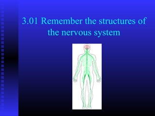
- 1. 3.01 Remember the structures of the nervous system
- 2. Essential Question What are the structures of the nervous system? 3.01 Remember the structures of the 2 nervous system
- 3. Introduction: Structures of the nervous system 3.01 Remember the structures of the 3 nervous system
- 4. Cellular structures of the nervous system Neuron- a nerve cell Extensions of the neuron Dendrites Axon 3.01 Remember the structures of the 4 nervous system
- 5. Cellular structures of the nervous system Neuron Myelin sheath Synapse 3.01 Remember the structures of the 5 nervous system
- 6. Structures of the nervous system Central nervous system Brain Spinal cord 3.01 Remember the structures of the 6 nervous system
- 7. Structures of the nervous system Peripheral nervous system Nerves Cranial Spinal Sensory receptors 3.01 Remember the structures of the 7 nervous system
- 8. Structures of the central nervous system: BRAIN 3.01 Remember the structures of the 8 nervous system
- 9. Structures of the central nervous system: BRAIN Cerebrum Frontal lobe Occipital lobe Parietal lobe Temporal lobe 3.01 Remember the structures of the 9 nervous system
- 10. Structures of the central nervous system: BRAIN Diencephalon Structures Thalamus Hypothalamus 3.01 Remember the structures of the 10 nervous system
- 11. Structures of the central nervous system: BRAIN Limbic system Olfactory bulb Amygdala Hippocampus Parahippocampus Fornix Mammillary body Cinginulated gyrus Septum pellucidum 3.01 Remember the structures of the 11 nervous system
- 12. Structures of the central nervous system: BRAIN Cerebellum 3.01 Remember the structures of the 12 nervous system
- 13. Structures of the central nervous system : BRAIN Brain stem Pons Midbrain Medulla oblongata 3.01 Remember the structures of the 13 nervous system
- 14. Structures of the central nervous system: BRAIN Meninges Dura mater Arachnoid mater Pia mater 3.01 Remember the structures of the 14 nervous system
- 15. Structures of the central nervous system: BRAIN Cerebral Ventricles Left lateral ventricle Right lateral ventricle Third ventricle Fourth ventricle 3.01 Remember the structures of the 15 nervous system
- 16. Structures of the central nervous system: Spinal Cord 3.01 Remember the structures of the 16 nervous system
- 17. Structures of the central nervous system: Spinal Cord Peripheral nervous system 12 cranial nerves 31 pairs of spinal nerves 3.01 Remember the structures of the 17 nervous system
- 18. Structures of the Central Nervous System I. Olfactory II. Optic III. Oculomotor IV. Trochlear V. Trigeminal VI. Abducens VII. Facial VIII. Vestibulocochlear IX. Glossopharyngeal X. Vagus XI. Accessory XII. Hypoglossal 3.01 Remember the structures of the 18 nervous system
- 19. Structures of the Central Nervous System Peripheral nervous system 31 pairs of spinal nerves 3.01 Remember the structures of the 19 nervous system
- 20. Structures of the Central Nervous System Motor system Autonomic nervous system Sympathetic nervous system Parasympathetic nervous system Somatic nervous system Sensory neurons Motor neurons 3.01 Remember the structures of the 20 nervous system
- 21. Structures of the Central Nervous System Autonomic Nervous System Sympathetic Nervous System Parasympathetic Nervous System 3.01 Remember the structures of the 21 nervous system
- 22. Structures of the Central Nervous System Somatic Nervous System 3.01 Remember the structures of the 22 nervous system
- 23. Structures of the central nervous system our c k y g e… he led C w k no 3.01 Remember the structures of the 23 nervous system
- 24. Structures of the nervous system ! er thi s Rememb 3.01 Remember the structures of the 24 nervous system
- 25. Essential question What are the structures of the nervous system? 3.01 Remember the structures of the 25 nervous system
Editor's Notes
- MAKE NEURON ACTIVITY Neuron has nuclues, cytoplasm, and cell membrane. Extensions (processes) of the cytoplasm from the cell body are dendrites and one axon. The processes are paths along which a nerve impulse can travel. Axons carry messages away from the cell body. Dendrites carry messages to the cell body.
- Myelin sheath- a special covering of the axon. The myelin sheath speeds up the nerve impulse as it travels along the axon. The myelin sheath produces a fatty substance called myelin which protects the axon Synapse= the space between neurons. Synapse is where the messages go from one cell to the next cell. Message will go from the axon of one cell to the dendrite of another cell. The cells never actually touch each other. This space is called the synaptic cleft. There are neurotransmitters (chemicals) which will actually carry the message from one cell to the next.
- Consists of two structures. Brain and Spinal cord
- Consists of the nerves of the body. 12 pairs of cranial nerves extending out from the brain 31 pairs of spinal nerves extending out from the spinal cord
- The adult brain weighs approximately 3 pounds and consists of 100 billion neurons. The brain is protected by the cranial cavity. It is also protected by the meninges (membranous covering) and the cerebrospinal fluid. The brain is made up of white and grey matter. The outer cortex (cerebral cortex) is grey matter. The deeper parts of the brain are white matter. The brain is divided into 4 major parts- cerebrum. Diencephalon, cerebellum, and the brain stem.
- The cerebrum is the largest part of the brain. It occupies the whole upper part of the skull and weighs about 2 pounds. Covering the upper and lower surfaces of the cerebrum is a layer of grey matter which is called the cerebral cortex. The cerebrum is divided into two hemispheres/sides (right and left) by a deep groove called the longitudinal fissure. The surface of the cerebrum is completely covered with grooves and ridges. The deeper grooves are called fissures and the shallower ones are called sulci. The elevated ridges between the sulci are called gyri (convulations). Each side is divided into a frontal parietal occipital and temporal lobe. The lobe is named for the bone that covers them.
- Located between the cerebrum and the midbrain. It contains two major structures- thalamus and hypothalamus. Thalamus is a spherical mass of grey matter. It is found deep inside each of the hemispheres. Hypothalamus lies beneath the thalamus.
- Located in the center of the brain beneath the four lobes and it encircles the top of the brain stem.
- Located below the cerebrum. It also consists of two hemispheres (sides)- right cerebellar hemisphere and the left cerebrellar hemisphere. The two hemispheres are connected by the vermis. The cerebellum consists of gray matter on the outside and white matter on the inside. The white matter is marked with a treelike pattern called arbor vitae (tree of life).
- Brain stem is made up of 3 parts: The pons (bridge) is located in front of the cerebullem between the mid brain and the medulla oblongata. The midbrain extends from the mammilary bodies to the pons The medulla oblongata is a bulb shaped structure found between the pons and the spinal cord. It lies inside the cranium and above the foramen magnum of the occipital bone.
- The 3 protective covers of the brain Dura mater Outermost layer which lines the inside of the skull. Tough dense mebrane of fibrous connective tissue that has a lot of blood vessels Arachnoid mater Middle layer which resembles a cobweb (spiderweb) Pia mater Innermost layer covers the brain surface. Consists of blood vessels held together by connective tissue Between the arachnoid and pia mater is where cerebrospinal fluid is located
- lined cavities deep within the brain and filled with cerebrospinal fluid. Left lateral ventricle & Right lateral ventricle located in the cerebral hemispheres Third ventricle- behind and below the laterals Fourth ventricle- located below the thrid and in front of the cerebellum and behind the pons and medulla oblongata Each of the ventricles have a rich network of blood vessels from the pia mater called Choroid plexus. This helps to produce cerebrospinal fluid.
- Continues down from the brain. It begins at the foramen magnum of the occipital bone and continues to the second lumbar vertebra (L2). It is soft white matter and lies within the vertebra of the spinal column. It is made up of a series of 31 segments. The spinal cord is protected by the vertebrae (spine) , three layers of meninges, and cerebrospinal fluid.
- Cranial nerves start in the brain. Most cranial nerves are mixed nerves- they carry nerve impulses to the brain (sensory) and nerve impulses to the organ/gland/muscle (motor) fibers. 1. Smell 2.Vision 3.Eyelid and eyeball movement 4.Turns eye downward and laterally 5.Face and mouth touch and chewing 6.Turns eyes laterally 7.Controls most facial expressions, secretion of tears and saliva, also responsible for taste 8.hearing, equilibrium and sensation 9. taste, senses carotid blood pressure 10. Senses aortic blood pressure, slows heart rate, stimulates digestive organs and also plays a role in taste 11. Controls Trapezius and Sternocleidomastoid muscles and controls swallowing movement 12. Movement of the tongue muscles. O n O ld O lympus' T owering T op, A F inn A nd G erman V iewed S ome H ops http://fc.units.it/ppb/neurobiol/Neuroscienze%20per%20tutti/cranial.html
- Start in the spinal cord and connect to each segment of the spinal cord. They exit through the openings in the vertebra. Each pair if spinal nerves is connected to that segment of the cord by two pairs of attachments called roots. The posterior/dorsal root is the sensory root and contains only sensory nerves. They conduct impulses from the periphery (ie: skin) to the spinal cord. The other point of attachment is the anterior/ventral root and is the motor root. It conducts impulses from the spinal cord to the periphery (ie: muscles). Cervical =8 Thoracic= 12 Lumbar= 5 Sacral=5 Coccygeal=1
- Sympathetic Nervous System is basically two cord beginning at the base of the brain, along both sides of the spinal column. These nerves connect to all vital internal organs (heart liver pancreas stomach intestines blood vessels iris of eyes sweat glands and the bladder) Referred to as the FLIGHT or FIGHT system. Parasympathetic Nervous System has two important active nerves The vagus nerve starts at the medulla oblongata and proceeds down the neck and sends branches to the chest and neck. The pelvic nerve emerges from the spinal cord around the hip region and connects with the organs of the lower body
- Conducts impulses from the brain and spinal cord to skeletal muscles These nerves are located along the spinal cord. Connect skeleton, muscles and joints to the brain.