SlideShare a Scribd company logo
1 of 54
Mega sporogenesis and gametogenesis
By SACHIN
Female gametophyte development
begins early in ovule development with
the formation of a diploid megaspore
mother cell that undergoes meiosis.
One resulting haploid megaspore then
develops into the female gametophyte.
Genetic and epigenetic processes
mediate specification of megaspore
mother cell identity and limit
megaspore mother cell formation to a
single cell per ovule.
Auxin gradients influence female
gametophyte polarity and a battery of
transcription factors mediate female
gametophyte cell specification and
Introduction
• During post-fertilization, the female gametophyte
influences seed development through maternal-
effect genes and by regulating parental
contributions.
• The mature female gametophyte secretes peptides
that guide the pollen tube to the embryo sac and
contains protein complexes that prevent seed
development before fertilization.
• Female gametophytes can form by an asexual
process called gametophytic apomixis, which
involves formation of a diploid female gametophyte
and fertilization-independent development of the
egg into the embryo.
Megasporangium
• The megasporangium is having the ovules
together with its protective coats,
integument , it is attached to the placenta,
on the inner wall of ovary by a stalk called
funiculus.
• An ovule ready for fertilization consists of
nucellar tissue enveloped almost completely
by one or two integuments, leaving a small
opening at the apical end, the opening is
known as micropyle.
• The basal region of the ovule, where
funiculus is attached is called chalaza.
Megasporogenesis
• Thus embryo sacs
maintain physical contact
with the parent
sporophyte throughout
their development, this
association of the female
gametophyte and the
sporophyte provides an
opportunity to examine
interactions between
cells, tissues, and
genomes.
The nucellus supports the female
lineage throughout
development.
o The egg cell in the female.
o Female central cell are shown
in green, as “companion cells,”
in view of their potential roles in
reinforcing gene silencing in
their neighbouring germ cells.
Female Reproductive Cell
Lineages in Arabidopsis thaliana
• (B) Phases of interaction between the
female reproductive lineage and its
surrounding cells.
– A signal peptide (TPD1) from the
megaspore mother cell seems to
stimulate the proliferation (blue
double-headed arrows) of dicot
nucellar cells in a pathway involving
EMS1; in monocots MAC1 (maize)
and TDL1a (rice) fulfill a similar role,
also restricting the proliferation of
the female cell line (red horizontal
“T”).
– In monocots, EAL1 (maize) is secreted
from the chalazal face of the egg (EC)
and determines central (CC) and
antipodal (A) cell fates.
– LIS and CLO expressed in the egg of
dicots are part of a signaling pathway
responsible for fate specification of the
synergid (SY), central, and antipodal
cells.
A
• The location of the
megasporocyte, directly below
the apex of the nucellus,
suggests that position may be an
important factor in
megasporocyte specification.
• SEEDSTICK (STK) and
SHATTERPROOF (SHP) 1 & 2 in
carpel are important
transductional factors.
Transcriptional factors
• Plant hormones, auxin in specifying
embryo sac cell fates ( Sundaresan and
Alandete-Saez, 2010; ).
• Auxin concentration is already established
in the nucellus at the distal end of the
very young ovule. Pagnussat et al. (2009)
have shown that the haploid nuclei
within the nucellus has different
developmental fates depending on the
concentration of auxin surrounding them.
Crosstalk between Cells of the
Gametophyte
• A model has been proposed in which
auxin acts as a morphogen gradient by
which nuclei exposed to the highest
levels of auxin cellularize and
differentiate into synergids and eggs,
whereas those experiencing low auxin
concentratation follow an antipodal
fate.
• Median auxin levels are proposed to be
required for correct central cell
development and the positioning of the
polar nuclei (Pagnussat et al., 2009).
The genes that have been characterised as playing key
roles have been positioned adjacent to the
developmental stage in which they are involved.
Genes that are involved in female gamete development
are shown in yellow, and those which impact on both
male and female gamete formation are in purple.
AP, antipodal cells
PN, polar nuclei
EC, egg cell
SY, synergids
VC, vegetative cell
GC, generative cells.
Schematic representation of the events leading to gamete formation in Arabidopsis.
Shortly after ovule initiation, a single
subdermal nucellar cell enlarges and
displays a prominent nucleus.
This cell represents the archesporium,
or spore-bearing tissue, and typically
occupies a position directly below the
apex of the nucellus.
• Although ovule
morphologies show
considerable diversity.
• Ovules are specialized
structures, derived from
the placenta of the
ovary wall, that
produce the
megasporocyte and are
the site of embryo sac
formation, fertilization,
and embryogenesis.
• The ovule consists of
Development of ovule
Ovule
• Events within individual ovules of the female carpel follow a similar
pattern, with the megaspore mother cell.
• The functional meiotic product requiring the presence of a normal
nucellar layer for correct development.
• Once the cells of the embryo sac are formed, their correct
positioning, division, and specification results from a complex
interplay involving hormone gradients.
• This is followed by the interactions between the egg and other cells
of the embryo sac.
• There is evidence that gametophytic central cell acts as a companion
cell to the egg. The polar nuclei may partially fuse with each other
before they are fertilized by a single sperm nucleus, generating the
triploid primary endosperm nucleus (Cass et al., 1985).
• The mature endosperm will provide nutrients for the developing embryo
or seedling.
Synergids
The synergids, which are
located on either side of the
egg cell, play an important
role in fertilization.
The pollen tube discharges its
contents into one of the
synergids prior to
incorporation of the
orientation of the embryo sac
with respect to the chalazal-
Antipodal Cells
• Three antipodal cells are located
opposite to the egg at the
chalazal end of the embryo sac.
• No specific function during
reproduction has been attributed
to the antipodals, but they are
thought to be involved in the
import of nutrients to the embryo
sac.
• Cytological characteristics of cells
within the embryo sac as well as
cytochemical localization of
proteins, starches, lipids, and
nucleic acids have been used to
assess the physiological state of
the embryo sac and suggest
relative rates of metabolic
• For example, the presence of
numerous ribosomes and
mitochondria in the synergids,
central cells, and antipodals suggests
a high metabolic activity.
• By contrast, the egg cell has fewer
ribosomes, plastids, and other
organelles and appears to be
relatively quiescent (Mansfield et al.,
1990).
• At the present time, there is little
data available about the regulation
of ovule and embryo sac
development and the temporal and
spatial patterns of gene expression
in the embryo sac.
Peter K. Endress Ann Bot 2011;107:1465-1489
© The Author 2011. Published by Oxford University Press on
behalf of the Annals of Botany Company. All rights reserved.
For Permissions, please email:
journals.permissions@oup.com
(A–F) Different nucellus shapes.
Meiocytes are shaded grey.
(A) Crassinucellar.
(B) Weakly crassinucellar.
(C) Pseudocrassinucellar.
(D) Incompletely tenuinucellar.
(E) Tenuinucellar.
(F) Reduced tenuinucellar.
(G–K) Integument differentiation. In
bitegmic ovules, the inner
integument is shaded red, the outer
blue.
(G) The outer integument is thicker
than the inner.
(H) The inner integument is thicker
than the outer.
(I) (I). Both integuments are equally
thick.
(II) (J) Unitegmic.
(III) (K) Ategmic.
Diversity of nucellus thickness
and integument number and
thickness
• The two integuments are considered to have
distinct evolutionary origins, it initiated at the
base of the nucellus during megasporogenesis.
• The inner integument is most often dermal (Ll) in
origin, whereas the outer integument is usually
derived from both dermal and subdermal layers.
• Periclinal divisions in the integuments generate an
increase in the number of cell layers, whereas
anticlinal divisions and cell elongation are
responsible for growth parallel to the nucellus.
Ramin Yadegari, and Gary N. Drews Plant Cell
2004;16:S133-S141
©2004 by American Society of Plant Biologists
Female Gametophyte Development in Arabidopsis
The steps are described by Christensen et al. (1997).
Category designations show the developmental stage affected in the
female gametophyte mutants.
Category 1= megaspores fail to undergo cell death;
Category 2= megaspores do not progress beyond stage FG1;
Category 3= pleiotropic defects during the nuclear division phase;
Category 4= failure to cellularize or abnormal cell shape;
Category 5= polar nuclei fail to fuse;
Category 6= antipodal cells fail to undergo cell death;
Category 7 (not shown), morphologically wild-type female
gametophytes at the terminal stage.
Ramin Yadegari, and Gary N. Drews Plant Cell
2004;16:S133-S141
©2004 by American Society of Plant Biologists
o
The mature female gametophyte in
Arabidopsis is 105 μm long and 25∼ ∼
μm wide.
(The gray areas represent cytoplasm,
the white areas represent vacuoles,
and the black areas represent nuclei).
ac, antipodal cells;
cc, central cell;
ch, chalazal region of the ovule;
ec, egg cell;
f, funiculus;
mp, micropyle;
sc, synergid cell;
sn, secondary nucleus.
The Arabidopsis Female Gametophyte
The view in (B) is perpendicular to that in (A)
Embryo Sac Development
Occurs along Chalazal-Micropylar Ovule Axis
The processes involved in megaspore selection may begin before
meiosis, as polarity of the developing megagametophyte reflects the
ovular polarity and suggests that the ovule plays a role in the selection
of a functional megaspore and in the organization of the embryo sac,
and expressed in the asymmetric distribution of cellular organelles and
plasmodesmata.
After meiosis, plastids are preferentially distributed at the micropylar
end of the functional megaspore, and plasmodesmata are usually
observed only between the functional megaspore and the nucellus,
thus the functional megaspore inherits a richer cytoplasm and nutrients
from the maternal tissues.
• The arrangement of microtubules indicated a role for the cytoskeleton
in distribution and positioning of cytoplasmic contents during
megasporogenesis and megagametogenesis, it is possible that
positional information, perhaps in the form of gradients, could be
inherited from the megasporocyte.
• The nature of specific nutritional or hormonal interactions between
the ovule and embryo sac are not known, hence connectivity between
the megagametophyte and the ovule presumed to establish a polar
nutrient flow.
• On the other side of the embryo sac, wall projections extend from the
antipodal cells into the chalazal nucellus and may represent another
site of metabolite flow from the nucellus to the embryo sac.
• In some species, cell wall projections are also found
between the central cell and the inner integuments;
however, in other species, a cutinized wall can be
found in this region.
• The cellular anatomy and the accumulation and
mobilization of starch reserves of the nucellus and
integuments also suggest a directional flow of
metabolites into the embryo sac.
• The micropylar portion of the egg cell is occupied by
a vacuole, and the nucleus, cytoplasm, and most of
the organelles are located at the chalazal end,
following fertilization, this polarity is also observed
in the zygote.
The Arabidopsis female gametophyte.
(A) Ovule. (B) Female gametophyte. (C) Synergid cells.
View in (B) and (C) is perpendicular to that in (A).
The dashed line at the chalazal ends of the synergid cells
in (C) represents a discontinuous or absent cell wall.
o Synergids are frequently observed to have elaborate
wall projections, the filiform apparatus that extends into
the nucellus, which may provide a mechanism for
nutrient flow from the ovule to the embryo sac.
ac, antipodal cells;
cc, central cell;
ch, chalazal region of the
ovule;
ec, egg cell;
f, funiculus;
fa, filiform apparatus;
mp, micropyle;
sc, synergid cell;
sn, synergid nucleus,
sv, synergid vacuole.
• At the micropylar pole, the synergid cell
wall is thickened and extensively
invaginated, forming a structure called
filiform apparatus.
• The filiform apparatus greatly increases
the surface area of the plasma membrane
in this region and contains a high
concentration of secretory organelles,
suggesting that it may facilitate transport
of substances into and out of the synergid
cells.
• Based on cytological staining properties in
species other than Arabidopsis, the
filiform apparatus appears to be
composed of a number of substances
including cellulose, hemicellulose, pectin,
callose, and protein.
Synergid cell wall is specialized structure
• Thus the filiform apparatus has at least
two functions associated with the
fertilization process.
• First, the synergid cells secrete pollen
tube attractants via the filiform
apparatus.
• In addition, the pollen tube enters the
synergid cell by growing through the
filiform apparatus.
• By contrast, the antipodal cells in
Arabidopsis have no dramatic
specializations and no known function.
• In cereals, the antipodal cells proliferate
into as many as 100 cells.
• Considerable diversity in the pattern
of embryo sac development was
found among plant species. Haig
(1990) proposed a model for embryo
sac development whereby different
patterns could be generated by
variations in meiosis, cytokinesis,
and the timing and number of
mitotic divisions
Some of the modes of embryo sac development that have
been observed
In Arabidopsis and most monocot
crops, three of the four linear
products of meiosis degenerate,
and the surviving haploid
“megaspore” divides three times to
form the cells that populate the
embryo sac—
o The single egg
o Two synergid cells that receive the
pollen tube
o Two polar nuclei that later fuse to
form the large diploid central cell,
o Species-specific number of
antipodal cells, which are held to be
secretory.
Development of the Polygonum-
Type Embryo Sac
• The Polygonum-
type pattern is the
most commonly
observed form of
embryo sac
development.
• Approximately
70% of the species
examined,
including
Arabidopsis and
• During the first meiotic
division, the spindle is
oriented parallel to the
micropylar-chalazal axis of the
nucellus.
• Wall formation occurs
perpendicular to this axis,
creating a dyad of
megaspores.
• Frequently, the dyad cell
closest to the micropyle
• The three non-functional megaspores
degenerate and are eventually crushed by
the expanding functional megaspore.
• Tetrahedral arrangements of megaspores
have also been observed in Arabidopsis
(Webb and Gunning, 1990), and T-shaped
tetrads have been seen in maize (Russell,
1978).
• The linear array is, however, most common.
Callose, a P-1,3-glucan, is thought to function in
the selection of a functional megaspore.
During megasporogenesis, callose accumulates
first in the cell walls of the megasporocyte and
then in the megaspore walls.
After meiosis, callose walls become thinner or
absent in the functional megaspore.
The presence of callose in the walls of the
nonfunctional megaspores probably ensures that
only the functional megaspore receives nutrients
from the nucellus.
The pattern of callose deposition is variable,
reflecting the pattern of megasporogenesis, for
example, in Oenothera, a monosporic species,
callose is thinner at the micropylar end of the ovule,
where the functional megaspore is located.
In tetrasporic species, meiosis occurs without
cytokinesis, and callose does not accumulate in the
walls of the single tetranucleate megaspore.
In most diplosporous species, callose level and distribution in
the megaspore mother cell differs from that in sexual
relatives.
For example, callose is absent in the megaspore mother cells
of diplosporous Tripsacum (Bicknell and Koltunow, 2004).
Both mitosis and cell specification seem to be disrupted in
some apomicts, for example, only two rounds of mitosis
occur in the cells of aposporous Pennisetum, leading to a
four-nucleate embryo sac, commonly containing an egg cell,
one polar nucleus and two synergid cells.
In aposporous Hieracium species, antipodals may not form
and multiple embryos may develop in an embryo sac (
Koltunow et al., 2000; Koltunow et al., 2011).
●
Click to edit the outline text
format
– Second Outline Level
●
Third Outline Level
– Fourth Outline
Level
●
Fifth
Outline
Level
●
Sixth
Outline
Level
Seventh Outline LevelClick to edit
Master text styles
I. Monosporic: Polygonum, Oenothera
II. Bisporic: Allium, Endymion
Tempera
ture
during
embryo
sac
develop
ment
Drusa
type
Adoxa
type
Other
type
15-19 C◦ 81 18 1
26-27 C◦ 89 6 5
III. Tetrasporic:
Adoxa
Penaea
Plumbago
Peperomia
Drusa
Fritillaria
Plumbagella
Effect of temperature on occurrence of two types of embryo sac in Ulmus glabra
Types of embryo sacs
Patterns of Female Gametophyte Development Exhibited by Angiosperms
Genera exhibiting these patterns are indicated in parentheses.
Patterns of Female Gametophyte Development Exhibited by Angiosperms.Genera exhibiting these
patterns are indicated in parentheses. More comprehensive descriptions of the variation among
angiosperms can be found in several reviews (Maheshwari, 1950; Willemse and van Went, 1984;
Haig, 1990; Huang and Russell, 1992; Russell, 2001). In this figure, the chalazal end of the female
gametophyte is up and the micropylar end is down. FG, female gametophyte.
• In -65% of the species examined, most of the nucelIus
degenerates before the embryo sac reaches maturity.
• The embryo sac is then in direct contact with the inner
integument.
• In these situations, the innermost cell layer of the inner
integument may differentiate into a unique cell layer termed the
endothelium.
• Radial cell expansion, endopolyploidy, and prominent nuclei are
observed in the endothelial cells and also the anther tapetum,
which is thought to be involved in secretion and nutrition of the
pollen speculated that the cytological features shared between
the endothelium and tapetum could indicate a similar function
for both tissues.
• In species in which the nucellus does not degenerate, the inner
integument does not differentiate an endothelium, and the
embryo sac may receive nutrients from the nucellus directly.
• Stalklike structure extending from the lowermost
part of the chalaza to the placenta, usually, a single
vascular strand runs through the funiculus from the
placenta terminating at the base of the embryo sac.
• The mature ovule displays a polarity with respect to
the axis determined by the location of the chalaza
and micropyle. Esau (1977) defined the chalaza as the
region extending from the base of the integuments to
the point of attachment of the funiculus.
• The micropyle is located at the point where the
integuments terminate and is the site where pollen
tubes enter the ovule.
As the embryo sac develops, the integuments continue
to enlarge, typically overgrowing the nucellus.
The amount of ovule curvature varies with the extent
of differential growth of the integuments and funiculus;
the degree of curvature forms a basis for classification
of ovule morphology.
Thus, the mature anatropous ovule shows extensive
curvature such that the long axis of the nucellus is
parallel to the axis of the funiculus.
Development of ovule
an anatropous bitegmic angiosperm ovule
Peter K. Endress Ann Bot 2011;107:1465-1489
© The Author 2011. Published by Oxford University Press on behalf of the Annals of Botany
Company. All rights reserved. For Permissions, please email:
journals.permissions@oup.com
Median longitudinal sections :
(A–E) Straight thin line drawn through
the middle of the nucellus. Arrowhead
indicating successive ovule curvature
from 0 to 180 °. (E, E′)
(A).Ovule before integument initiation.
(B).Ovule at initiation of inner
integument.
(C) Slightly older ovule.
(D) Ovule with both integuments
formed.
(E) Mature ovule. Raphal side blue,
antiraphal side red.
(E′) Mature ovule. Concave side blue,
convex side red.
Different orientations of curved ovules with respect to
carpel curvature (denoted by a red line).
Peter K. Endress Ann Bot 2011;107:1465-1489
© The Author 2011. Published by Oxford University Press on behalf of the Annals of Botany
Company. All rights reserved. For Permissions, please email: journals.permissions@oup.com
Different orientations of curved
ovules with respect to carpel
curvature (denoted by a red
line).
(A) Syntropous: Curvature of the
ovule in the same direction as
the curvature of the carpel.
(B) Antitropous: Curvature of the
ovule in the opposite direction
to that of the carpel.
(A–C) Ovary or locules not filled with secretion.
(A) Single ovule with basal placenta (LS gynoecium)
(e.g. Piperaceae, Juglandaceae, Urticaceae).
(A) Ovules on parietal placentae with the micropyle
directed toward another placenta (TS ovary)
(e.g. Casearia, Salicaceae; Mayaca,
Mayacaceae).
(A) Ovules with long funiculi curved to their own
placenta (LS gynoecium) (e.g. Helianthemum,
Cistaceae).
(A) (D–F) Ovary or locules filled with secretion
(secretion shaded blue).
(A) Basal diffuse placenta (LS gynoecium)
(e.g. Pistia, Araceae).
(A) Laminar-diffuse placenta (TS carpel/ovary)
(e.g. Barclaya, Nymphaeaceae; Hydrocharis,
Hydrocharitaceae; Akebia.
Orthotropous ovules and conditions of
ovary locule architectures under which they
occur (schematic, only one integument is
drawn in each ovule for simplicity;
augmented and modified from (Endress
, 1994).
Genetic and molecular analysis of ovule and embryo sac
development
• Mutations are used to study the effect of embryo sac
development which are expressed in the parent
sporophyte or in the gametophyte.
• In the case of a recessive mutation, 25% of the progeny
from selfed heterozygous plants will display the sterile
phenotype.
• Defects in fertility that are specific to the female are
distinguished from male sterile by reciprocal crosses
with wild-type plants.
• Mutations that abolish or reduce female fertility can be
transmitted and recovered through the pollen.
• Several female-sterile mutations have been identified
among populations of Arabidopsis mutagenized with
ethylmethane sulfonate.
• Representative female-sterile mutants of
Arabidopsis include short integuments-1
(sinl).
• Each of these mutants has a defect in a
discrete aspect of ovule and embryo sac
development, such as initiation of the
integuments, development of the nucellus,
megasporogenesis, and
Illustration of the utility of a genetic approach
for understanding how the ovule develops and
interacts with the developing embryo sac
Gametophytic Mutants
• The indeterminate gametophyte (ig) mutation of maize
has pleiotropic effects on embryo sac development but
primarily affects the number of mitotic divisions.
• It is associated with the occurrence of multiple
functional egg cells and embryos within an embryo sac
and with the formation of defective seed.
• Another mutation with gametophytic expression, Gf,
was described in Arabidopsis, showed reduced
transmission through the female gametophyte of
genes linked to Gf.
• In maize, heterozygous plants that contain deletions
frequently display reduction in pollen viability
(Patterson, 1978).
Practical
Pollen tube subgroup 18 (S18) MYB transcription factors respond to growth through the pistilA
schematic diagram of an A. thaliana ovary with 12 ovules (instead of the actual 50–60). Pollen grains
(red) are germinating pollen tubes on the stigma. Pollen tubes target the female gametophyte (grey).
Mature pollen grains [represented with two sperm (black) and a nucleus (grey)] express MYB101, and
lower levels of both MYB97 and MYB120. Transcription of MYB targets is low in, or absent from,
pollen grains. Growth through the stigma and style activate transcription of MYB97 and MYB120; the
three MYBs activate target genes in the pollen tube. The pollen tube factors that sense the pistil
environment and activate MYB expression are not yet known.
Subgroup 18 (S18) MYB-regulated gene classes and their putative roles during pollen tube receptions(A)
Schematic diagram of the pollen tube and female gametophyte at the beginning of pollen tube reception.
Pollen tube contact with one of the synergid cells is indicated (broken line). Proteins with roles in pollen tube
reception and MYB-regulated gene products are highlighted. Abbreviations: sn, synergid nucleus; vn,
vegetative nucleus. (B andC) Schematic diagram of pollen tube reception between a pollen tube and the
synergid that will degenerate. Cytosolic Ca2+ concentrations are symbolized with a colour spectrum (red, high
Ca2+; green, low Ca2+). (B) FER-dependent NTA relocalization occurs after pollen tube arrival. (C) Synergid
degeneration is accompanied by loss of synergid nuclear integrity. (D) Pollen tube burst, sperm release and
double fertilization.
Pollen tubes differentiate in response to growth through the pistil
Pollen tube physiology changes when it grows through floral tissue. For example, pollen
tubes must grow through pistil explants to be able to respond to attractants in vitro [8–10
]. These experiments use a SIV (semi-in vitro) pollen-tube-guidance assay in which pollen
tubes grow through the stigma and a portion of style before growing on to the surface of
pollen growth media towards ovules [10]. In Torenia, where LURE pollen tube attractants
were first described [11], the ability to respond to attractants in vitro increases with
prolonged growth through pistil tissue [12]. However, when LURE-binding sites were
assessed by incubating pollen tubes with LUREs followed by detection with anti-LURE
antibodies, it was observed that, whereas growth in the pistil was required to generate
LURE-binding sites, prolonged exposure to the pistil did not increase the detection of
binding sites further [12]. These data suggest that the pollen tube senses the pistil
environment, that LURE receptor (not yet identified) expression reaches a maximum as a
consequence, but that prolonged exposure to the pistil environment may activate the
receptor.
Interestingly, a pair of A. thaliana pollen-tube-expressed membrane-associated receptor-
like cytoplasmic kinases (LIP1 and LIP2) were recently shown to be induced by growth in
the pistil and required for the full response to purified LURE proteins in the SIV assay and
for pollen tube guidance in the pistil [13]. It is not yet clear whether LIP1 and LIP2 are
involved directly in LURE perception; however, these findings underscore the idea that
pollen tubes differentiate during growth in the pistil, resulting in expression of proteins
required to respond to pollen tube guidance cues.
Pollen tube differentiation for sperm release is controlled by three MYBs
Over 1000 genes were detected in the transcriptome of pollen tubes grown in the SIV
assay that were not detected in pollen tubes grown in vitro [14]. To identify regulators
that control pollen tube gene expression in response to the pistil, 26 SIV-induced
transcription factors were identified [14]. This list included three closely related R2R3-
MYB type transcription factors [subgroup 18: MYB97,MYB101 and MYB120] [14]. Single
and double myb mutants did not display seed set defects; however, triple mutants had
a ~70% reduction in seed production [15]. myb triple mutant pollen grains and tubes
develop normally, grow through the pistil and target ovules nearly as efficiently as wild-
type. These data suggest that MYB97, MYB101 and MYB120 do not regulate expression
of the LURE receptors. However, upon interaction with the female
gametophyte, myb triple mutants fail to arrest their growth and ~72% of ovules contain
coils of pollen tubes within the female gametophyte [15]. Synergid cells targeted by
these mutant pollen tubes fail to degenerate normally, suggesting a loss of
communication between pollen tube and synergid cells during pollen tube reception [
15]. Furthermore, myb triple mutant pollen tubes fail to burst and release sperm. These
data suggest that MYB97, MYB101 and MYB120 are critical for the pollen tube to
exchange signals with the female gametophyte required for successful fertilization.
Pollen tube reception signalling by the female gametophyte
Pollen tube reception requires a number of synergid-expressed genes
including FER (FERONIA) [16], NTA (NORTIA) [17] and LRE (LORELEI) [18] (Figure 2A).
Loss-of-function mutations in of each of these genes cause the same phenotype: wild-
type pollen tubes coil in mutant ovules and fail to release sperm. FER is a
transmembrane receptor-like kinase of the CrRLK1L family predicted to have pollen
tube or female-gametophyte ligand(s) important for pollen tube reception. FER has a
malectin-like extracellular domain, so its ligand could be a carbohydrate or glycoprotein
[19] (Figure 2A). FER is localized to the filiform apparatus, and is required for NTA, a
seven-pass transmembrane mildew-resistance locus O family protein, to be relocalized
from the secretory system to the filiform apparatus upon pollen tube arrival [17] (
Figures 2A and 2B). NTA may interact with synergid- or pollen-tube-expressed proteins
upon relocalization and also contains a calmodulin-binding domain in its cytoplasmic C-
terminus, possibly allowing synergid cell perception of Ca2+ oscillations during
reception [20] (Figures 2B–2D). LRE encodes a glycosylphosphatidylinositol-anchored
protein predicted to be associated with the synergid membrane [18] (Figure 2A). The
mechanisms by which FER, NTA and LRE direct pollen tube reception are unknown, and
it will be important to define how they sense pollen tube arrival to initiate synergid
degeneration and pollen tube burst.
Relatively little is known about the pollen-tube-expressed genes involved in pollen
tube reception, and no direct connections between pollen tube and synergid genes
have been made. Interestingly, a pair of pollen-tube-expressed members of
the FER family members [CrRLK receptor-like kinases, ANX1 (ANXUR1)
and ANX2 (ANXUR2)] may be negative regulators of pollen tube
burst. anx1,anx2 double mutants produce pollen tubes that burst very soon after
germination [21,22]. Like FER, the ligands for ANX1 and ANX2 are not yet known,
and the nature of ANX contribution to pollen tube reception is unclear because the
double mutant phenotype is not informative about their role during synergid
interactions.
ACA9 (autoinhibited Ca2+-ATPase 9) encodes a pollen-specific calmodulin-binding
Ca2+ pump localized to the pollen tube plasma membrane [23]. aca9 pollen tubes
have growth defects; however, they can reach ovules in the upper portion of the
pistil and ~50% of these enter the ovule micropyle and arrest, but fail to burst [23].
This result suggests that ACA9 is important for regulating pollen tube
Ca2+ dynamics required for pollen tube burst. Recent live-imaging experiments
have shown that Ca2+ concentrations spike in the pollen tube and the synergids as
pollen tube burst occurs [20] (Figures 2B–2D). It will be very interesting to
determine how aca9pollen and nta (which contains a predicted calmodulin-binding
domain) synergid mutants affect these dynamics.
Do MYB target genes interact with the pollen tube reception signalling
components?
myb97,myb101,myb120 triple mutant pollen tubes fail to stop growing and burst
within the female gametophyte, so the genes regulated by these transcription
factors are candidates for pollen tube components of the pollen tube reception
mechanism. By comparing the transcriptomes of pistils pollinated with either wild-
type or myb triple mutant pollen, three main categories of MYB-regulated genes
were identified: transporters, small proteins and peptides, and carbohydrate-
active enzymes [15] (Figure 2A). Many of these genes were found to be in large
gene families, potentially explaining why extensive genetic screening in A. 
thaliana has not identified male pollen tube reception mutants.
Several sugar/proton symporters in the major facilitator family [24] were
identified as being MYB-regulated. AtSUC7 (At indicates A. 
thaliana), AtSUC8 and AtSUC9 are highly induced in wild-type pollen tubes grown
in SIV conditions, but are absent from myb triple mutants [15]. myb triple mutants
have no pollen tube growth defect in vitro or in the pistil, suggesting that these
sucrose transporters are not required to import sugars to fuel growth, but may
have a specialized function in regulating the pollen tube osmotic state during
pollen tube reception.
Multiple small proteins and peptides were found to be potential targets of MYB activation during
pollen tube growth (Figure 2A). One of these is a thionin, an 87-amino-acid secreted cysteine-rich
protein (CRP2460) [25]. Thionins have been shown to nucleate membrane pores in artificial lipid
bilyers and rat neuronal cells [26]; this is an intriguing potential function for pollen-tube-expressed
thionins given that myb triple mutant pollen tubes fail to express these peptides and also have
defects in pollen tube burst and initiating synergid degeneration [15]. Interestingly, small (130–152
amino acids) secreted proteins with domains homologous with stigmatic Papaver rhoeas SI (self-
incompatibility) protein PrsS1 [27] were also found to require pollen tube MYBs for expression in
the pollen tube [15]. In Papaver, PrsS1 is secreted from the stigma and is bound by PrpS, its pollen-
expressed receptor [28]. Ligand–receptor interaction blocks germination of self pollen through a
mechanism involving Ca2+ as a second messenger and PCD (programmed cell death) [29]. The
function of A. thaliana SPHs (S-protein homologues) is unclear [30] and it is surprising to find that
they are expressed in pollen tubes in response to growth through the pistil. It will be interesting to
test whether a signalling module similar to that described in Papaver is involved in gametophyte
interactions that lead to synergid degeneration and pollen tube burst.
The third category of pollen tube MYB-regulated genes encode proteins that interact with
carbohydrates, many of which are predicted to be extracellular (Figure 2A). These include
hydrolases (i.e. glycoamylase, O-glycosylhydrolase, β-1,3-glucanase, pectin lyase-like) and pectin
methylesterase inhibitors [15]. These proteins may be expressed by the pollen tube to modify the
cell wall in preparation for pollen tube reception. Alternatively, these proteins may modify the cell
wall or other pollen-derived carbohydrates for recognition by the female gametophyte. As
mentioned above, FER contains an extracellular malectin domain that could interact with a pollen-
tube-derived carbohydrate, and, since myb triple mutant pollen tubes behave as though FER
signalling is defective, it will be interesting to determine whether pollen tube MYBs regulate
production of FER ligands.
MYB-mediated pollen tube differentiation is important for species recognition
during pollen tube reception
Pollen tube reception fails in interspecific crosses of Rhododendron [31] or A. 
thaliana [16]; pollen tubes of one species overgrow without releasing sperm in
ovules of the exotic species. These data suggest that pollen tube reception is an
important pre-zygotic barrier to interspecific hybridization. When A. thaliana 
myb triple mutant pollen tubes enter a wild-type A. thaliana ovule, they overgrow
and fail to release sperm [15]. These observations led to the hypothesis that pollen
tube MYBs may control expression of the determinants of pollen tube identity
during interspecific recognition.
To begin to assess the role of pollen tube MYBs in determining pollen tube
recognition, pollen tube reception was investigated in interaccession and
interspecies crosses (Figure 3). The rates of pollen tube overgrowth
when myb triple and myb double mutants were used to pollinate A. thaliana pistils
were higher than in any of the interaccession crosses tested (Figure 3
M).Arabidopsis korshinskyi and Olimarabidopsis pumila pollen tubes overgrew in A. 
thaliana ovules more frequently than any of the accessions tested, but the A. 
thaliana myb triple mutant phenotype was even more pronounced (Figure 3M).
These results are consistent with the idea that pollen tube MYBs control pollen
tube identity as recognized by the female gametophyte.
Female gametophyte development in diplosporous
and aposporous apomicts compared with Arabidopsis.
(A) Steps of megasporogenesis and
megagametogenesis in
Arabidopsis ovules. Red and blue
colors represent diploid and
haploid cells, respectively.
(B) Steps in diplosporous female
gametophyte development. The
megaspore mother cell enters
meiosis and the process fails with
the resultant diploid cell
undergoing mitosis to form a
diploid female gametophyte (red).
Alternatively, the megaspore
mother cell may directly undergo
mitosis to form the diploid female
gametophyte.
Abbreviations: ai, aposporous initial cells; dfg, diploid female
gametophyte; dm, degenerating megaspores; fm, functional
megaspore; hfg, haploid female gametophyte; mmc, megaspore
mother cell; mt, meiotic tetrad; (+/-), may be present or absent.
• Megaspore mother cell differentiatiation (red) occurs and it can undergo megasporogenesis
and megagametogenesis to form a haploid female gametophyte (blue).
• Diploid aposporous initial cells differentiate during the events of megasporogenesis close to
sexually programmed cells and undergo mitosis forming a diploid female gametophyte
(yellow).
• Aposporous initial cell formation begins at different times in different apomictic species,
either soon after megaspore mother cell formation, during meiotic tetrad development or
functional megaspore selection.
• In some species, both haploid and aposporous gametophytes can co-exist in ovules while in
others the sexual pathway terminates, usually during early mitotic divisions of the
aposporous initial cell.
(C) Steps in aposporous female gametophyte development
Abbreviations:
ai, aposporous initial cells; dfg, diploid female gametophyte; dm,
degenerating megaspores; fm, functional megaspore; hfg, haploid
female gametophyte; mmc, megaspore mother cell; mt, meiotic
tetrad; (+/-), may be present or absent.

More Related Content

What's hot

The Embryo sac Structure of Angiosperms
The Embryo sac Structure of AngiospermsThe Embryo sac Structure of Angiosperms
The Embryo sac Structure of AngiospermsMallikharjuna Palle B
 
Development of female gametophyte
Development of female gametophyteDevelopment of female gametophyte
Development of female gametophyteDrSwatiPundkar
 
APOGAMY & APOSPORY.pptx
APOGAMY & APOSPORY.pptxAPOGAMY & APOSPORY.pptx
APOGAMY & APOSPORY.pptxPravinKorePk
 
polyembryony.pptx
polyembryony.pptxpolyembryony.pptx
polyembryony.pptxmani309693
 
PALYNOLOGY IN RELATION TO TAXONOMY
PALYNOLOGY IN RELATION TO TAXONOMY PALYNOLOGY IN RELATION TO TAXONOMY
PALYNOLOGY IN RELATION TO TAXONOMY anjusha suki
 
Fertilisation in _angiosperms
Fertilisation in _angiospermsFertilisation in _angiosperms
Fertilisation in _angiospermsJayakara Bhandary
 
Embryology and taxonomy
Embryology and taxonomyEmbryology and taxonomy
Embryology and taxonomyNavneetkaur678
 

What's hot (20)

Structure of ovule
Structure of ovuleStructure of ovule
Structure of ovule
 
The Embryo sac Structure of Angiosperms
The Embryo sac Structure of AngiospermsThe Embryo sac Structure of Angiosperms
The Embryo sac Structure of Angiosperms
 
Pollen viability, storage & germination -A basic idea
Pollen viability, storage & germination -A basic ideaPollen viability, storage & germination -A basic idea
Pollen viability, storage & germination -A basic idea
 
Development of female gametophyte
Development of female gametophyteDevelopment of female gametophyte
Development of female gametophyte
 
Dicot embryogenesis
Dicot embryogenesisDicot embryogenesis
Dicot embryogenesis
 
APOGAMY & APOSPORY.pptx
APOGAMY & APOSPORY.pptxAPOGAMY & APOSPORY.pptx
APOGAMY & APOSPORY.pptx
 
polyembryony.pptx
polyembryony.pptxpolyembryony.pptx
polyembryony.pptx
 
Megasporogenesis
MegasporogenesisMegasporogenesis
Megasporogenesis
 
Pollen pistil interaction
Pollen pistil interactionPollen pistil interaction
Pollen pistil interaction
 
Embryogenesis
EmbryogenesisEmbryogenesis
Embryogenesis
 
PALYNOLOGY IN RELATION TO TAXONOMY
PALYNOLOGY IN RELATION TO TAXONOMY PALYNOLOGY IN RELATION TO TAXONOMY
PALYNOLOGY IN RELATION TO TAXONOMY
 
Welcome to sem5(hons) microgemetogenesis
Welcome to sem5(hons) microgemetogenesisWelcome to sem5(hons) microgemetogenesis
Welcome to sem5(hons) microgemetogenesis
 
Microsporogenesis
MicrosporogenesisMicrosporogenesis
Microsporogenesis
 
Fertilisation in _angiosperms
Fertilisation in _angiospermsFertilisation in _angiosperms
Fertilisation in _angiosperms
 
Taxonomic evidences
Taxonomic evidencesTaxonomic evidences
Taxonomic evidences
 
Endosperm development
Endosperm developmentEndosperm development
Endosperm development
 
Microsporogenesis
MicrosporogenesisMicrosporogenesis
Microsporogenesis
 
Embryology and taxonomy
Embryology and taxonomyEmbryology and taxonomy
Embryology and taxonomy
 
Structure of Anther
Structure of  AntherStructure of  Anther
Structure of Anther
 
Ovule structure & types
Ovule structure & typesOvule structure & types
Ovule structure & types
 

Similar to Megasporogenesis and megagametogenesis

Early development of invertebrates
Early development of invertebratesEarly development of invertebrates
Early development of invertebratesMerlyn Denesia
 
Cell proliferation & differentiation
Cell proliferation & differentiationCell proliferation & differentiation
Cell proliferation & differentiationtamoohy
 
Somatic embryogenesis .pptx 1
Somatic embryogenesis .pptx 1Somatic embryogenesis .pptx 1
Somatic embryogenesis .pptx 1PARDEEP KUMAR
 
Sporogenesis, Gametogenesis, Fertilization & Seed Development.pdf
Sporogenesis, Gametogenesis, Fertilization & Seed Development.pdfSporogenesis, Gametogenesis, Fertilization & Seed Development.pdf
Sporogenesis, Gametogenesis, Fertilization & Seed Development.pdfVanangamudiK1
 
Drosophila development.pdf
Drosophila development.pdfDrosophila development.pdf
Drosophila development.pdfssuser880f82
 
Gametogenesis gah
Gametogenesis gahGametogenesis gah
Gametogenesis gahGauri Haval
 
Fabrizio 2003 dev biol
Fabrizio 2003 dev biolFabrizio 2003 dev biol
Fabrizio 2003 dev biolJames Fabrizio
 
L12 gametogenesis
L12 gametogenesisL12 gametogenesis
L12 gametogenesisMUBOSScz
 
Presentation 01 - The Cell Cycle
Presentation 01 - The Cell CyclePresentation 01 - The Cell Cycle
Presentation 01 - The Cell CycleMa'am Dawn
 
Processes on animal development
Processes on animal developmentProcesses on animal development
Processes on animal developmentElaine Sebastian
 
IVF - Training in India -Dr.VM.Thomas,PhD,FSAB:chennai fertility center and r...
IVF - Training in India -Dr.VM.Thomas,PhD,FSAB:chennai fertility center and r...IVF - Training in India -Dr.VM.Thomas,PhD,FSAB:chennai fertility center and r...
IVF - Training in India -Dr.VM.Thomas,PhD,FSAB:chennai fertility center and r...Thomas Vadingadu
 
Gametogenesis seminar
Gametogenesis seminarGametogenesis seminar
Gametogenesis seminarKomal Parmar
 
Biochemical changes during oogenesis
Biochemical changes during oogenesisBiochemical changes during oogenesis
Biochemical changes during oogenesisGomathi Gomu
 
making sperm reproductive Physiology zoology
making sperm reproductive Physiology  zoologymaking sperm reproductive Physiology  zoology
making sperm reproductive Physiology zoologyblochsaeed129
 
03 gametogenesis.ppt
03 gametogenesis.ppt03 gametogenesis.ppt
03 gametogenesis.pptjaludax
 
Lifehistoryandembryogenesis(revised)
Lifehistoryandembryogenesis(revised)Lifehistoryandembryogenesis(revised)
Lifehistoryandembryogenesis(revised)Geonyzl Alviola
 
3_GAMETOGENESIS,_FERTILIZATION,[1].pptx
3_GAMETOGENESIS,_FERTILIZATION,[1].pptx3_GAMETOGENESIS,_FERTILIZATION,[1].pptx
3_GAMETOGENESIS,_FERTILIZATION,[1].pptxCALEBKIRUI1
 

Similar to Megasporogenesis and megagametogenesis (20)

Embryogenesis
EmbryogenesisEmbryogenesis
Embryogenesis
 
Early development of invertebrates
Early development of invertebratesEarly development of invertebrates
Early development of invertebrates
 
Cell proliferation & differentiation
Cell proliferation & differentiationCell proliferation & differentiation
Cell proliferation & differentiation
 
Somatic embryogenesis .pptx 1
Somatic embryogenesis .pptx 1Somatic embryogenesis .pptx 1
Somatic embryogenesis .pptx 1
 
Sporogenesis, Gametogenesis, Fertilization & Seed Development.pdf
Sporogenesis, Gametogenesis, Fertilization & Seed Development.pdfSporogenesis, Gametogenesis, Fertilization & Seed Development.pdf
Sporogenesis, Gametogenesis, Fertilization & Seed Development.pdf
 
Drosophila development.pdf
Drosophila development.pdfDrosophila development.pdf
Drosophila development.pdf
 
Gametogenesis gah
Gametogenesis gahGametogenesis gah
Gametogenesis gah
 
Fabrizio 2003 dev biol
Fabrizio 2003 dev biolFabrizio 2003 dev biol
Fabrizio 2003 dev biol
 
L12 gametogenesis
L12 gametogenesisL12 gametogenesis
L12 gametogenesis
 
Presentation 01 - The Cell Cycle
Presentation 01 - The Cell CyclePresentation 01 - The Cell Cycle
Presentation 01 - The Cell Cycle
 
Processes on animal development
Processes on animal developmentProcesses on animal development
Processes on animal development
 
Developmental genetics
Developmental genetics Developmental genetics
Developmental genetics
 
IVF - Training in India -Dr.VM.Thomas,PhD,FSAB:chennai fertility center and r...
IVF - Training in India -Dr.VM.Thomas,PhD,FSAB:chennai fertility center and r...IVF - Training in India -Dr.VM.Thomas,PhD,FSAB:chennai fertility center and r...
IVF - Training in India -Dr.VM.Thomas,PhD,FSAB:chennai fertility center and r...
 
Chap20
Chap20Chap20
Chap20
 
Gametogenesis seminar
Gametogenesis seminarGametogenesis seminar
Gametogenesis seminar
 
Biochemical changes during oogenesis
Biochemical changes during oogenesisBiochemical changes during oogenesis
Biochemical changes during oogenesis
 
making sperm reproductive Physiology zoology
making sperm reproductive Physiology  zoologymaking sperm reproductive Physiology  zoology
making sperm reproductive Physiology zoology
 
03 gametogenesis.ppt
03 gametogenesis.ppt03 gametogenesis.ppt
03 gametogenesis.ppt
 
Lifehistoryandembryogenesis(revised)
Lifehistoryandembryogenesis(revised)Lifehistoryandembryogenesis(revised)
Lifehistoryandembryogenesis(revised)
 
3_GAMETOGENESIS,_FERTILIZATION,[1].pptx
3_GAMETOGENESIS,_FERTILIZATION,[1].pptx3_GAMETOGENESIS,_FERTILIZATION,[1].pptx
3_GAMETOGENESIS,_FERTILIZATION,[1].pptx
 

Recently uploaded

Integumentary System SMP B. Pharm Sem I.ppt
Integumentary System SMP B. Pharm Sem I.pptIntegumentary System SMP B. Pharm Sem I.ppt
Integumentary System SMP B. Pharm Sem I.pptshraddhaparab530
 
ANG SEKTOR NG agrikultura.pptx QUARTER 4
ANG SEKTOR NG agrikultura.pptx QUARTER 4ANG SEKTOR NG agrikultura.pptx QUARTER 4
ANG SEKTOR NG agrikultura.pptx QUARTER 4MiaBumagat1
 
4.16.24 Poverty and Precarity--Desmond.pptx
4.16.24 Poverty and Precarity--Desmond.pptx4.16.24 Poverty and Precarity--Desmond.pptx
4.16.24 Poverty and Precarity--Desmond.pptxmary850239
 
Inclusivity Essentials_ Creating Accessible Websites for Nonprofits .pdf
Inclusivity Essentials_ Creating Accessible Websites for Nonprofits .pdfInclusivity Essentials_ Creating Accessible Websites for Nonprofits .pdf
Inclusivity Essentials_ Creating Accessible Websites for Nonprofits .pdfTechSoup
 
GRADE 4 - SUMMATIVE TEST QUARTER 4 ALL SUBJECTS
GRADE 4 - SUMMATIVE TEST QUARTER 4 ALL SUBJECTSGRADE 4 - SUMMATIVE TEST QUARTER 4 ALL SUBJECTS
GRADE 4 - SUMMATIVE TEST QUARTER 4 ALL SUBJECTSJoshuaGantuangco2
 
Field Attribute Index Feature in Odoo 17
Field Attribute Index Feature in Odoo 17Field Attribute Index Feature in Odoo 17
Field Attribute Index Feature in Odoo 17Celine George
 
ENG 5 Q4 WEEk 1 DAY 1 Restate sentences heard in one’s own words. Use appropr...
ENG 5 Q4 WEEk 1 DAY 1 Restate sentences heard in one’s own words. Use appropr...ENG 5 Q4 WEEk 1 DAY 1 Restate sentences heard in one’s own words. Use appropr...
ENG 5 Q4 WEEk 1 DAY 1 Restate sentences heard in one’s own words. Use appropr...JojoEDelaCruz
 
Expanded definition: technical and operational
Expanded definition: technical and operationalExpanded definition: technical and operational
Expanded definition: technical and operationalssuser3e220a
 
Incoming and Outgoing Shipments in 3 STEPS Using Odoo 17
Incoming and Outgoing Shipments in 3 STEPS Using Odoo 17Incoming and Outgoing Shipments in 3 STEPS Using Odoo 17
Incoming and Outgoing Shipments in 3 STEPS Using Odoo 17Celine George
 
Oppenheimer Film Discussion for Philosophy and Film
Oppenheimer Film Discussion for Philosophy and FilmOppenheimer Film Discussion for Philosophy and Film
Oppenheimer Film Discussion for Philosophy and FilmStan Meyer
 
Dust Of Snow By Robert Frost Class-X English CBSE
Dust Of Snow By Robert Frost Class-X English CBSEDust Of Snow By Robert Frost Class-X English CBSE
Dust Of Snow By Robert Frost Class-X English CBSEaurabinda banchhor
 
Grade 9 Quarter 4 Dll Grade 9 Quarter 4 DLL.pdf
Grade 9 Quarter 4 Dll Grade 9 Quarter 4 DLL.pdfGrade 9 Quarter 4 Dll Grade 9 Quarter 4 DLL.pdf
Grade 9 Quarter 4 Dll Grade 9 Quarter 4 DLL.pdfJemuel Francisco
 
Choosing the Right CBSE School A Comprehensive Guide for Parents
Choosing the Right CBSE School A Comprehensive Guide for ParentsChoosing the Right CBSE School A Comprehensive Guide for Parents
Choosing the Right CBSE School A Comprehensive Guide for Parentsnavabharathschool99
 
Influencing policy (training slides from Fast Track Impact)
Influencing policy (training slides from Fast Track Impact)Influencing policy (training slides from Fast Track Impact)
Influencing policy (training slides from Fast Track Impact)Mark Reed
 
Millenials and Fillennials (Ethical Challenge and Responses).pptx
Millenials and Fillennials (Ethical Challenge and Responses).pptxMillenials and Fillennials (Ethical Challenge and Responses).pptx
Millenials and Fillennials (Ethical Challenge and Responses).pptxJanEmmanBrigoli
 
EMBODO Lesson Plan Grade 9 Law of Sines.docx
EMBODO Lesson Plan Grade 9 Law of Sines.docxEMBODO Lesson Plan Grade 9 Law of Sines.docx
EMBODO Lesson Plan Grade 9 Law of Sines.docxElton John Embodo
 
MULTIDISCIPLINRY NATURE OF THE ENVIRONMENTAL STUDIES.pptx
MULTIDISCIPLINRY NATURE OF THE ENVIRONMENTAL STUDIES.pptxMULTIDISCIPLINRY NATURE OF THE ENVIRONMENTAL STUDIES.pptx
MULTIDISCIPLINRY NATURE OF THE ENVIRONMENTAL STUDIES.pptxAnupkumar Sharma
 

Recently uploaded (20)

Integumentary System SMP B. Pharm Sem I.ppt
Integumentary System SMP B. Pharm Sem I.pptIntegumentary System SMP B. Pharm Sem I.ppt
Integumentary System SMP B. Pharm Sem I.ppt
 
ANG SEKTOR NG agrikultura.pptx QUARTER 4
ANG SEKTOR NG agrikultura.pptx QUARTER 4ANG SEKTOR NG agrikultura.pptx QUARTER 4
ANG SEKTOR NG agrikultura.pptx QUARTER 4
 
INCLUSIVE EDUCATION PRACTICES FOR TEACHERS AND TRAINERS.pptx
INCLUSIVE EDUCATION PRACTICES FOR TEACHERS AND TRAINERS.pptxINCLUSIVE EDUCATION PRACTICES FOR TEACHERS AND TRAINERS.pptx
INCLUSIVE EDUCATION PRACTICES FOR TEACHERS AND TRAINERS.pptx
 
4.16.24 Poverty and Precarity--Desmond.pptx
4.16.24 Poverty and Precarity--Desmond.pptx4.16.24 Poverty and Precarity--Desmond.pptx
4.16.24 Poverty and Precarity--Desmond.pptx
 
Inclusivity Essentials_ Creating Accessible Websites for Nonprofits .pdf
Inclusivity Essentials_ Creating Accessible Websites for Nonprofits .pdfInclusivity Essentials_ Creating Accessible Websites for Nonprofits .pdf
Inclusivity Essentials_ Creating Accessible Websites for Nonprofits .pdf
 
GRADE 4 - SUMMATIVE TEST QUARTER 4 ALL SUBJECTS
GRADE 4 - SUMMATIVE TEST QUARTER 4 ALL SUBJECTSGRADE 4 - SUMMATIVE TEST QUARTER 4 ALL SUBJECTS
GRADE 4 - SUMMATIVE TEST QUARTER 4 ALL SUBJECTS
 
Field Attribute Index Feature in Odoo 17
Field Attribute Index Feature in Odoo 17Field Attribute Index Feature in Odoo 17
Field Attribute Index Feature in Odoo 17
 
ENG 5 Q4 WEEk 1 DAY 1 Restate sentences heard in one’s own words. Use appropr...
ENG 5 Q4 WEEk 1 DAY 1 Restate sentences heard in one’s own words. Use appropr...ENG 5 Q4 WEEk 1 DAY 1 Restate sentences heard in one’s own words. Use appropr...
ENG 5 Q4 WEEk 1 DAY 1 Restate sentences heard in one’s own words. Use appropr...
 
Expanded definition: technical and operational
Expanded definition: technical and operationalExpanded definition: technical and operational
Expanded definition: technical and operational
 
Incoming and Outgoing Shipments in 3 STEPS Using Odoo 17
Incoming and Outgoing Shipments in 3 STEPS Using Odoo 17Incoming and Outgoing Shipments in 3 STEPS Using Odoo 17
Incoming and Outgoing Shipments in 3 STEPS Using Odoo 17
 
Oppenheimer Film Discussion for Philosophy and Film
Oppenheimer Film Discussion for Philosophy and FilmOppenheimer Film Discussion for Philosophy and Film
Oppenheimer Film Discussion for Philosophy and Film
 
Dust Of Snow By Robert Frost Class-X English CBSE
Dust Of Snow By Robert Frost Class-X English CBSEDust Of Snow By Robert Frost Class-X English CBSE
Dust Of Snow By Robert Frost Class-X English CBSE
 
FINALS_OF_LEFT_ON_C'N_EL_DORADO_2024.pptx
FINALS_OF_LEFT_ON_C'N_EL_DORADO_2024.pptxFINALS_OF_LEFT_ON_C'N_EL_DORADO_2024.pptx
FINALS_OF_LEFT_ON_C'N_EL_DORADO_2024.pptx
 
Grade 9 Quarter 4 Dll Grade 9 Quarter 4 DLL.pdf
Grade 9 Quarter 4 Dll Grade 9 Quarter 4 DLL.pdfGrade 9 Quarter 4 Dll Grade 9 Quarter 4 DLL.pdf
Grade 9 Quarter 4 Dll Grade 9 Quarter 4 DLL.pdf
 
Choosing the Right CBSE School A Comprehensive Guide for Parents
Choosing the Right CBSE School A Comprehensive Guide for ParentsChoosing the Right CBSE School A Comprehensive Guide for Parents
Choosing the Right CBSE School A Comprehensive Guide for Parents
 
Influencing policy (training slides from Fast Track Impact)
Influencing policy (training slides from Fast Track Impact)Influencing policy (training slides from Fast Track Impact)
Influencing policy (training slides from Fast Track Impact)
 
Millenials and Fillennials (Ethical Challenge and Responses).pptx
Millenials and Fillennials (Ethical Challenge and Responses).pptxMillenials and Fillennials (Ethical Challenge and Responses).pptx
Millenials and Fillennials (Ethical Challenge and Responses).pptx
 
EMBODO Lesson Plan Grade 9 Law of Sines.docx
EMBODO Lesson Plan Grade 9 Law of Sines.docxEMBODO Lesson Plan Grade 9 Law of Sines.docx
EMBODO Lesson Plan Grade 9 Law of Sines.docx
 
LEFT_ON_C'N_ PRELIMS_EL_DORADO_2024.pptx
LEFT_ON_C'N_ PRELIMS_EL_DORADO_2024.pptxLEFT_ON_C'N_ PRELIMS_EL_DORADO_2024.pptx
LEFT_ON_C'N_ PRELIMS_EL_DORADO_2024.pptx
 
MULTIDISCIPLINRY NATURE OF THE ENVIRONMENTAL STUDIES.pptx
MULTIDISCIPLINRY NATURE OF THE ENVIRONMENTAL STUDIES.pptxMULTIDISCIPLINRY NATURE OF THE ENVIRONMENTAL STUDIES.pptx
MULTIDISCIPLINRY NATURE OF THE ENVIRONMENTAL STUDIES.pptx
 

Megasporogenesis and megagametogenesis

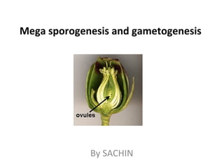
  • 1. Mega sporogenesis and gametogenesis By SACHIN
  • 2. Female gametophyte development begins early in ovule development with the formation of a diploid megaspore mother cell that undergoes meiosis. One resulting haploid megaspore then develops into the female gametophyte. Genetic and epigenetic processes mediate specification of megaspore mother cell identity and limit megaspore mother cell formation to a single cell per ovule. Auxin gradients influence female gametophyte polarity and a battery of transcription factors mediate female gametophyte cell specification and Introduction
  • 3. • During post-fertilization, the female gametophyte influences seed development through maternal- effect genes and by regulating parental contributions. • The mature female gametophyte secretes peptides that guide the pollen tube to the embryo sac and contains protein complexes that prevent seed development before fertilization. • Female gametophytes can form by an asexual process called gametophytic apomixis, which involves formation of a diploid female gametophyte and fertilization-independent development of the egg into the embryo.
  • 4. Megasporangium • The megasporangium is having the ovules together with its protective coats, integument , it is attached to the placenta, on the inner wall of ovary by a stalk called funiculus. • An ovule ready for fertilization consists of nucellar tissue enveloped almost completely by one or two integuments, leaving a small opening at the apical end, the opening is known as micropyle. • The basal region of the ovule, where funiculus is attached is called chalaza.
  • 5. Megasporogenesis • Thus embryo sacs maintain physical contact with the parent sporophyte throughout their development, this association of the female gametophyte and the sporophyte provides an opportunity to examine interactions between cells, tissues, and genomes.
  • 6. The nucellus supports the female lineage throughout development. o The egg cell in the female. o Female central cell are shown in green, as “companion cells,” in view of their potential roles in reinforcing gene silencing in their neighbouring germ cells. Female Reproductive Cell Lineages in Arabidopsis thaliana
  • 7. • (B) Phases of interaction between the female reproductive lineage and its surrounding cells. – A signal peptide (TPD1) from the megaspore mother cell seems to stimulate the proliferation (blue double-headed arrows) of dicot nucellar cells in a pathway involving EMS1; in monocots MAC1 (maize) and TDL1a (rice) fulfill a similar role, also restricting the proliferation of the female cell line (red horizontal “T”). – In monocots, EAL1 (maize) is secreted from the chalazal face of the egg (EC) and determines central (CC) and antipodal (A) cell fates. – LIS and CLO expressed in the egg of dicots are part of a signaling pathway responsible for fate specification of the synergid (SY), central, and antipodal cells. A
  • 8. • The location of the megasporocyte, directly below the apex of the nucellus, suggests that position may be an important factor in megasporocyte specification. • SEEDSTICK (STK) and SHATTERPROOF (SHP) 1 & 2 in carpel are important transductional factors. Transcriptional factors • Plant hormones, auxin in specifying embryo sac cell fates ( Sundaresan and Alandete-Saez, 2010; ). • Auxin concentration is already established in the nucellus at the distal end of the very young ovule. Pagnussat et al. (2009) have shown that the haploid nuclei within the nucellus has different developmental fates depending on the concentration of auxin surrounding them. Crosstalk between Cells of the Gametophyte
  • 9. • A model has been proposed in which auxin acts as a morphogen gradient by which nuclei exposed to the highest levels of auxin cellularize and differentiate into synergids and eggs, whereas those experiencing low auxin concentratation follow an antipodal fate. • Median auxin levels are proposed to be required for correct central cell development and the positioning of the polar nuclei (Pagnussat et al., 2009).
  • 10. The genes that have been characterised as playing key roles have been positioned adjacent to the developmental stage in which they are involved. Genes that are involved in female gamete development are shown in yellow, and those which impact on both male and female gamete formation are in purple. AP, antipodal cells PN, polar nuclei EC, egg cell SY, synergids VC, vegetative cell GC, generative cells. Schematic representation of the events leading to gamete formation in Arabidopsis.
  • 11. Shortly after ovule initiation, a single subdermal nucellar cell enlarges and displays a prominent nucleus. This cell represents the archesporium, or spore-bearing tissue, and typically occupies a position directly below the apex of the nucellus. • Although ovule morphologies show considerable diversity. • Ovules are specialized structures, derived from the placenta of the ovary wall, that produce the megasporocyte and are the site of embryo sac formation, fertilization, and embryogenesis. • The ovule consists of Development of ovule
  • 12. Ovule • Events within individual ovules of the female carpel follow a similar pattern, with the megaspore mother cell. • The functional meiotic product requiring the presence of a normal nucellar layer for correct development. • Once the cells of the embryo sac are formed, their correct positioning, division, and specification results from a complex interplay involving hormone gradients. • This is followed by the interactions between the egg and other cells of the embryo sac. • There is evidence that gametophytic central cell acts as a companion cell to the egg. The polar nuclei may partially fuse with each other before they are fertilized by a single sperm nucleus, generating the triploid primary endosperm nucleus (Cass et al., 1985). • The mature endosperm will provide nutrients for the developing embryo or seedling.
  • 13. Synergids The synergids, which are located on either side of the egg cell, play an important role in fertilization. The pollen tube discharges its contents into one of the synergids prior to incorporation of the orientation of the embryo sac with respect to the chalazal-
  • 14. Antipodal Cells • Three antipodal cells are located opposite to the egg at the chalazal end of the embryo sac. • No specific function during reproduction has been attributed to the antipodals, but they are thought to be involved in the import of nutrients to the embryo sac. • Cytological characteristics of cells within the embryo sac as well as cytochemical localization of proteins, starches, lipids, and nucleic acids have been used to assess the physiological state of the embryo sac and suggest relative rates of metabolic • For example, the presence of numerous ribosomes and mitochondria in the synergids, central cells, and antipodals suggests a high metabolic activity. • By contrast, the egg cell has fewer ribosomes, plastids, and other organelles and appears to be relatively quiescent (Mansfield et al., 1990). • At the present time, there is little data available about the regulation of ovule and embryo sac development and the temporal and spatial patterns of gene expression in the embryo sac.
  • 15. Peter K. Endress Ann Bot 2011;107:1465-1489 © The Author 2011. Published by Oxford University Press on behalf of the Annals of Botany Company. All rights reserved. For Permissions, please email: journals.permissions@oup.com (A–F) Different nucellus shapes. Meiocytes are shaded grey. (A) Crassinucellar. (B) Weakly crassinucellar. (C) Pseudocrassinucellar. (D) Incompletely tenuinucellar. (E) Tenuinucellar. (F) Reduced tenuinucellar. (G–K) Integument differentiation. In bitegmic ovules, the inner integument is shaded red, the outer blue. (G) The outer integument is thicker than the inner. (H) The inner integument is thicker than the outer. (I) (I). Both integuments are equally thick. (II) (J) Unitegmic. (III) (K) Ategmic. Diversity of nucellus thickness and integument number and thickness
  • 16. • The two integuments are considered to have distinct evolutionary origins, it initiated at the base of the nucellus during megasporogenesis. • The inner integument is most often dermal (Ll) in origin, whereas the outer integument is usually derived from both dermal and subdermal layers. • Periclinal divisions in the integuments generate an increase in the number of cell layers, whereas anticlinal divisions and cell elongation are responsible for growth parallel to the nucellus.
  • 17. Ramin Yadegari, and Gary N. Drews Plant Cell 2004;16:S133-S141 ©2004 by American Society of Plant Biologists Female Gametophyte Development in Arabidopsis The steps are described by Christensen et al. (1997). Category designations show the developmental stage affected in the female gametophyte mutants. Category 1= megaspores fail to undergo cell death; Category 2= megaspores do not progress beyond stage FG1; Category 3= pleiotropic defects during the nuclear division phase; Category 4= failure to cellularize or abnormal cell shape; Category 5= polar nuclei fail to fuse; Category 6= antipodal cells fail to undergo cell death; Category 7 (not shown), morphologically wild-type female gametophytes at the terminal stage.
  • 18. Ramin Yadegari, and Gary N. Drews Plant Cell 2004;16:S133-S141 ©2004 by American Society of Plant Biologists o The mature female gametophyte in Arabidopsis is 105 μm long and 25∼ ∼ μm wide. (The gray areas represent cytoplasm, the white areas represent vacuoles, and the black areas represent nuclei). ac, antipodal cells; cc, central cell; ch, chalazal region of the ovule; ec, egg cell; f, funiculus; mp, micropyle; sc, synergid cell; sn, secondary nucleus. The Arabidopsis Female Gametophyte The view in (B) is perpendicular to that in (A)
  • 19. Embryo Sac Development Occurs along Chalazal-Micropylar Ovule Axis The processes involved in megaspore selection may begin before meiosis, as polarity of the developing megagametophyte reflects the ovular polarity and suggests that the ovule plays a role in the selection of a functional megaspore and in the organization of the embryo sac, and expressed in the asymmetric distribution of cellular organelles and plasmodesmata. After meiosis, plastids are preferentially distributed at the micropylar end of the functional megaspore, and plasmodesmata are usually observed only between the functional megaspore and the nucellus, thus the functional megaspore inherits a richer cytoplasm and nutrients from the maternal tissues.
  • 20. • The arrangement of microtubules indicated a role for the cytoskeleton in distribution and positioning of cytoplasmic contents during megasporogenesis and megagametogenesis, it is possible that positional information, perhaps in the form of gradients, could be inherited from the megasporocyte. • The nature of specific nutritional or hormonal interactions between the ovule and embryo sac are not known, hence connectivity between the megagametophyte and the ovule presumed to establish a polar nutrient flow. • On the other side of the embryo sac, wall projections extend from the antipodal cells into the chalazal nucellus and may represent another site of metabolite flow from the nucellus to the embryo sac.
  • 21. • In some species, cell wall projections are also found between the central cell and the inner integuments; however, in other species, a cutinized wall can be found in this region. • The cellular anatomy and the accumulation and mobilization of starch reserves of the nucellus and integuments also suggest a directional flow of metabolites into the embryo sac. • The micropylar portion of the egg cell is occupied by a vacuole, and the nucleus, cytoplasm, and most of the organelles are located at the chalazal end, following fertilization, this polarity is also observed in the zygote.
  • 22. The Arabidopsis female gametophyte. (A) Ovule. (B) Female gametophyte. (C) Synergid cells. View in (B) and (C) is perpendicular to that in (A). The dashed line at the chalazal ends of the synergid cells in (C) represents a discontinuous or absent cell wall. o Synergids are frequently observed to have elaborate wall projections, the filiform apparatus that extends into the nucellus, which may provide a mechanism for nutrient flow from the ovule to the embryo sac. ac, antipodal cells; cc, central cell; ch, chalazal region of the ovule; ec, egg cell; f, funiculus; fa, filiform apparatus; mp, micropyle; sc, synergid cell; sn, synergid nucleus, sv, synergid vacuole.
  • 23. • At the micropylar pole, the synergid cell wall is thickened and extensively invaginated, forming a structure called filiform apparatus. • The filiform apparatus greatly increases the surface area of the plasma membrane in this region and contains a high concentration of secretory organelles, suggesting that it may facilitate transport of substances into and out of the synergid cells. • Based on cytological staining properties in species other than Arabidopsis, the filiform apparatus appears to be composed of a number of substances including cellulose, hemicellulose, pectin, callose, and protein. Synergid cell wall is specialized structure
  • 24. • Thus the filiform apparatus has at least two functions associated with the fertilization process. • First, the synergid cells secrete pollen tube attractants via the filiform apparatus. • In addition, the pollen tube enters the synergid cell by growing through the filiform apparatus. • By contrast, the antipodal cells in Arabidopsis have no dramatic specializations and no known function. • In cereals, the antipodal cells proliferate into as many as 100 cells.
  • 25. • Considerable diversity in the pattern of embryo sac development was found among plant species. Haig (1990) proposed a model for embryo sac development whereby different patterns could be generated by variations in meiosis, cytokinesis, and the timing and number of mitotic divisions Some of the modes of embryo sac development that have been observed In Arabidopsis and most monocot crops, three of the four linear products of meiosis degenerate, and the surviving haploid “megaspore” divides three times to form the cells that populate the embryo sac— o The single egg o Two synergid cells that receive the pollen tube o Two polar nuclei that later fuse to form the large diploid central cell, o Species-specific number of antipodal cells, which are held to be secretory.
  • 26. Development of the Polygonum- Type Embryo Sac • The Polygonum- type pattern is the most commonly observed form of embryo sac development. • Approximately 70% of the species examined, including Arabidopsis and
  • 27. • During the first meiotic division, the spindle is oriented parallel to the micropylar-chalazal axis of the nucellus. • Wall formation occurs perpendicular to this axis, creating a dyad of megaspores. • Frequently, the dyad cell closest to the micropyle
  • 28. • The three non-functional megaspores degenerate and are eventually crushed by the expanding functional megaspore. • Tetrahedral arrangements of megaspores have also been observed in Arabidopsis (Webb and Gunning, 1990), and T-shaped tetrads have been seen in maize (Russell, 1978). • The linear array is, however, most common.
  • 29. Callose, a P-1,3-glucan, is thought to function in the selection of a functional megaspore. During megasporogenesis, callose accumulates first in the cell walls of the megasporocyte and then in the megaspore walls. After meiosis, callose walls become thinner or absent in the functional megaspore. The presence of callose in the walls of the nonfunctional megaspores probably ensures that only the functional megaspore receives nutrients from the nucellus.
  • 30. The pattern of callose deposition is variable, reflecting the pattern of megasporogenesis, for example, in Oenothera, a monosporic species, callose is thinner at the micropylar end of the ovule, where the functional megaspore is located. In tetrasporic species, meiosis occurs without cytokinesis, and callose does not accumulate in the walls of the single tetranucleate megaspore.
  • 31. In most diplosporous species, callose level and distribution in the megaspore mother cell differs from that in sexual relatives. For example, callose is absent in the megaspore mother cells of diplosporous Tripsacum (Bicknell and Koltunow, 2004). Both mitosis and cell specification seem to be disrupted in some apomicts, for example, only two rounds of mitosis occur in the cells of aposporous Pennisetum, leading to a four-nucleate embryo sac, commonly containing an egg cell, one polar nucleus and two synergid cells. In aposporous Hieracium species, antipodals may not form and multiple embryos may develop in an embryo sac ( Koltunow et al., 2000; Koltunow et al., 2011).
  • 32. ● Click to edit the outline text format – Second Outline Level ● Third Outline Level – Fourth Outline Level ● Fifth Outline Level ● Sixth Outline Level Seventh Outline LevelClick to edit Master text styles I. Monosporic: Polygonum, Oenothera II. Bisporic: Allium, Endymion Tempera ture during embryo sac develop ment Drusa type Adoxa type Other type 15-19 C◦ 81 18 1 26-27 C◦ 89 6 5 III. Tetrasporic: Adoxa Penaea Plumbago Peperomia Drusa Fritillaria Plumbagella Effect of temperature on occurrence of two types of embryo sac in Ulmus glabra Types of embryo sacs
  • 33. Patterns of Female Gametophyte Development Exhibited by Angiosperms Genera exhibiting these patterns are indicated in parentheses. Patterns of Female Gametophyte Development Exhibited by Angiosperms.Genera exhibiting these patterns are indicated in parentheses. More comprehensive descriptions of the variation among angiosperms can be found in several reviews (Maheshwari, 1950; Willemse and van Went, 1984; Haig, 1990; Huang and Russell, 1992; Russell, 2001). In this figure, the chalazal end of the female gametophyte is up and the micropylar end is down. FG, female gametophyte.
  • 34. • In -65% of the species examined, most of the nucelIus degenerates before the embryo sac reaches maturity. • The embryo sac is then in direct contact with the inner integument. • In these situations, the innermost cell layer of the inner integument may differentiate into a unique cell layer termed the endothelium. • Radial cell expansion, endopolyploidy, and prominent nuclei are observed in the endothelial cells and also the anther tapetum, which is thought to be involved in secretion and nutrition of the pollen speculated that the cytological features shared between the endothelium and tapetum could indicate a similar function for both tissues. • In species in which the nucellus does not degenerate, the inner integument does not differentiate an endothelium, and the embryo sac may receive nutrients from the nucellus directly.
  • 35. • Stalklike structure extending from the lowermost part of the chalaza to the placenta, usually, a single vascular strand runs through the funiculus from the placenta terminating at the base of the embryo sac. • The mature ovule displays a polarity with respect to the axis determined by the location of the chalaza and micropyle. Esau (1977) defined the chalaza as the region extending from the base of the integuments to the point of attachment of the funiculus. • The micropyle is located at the point where the integuments terminate and is the site where pollen tubes enter the ovule.
  • 36. As the embryo sac develops, the integuments continue to enlarge, typically overgrowing the nucellus. The amount of ovule curvature varies with the extent of differential growth of the integuments and funiculus; the degree of curvature forms a basis for classification of ovule morphology. Thus, the mature anatropous ovule shows extensive curvature such that the long axis of the nucellus is parallel to the axis of the funiculus.
  • 37. Development of ovule an anatropous bitegmic angiosperm ovule Peter K. Endress Ann Bot 2011;107:1465-1489 © The Author 2011. Published by Oxford University Press on behalf of the Annals of Botany Company. All rights reserved. For Permissions, please email: journals.permissions@oup.com Median longitudinal sections : (A–E) Straight thin line drawn through the middle of the nucellus. Arrowhead indicating successive ovule curvature from 0 to 180 °. (E, E′) (A).Ovule before integument initiation. (B).Ovule at initiation of inner integument. (C) Slightly older ovule. (D) Ovule with both integuments formed. (E) Mature ovule. Raphal side blue, antiraphal side red. (E′) Mature ovule. Concave side blue, convex side red.
  • 38. Different orientations of curved ovules with respect to carpel curvature (denoted by a red line). Peter K. Endress Ann Bot 2011;107:1465-1489 © The Author 2011. Published by Oxford University Press on behalf of the Annals of Botany Company. All rights reserved. For Permissions, please email: journals.permissions@oup.com Different orientations of curved ovules with respect to carpel curvature (denoted by a red line). (A) Syntropous: Curvature of the ovule in the same direction as the curvature of the carpel. (B) Antitropous: Curvature of the ovule in the opposite direction to that of the carpel.
  • 39. (A–C) Ovary or locules not filled with secretion. (A) Single ovule with basal placenta (LS gynoecium) (e.g. Piperaceae, Juglandaceae, Urticaceae). (A) Ovules on parietal placentae with the micropyle directed toward another placenta (TS ovary) (e.g. Casearia, Salicaceae; Mayaca, Mayacaceae). (A) Ovules with long funiculi curved to their own placenta (LS gynoecium) (e.g. Helianthemum, Cistaceae). (A) (D–F) Ovary or locules filled with secretion (secretion shaded blue). (A) Basal diffuse placenta (LS gynoecium) (e.g. Pistia, Araceae). (A) Laminar-diffuse placenta (TS carpel/ovary) (e.g. Barclaya, Nymphaeaceae; Hydrocharis, Hydrocharitaceae; Akebia. Orthotropous ovules and conditions of ovary locule architectures under which they occur (schematic, only one integument is drawn in each ovule for simplicity; augmented and modified from (Endress , 1994).
  • 40. Genetic and molecular analysis of ovule and embryo sac development • Mutations are used to study the effect of embryo sac development which are expressed in the parent sporophyte or in the gametophyte. • In the case of a recessive mutation, 25% of the progeny from selfed heterozygous plants will display the sterile phenotype. • Defects in fertility that are specific to the female are distinguished from male sterile by reciprocal crosses with wild-type plants. • Mutations that abolish or reduce female fertility can be transmitted and recovered through the pollen. • Several female-sterile mutations have been identified among populations of Arabidopsis mutagenized with ethylmethane sulfonate.
  • 41. • Representative female-sterile mutants of Arabidopsis include short integuments-1 (sinl). • Each of these mutants has a defect in a discrete aspect of ovule and embryo sac development, such as initiation of the integuments, development of the nucellus, megasporogenesis, and Illustration of the utility of a genetic approach for understanding how the ovule develops and interacts with the developing embryo sac
  • 42. Gametophytic Mutants • The indeterminate gametophyte (ig) mutation of maize has pleiotropic effects on embryo sac development but primarily affects the number of mitotic divisions. • It is associated with the occurrence of multiple functional egg cells and embryos within an embryo sac and with the formation of defective seed. • Another mutation with gametophytic expression, Gf, was described in Arabidopsis, showed reduced transmission through the female gametophyte of genes linked to Gf. • In maize, heterozygous plants that contain deletions frequently display reduction in pollen viability (Patterson, 1978).
  • 44. Pollen tube subgroup 18 (S18) MYB transcription factors respond to growth through the pistilA schematic diagram of an A. thaliana ovary with 12 ovules (instead of the actual 50–60). Pollen grains (red) are germinating pollen tubes on the stigma. Pollen tubes target the female gametophyte (grey). Mature pollen grains [represented with two sperm (black) and a nucleus (grey)] express MYB101, and lower levels of both MYB97 and MYB120. Transcription of MYB targets is low in, or absent from, pollen grains. Growth through the stigma and style activate transcription of MYB97 and MYB120; the three MYBs activate target genes in the pollen tube. The pollen tube factors that sense the pistil environment and activate MYB expression are not yet known.
  • 45. Subgroup 18 (S18) MYB-regulated gene classes and their putative roles during pollen tube receptions(A) Schematic diagram of the pollen tube and female gametophyte at the beginning of pollen tube reception. Pollen tube contact with one of the synergid cells is indicated (broken line). Proteins with roles in pollen tube reception and MYB-regulated gene products are highlighted. Abbreviations: sn, synergid nucleus; vn, vegetative nucleus. (B andC) Schematic diagram of pollen tube reception between a pollen tube and the synergid that will degenerate. Cytosolic Ca2+ concentrations are symbolized with a colour spectrum (red, high Ca2+; green, low Ca2+). (B) FER-dependent NTA relocalization occurs after pollen tube arrival. (C) Synergid degeneration is accompanied by loss of synergid nuclear integrity. (D) Pollen tube burst, sperm release and double fertilization.
  • 46. Pollen tubes differentiate in response to growth through the pistil Pollen tube physiology changes when it grows through floral tissue. For example, pollen tubes must grow through pistil explants to be able to respond to attractants in vitro [8–10 ]. These experiments use a SIV (semi-in vitro) pollen-tube-guidance assay in which pollen tubes grow through the stigma and a portion of style before growing on to the surface of pollen growth media towards ovules [10]. In Torenia, where LURE pollen tube attractants were first described [11], the ability to respond to attractants in vitro increases with prolonged growth through pistil tissue [12]. However, when LURE-binding sites were assessed by incubating pollen tubes with LUREs followed by detection with anti-LURE antibodies, it was observed that, whereas growth in the pistil was required to generate LURE-binding sites, prolonged exposure to the pistil did not increase the detection of binding sites further [12]. These data suggest that the pollen tube senses the pistil environment, that LURE receptor (not yet identified) expression reaches a maximum as a consequence, but that prolonged exposure to the pistil environment may activate the receptor. Interestingly, a pair of A. thaliana pollen-tube-expressed membrane-associated receptor- like cytoplasmic kinases (LIP1 and LIP2) were recently shown to be induced by growth in the pistil and required for the full response to purified LURE proteins in the SIV assay and for pollen tube guidance in the pistil [13]. It is not yet clear whether LIP1 and LIP2 are involved directly in LURE perception; however, these findings underscore the idea that pollen tubes differentiate during growth in the pistil, resulting in expression of proteins required to respond to pollen tube guidance cues.
  • 47. Pollen tube differentiation for sperm release is controlled by three MYBs Over 1000 genes were detected in the transcriptome of pollen tubes grown in the SIV assay that were not detected in pollen tubes grown in vitro [14]. To identify regulators that control pollen tube gene expression in response to the pistil, 26 SIV-induced transcription factors were identified [14]. This list included three closely related R2R3- MYB type transcription factors [subgroup 18: MYB97,MYB101 and MYB120] [14]. Single and double myb mutants did not display seed set defects; however, triple mutants had a ~70% reduction in seed production [15]. myb triple mutant pollen grains and tubes develop normally, grow through the pistil and target ovules nearly as efficiently as wild- type. These data suggest that MYB97, MYB101 and MYB120 do not regulate expression of the LURE receptors. However, upon interaction with the female gametophyte, myb triple mutants fail to arrest their growth and ~72% of ovules contain coils of pollen tubes within the female gametophyte [15]. Synergid cells targeted by these mutant pollen tubes fail to degenerate normally, suggesting a loss of communication between pollen tube and synergid cells during pollen tube reception [ 15]. Furthermore, myb triple mutant pollen tubes fail to burst and release sperm. These data suggest that MYB97, MYB101 and MYB120 are critical for the pollen tube to exchange signals with the female gametophyte required for successful fertilization.
  • 48. Pollen tube reception signalling by the female gametophyte Pollen tube reception requires a number of synergid-expressed genes including FER (FERONIA) [16], NTA (NORTIA) [17] and LRE (LORELEI) [18] (Figure 2A). Loss-of-function mutations in of each of these genes cause the same phenotype: wild- type pollen tubes coil in mutant ovules and fail to release sperm. FER is a transmembrane receptor-like kinase of the CrRLK1L family predicted to have pollen tube or female-gametophyte ligand(s) important for pollen tube reception. FER has a malectin-like extracellular domain, so its ligand could be a carbohydrate or glycoprotein [19] (Figure 2A). FER is localized to the filiform apparatus, and is required for NTA, a seven-pass transmembrane mildew-resistance locus O family protein, to be relocalized from the secretory system to the filiform apparatus upon pollen tube arrival [17] ( Figures 2A and 2B). NTA may interact with synergid- or pollen-tube-expressed proteins upon relocalization and also contains a calmodulin-binding domain in its cytoplasmic C- terminus, possibly allowing synergid cell perception of Ca2+ oscillations during reception [20] (Figures 2B–2D). LRE encodes a glycosylphosphatidylinositol-anchored protein predicted to be associated with the synergid membrane [18] (Figure 2A). The mechanisms by which FER, NTA and LRE direct pollen tube reception are unknown, and it will be important to define how they sense pollen tube arrival to initiate synergid degeneration and pollen tube burst.
  • 49. Relatively little is known about the pollen-tube-expressed genes involved in pollen tube reception, and no direct connections between pollen tube and synergid genes have been made. Interestingly, a pair of pollen-tube-expressed members of the FER family members [CrRLK receptor-like kinases, ANX1 (ANXUR1) and ANX2 (ANXUR2)] may be negative regulators of pollen tube burst. anx1,anx2 double mutants produce pollen tubes that burst very soon after germination [21,22]. Like FER, the ligands for ANX1 and ANX2 are not yet known, and the nature of ANX contribution to pollen tube reception is unclear because the double mutant phenotype is not informative about their role during synergid interactions. ACA9 (autoinhibited Ca2+-ATPase 9) encodes a pollen-specific calmodulin-binding Ca2+ pump localized to the pollen tube plasma membrane [23]. aca9 pollen tubes have growth defects; however, they can reach ovules in the upper portion of the pistil and ~50% of these enter the ovule micropyle and arrest, but fail to burst [23]. This result suggests that ACA9 is important for regulating pollen tube Ca2+ dynamics required for pollen tube burst. Recent live-imaging experiments have shown that Ca2+ concentrations spike in the pollen tube and the synergids as pollen tube burst occurs [20] (Figures 2B–2D). It will be very interesting to determine how aca9pollen and nta (which contains a predicted calmodulin-binding domain) synergid mutants affect these dynamics.
  • 50. Do MYB target genes interact with the pollen tube reception signalling components? myb97,myb101,myb120 triple mutant pollen tubes fail to stop growing and burst within the female gametophyte, so the genes regulated by these transcription factors are candidates for pollen tube components of the pollen tube reception mechanism. By comparing the transcriptomes of pistils pollinated with either wild- type or myb triple mutant pollen, three main categories of MYB-regulated genes were identified: transporters, small proteins and peptides, and carbohydrate- active enzymes [15] (Figure 2A). Many of these genes were found to be in large gene families, potentially explaining why extensive genetic screening in A.  thaliana has not identified male pollen tube reception mutants. Several sugar/proton symporters in the major facilitator family [24] were identified as being MYB-regulated. AtSUC7 (At indicates A.  thaliana), AtSUC8 and AtSUC9 are highly induced in wild-type pollen tubes grown in SIV conditions, but are absent from myb triple mutants [15]. myb triple mutants have no pollen tube growth defect in vitro or in the pistil, suggesting that these sucrose transporters are not required to import sugars to fuel growth, but may have a specialized function in regulating the pollen tube osmotic state during pollen tube reception.
  • 51. Multiple small proteins and peptides were found to be potential targets of MYB activation during pollen tube growth (Figure 2A). One of these is a thionin, an 87-amino-acid secreted cysteine-rich protein (CRP2460) [25]. Thionins have been shown to nucleate membrane pores in artificial lipid bilyers and rat neuronal cells [26]; this is an intriguing potential function for pollen-tube-expressed thionins given that myb triple mutant pollen tubes fail to express these peptides and also have defects in pollen tube burst and initiating synergid degeneration [15]. Interestingly, small (130–152 amino acids) secreted proteins with domains homologous with stigmatic Papaver rhoeas SI (self- incompatibility) protein PrsS1 [27] were also found to require pollen tube MYBs for expression in the pollen tube [15]. In Papaver, PrsS1 is secreted from the stigma and is bound by PrpS, its pollen- expressed receptor [28]. Ligand–receptor interaction blocks germination of self pollen through a mechanism involving Ca2+ as a second messenger and PCD (programmed cell death) [29]. The function of A. thaliana SPHs (S-protein homologues) is unclear [30] and it is surprising to find that they are expressed in pollen tubes in response to growth through the pistil. It will be interesting to test whether a signalling module similar to that described in Papaver is involved in gametophyte interactions that lead to synergid degeneration and pollen tube burst. The third category of pollen tube MYB-regulated genes encode proteins that interact with carbohydrates, many of which are predicted to be extracellular (Figure 2A). These include hydrolases (i.e. glycoamylase, O-glycosylhydrolase, β-1,3-glucanase, pectin lyase-like) and pectin methylesterase inhibitors [15]. These proteins may be expressed by the pollen tube to modify the cell wall in preparation for pollen tube reception. Alternatively, these proteins may modify the cell wall or other pollen-derived carbohydrates for recognition by the female gametophyte. As mentioned above, FER contains an extracellular malectin domain that could interact with a pollen- tube-derived carbohydrate, and, since myb triple mutant pollen tubes behave as though FER signalling is defective, it will be interesting to determine whether pollen tube MYBs regulate production of FER ligands.
  • 52. MYB-mediated pollen tube differentiation is important for species recognition during pollen tube reception Pollen tube reception fails in interspecific crosses of Rhododendron [31] or A.  thaliana [16]; pollen tubes of one species overgrow without releasing sperm in ovules of the exotic species. These data suggest that pollen tube reception is an important pre-zygotic barrier to interspecific hybridization. When A. thaliana  myb triple mutant pollen tubes enter a wild-type A. thaliana ovule, they overgrow and fail to release sperm [15]. These observations led to the hypothesis that pollen tube MYBs may control expression of the determinants of pollen tube identity during interspecific recognition. To begin to assess the role of pollen tube MYBs in determining pollen tube recognition, pollen tube reception was investigated in interaccession and interspecies crosses (Figure 3). The rates of pollen tube overgrowth when myb triple and myb double mutants were used to pollinate A. thaliana pistils were higher than in any of the interaccession crosses tested (Figure 3 M).Arabidopsis korshinskyi and Olimarabidopsis pumila pollen tubes overgrew in A.  thaliana ovules more frequently than any of the accessions tested, but the A.  thaliana myb triple mutant phenotype was even more pronounced (Figure 3M). These results are consistent with the idea that pollen tube MYBs control pollen tube identity as recognized by the female gametophyte.
  • 53. Female gametophyte development in diplosporous and aposporous apomicts compared with Arabidopsis. (A) Steps of megasporogenesis and megagametogenesis in Arabidopsis ovules. Red and blue colors represent diploid and haploid cells, respectively. (B) Steps in diplosporous female gametophyte development. The megaspore mother cell enters meiosis and the process fails with the resultant diploid cell undergoing mitosis to form a diploid female gametophyte (red). Alternatively, the megaspore mother cell may directly undergo mitosis to form the diploid female gametophyte. Abbreviations: ai, aposporous initial cells; dfg, diploid female gametophyte; dm, degenerating megaspores; fm, functional megaspore; hfg, haploid female gametophyte; mmc, megaspore mother cell; mt, meiotic tetrad; (+/-), may be present or absent.
  • 54. • Megaspore mother cell differentiatiation (red) occurs and it can undergo megasporogenesis and megagametogenesis to form a haploid female gametophyte (blue). • Diploid aposporous initial cells differentiate during the events of megasporogenesis close to sexually programmed cells and undergo mitosis forming a diploid female gametophyte (yellow). • Aposporous initial cell formation begins at different times in different apomictic species, either soon after megaspore mother cell formation, during meiotic tetrad development or functional megaspore selection. • In some species, both haploid and aposporous gametophytes can co-exist in ovules while in others the sexual pathway terminates, usually during early mitotic divisions of the aposporous initial cell. (C) Steps in aposporous female gametophyte development Abbreviations: ai, aposporous initial cells; dfg, diploid female gametophyte; dm, degenerating megaspores; fm, functional megaspore; hfg, haploid female gametophyte; mmc, megaspore mother cell; mt, meiotic tetrad; (+/-), may be present or absent.

Editor's Notes

  1. Different orientations of curved ovules with respect to carpel curvature (denoted by a red line). (A) Syntropous. Curvature of the ovule in the same direction as the curvature of the carpel. (B) Antitropous. Curvature of the ovule in the opposite direction to that of the carpel.