SlideShare a Scribd company logo
1 of 12
Download to read offline
Figure:
Immunohistochemical
analysis of paraffin
embedded Human
Tonsil tissue. 1: c-Src
Polyclonal Antibody
was diluted at 1:200 (4
degree
Celsius,overnight). 2:
Sodium citrate pH 6.0
was used for antibody
retrieval (>98 degree
Celsius,20min). 3:
Secondary antibody was
diluted at 1:200 (room
temperature, 30min).
Negative control was
used by secondary
antibody only.
Report Number 92501-a Host Rabbit
Application IHC-P Clonality Polyclonal
Model Number STJ92501 Clone ID NA
Antibody Name Anti-c-Src antibody
Testing Species HUMAN Testing Tissue TONSIL
ANTIBODY VALIDATION REPORT
a. (A small amount of distilled water was added into the incubation
box to prevent evaporation of antibody).
129. Secondary antibody incubation
a. Slides were washed 3 times, with PBS on a shaker for 5min.
Shortly after the slides were dried the corresponding secondary
antibody solution was added (HRP labelled), covering the
tissues, and incubated at room temperature for 30min.
b.
130. DAB staining
a. Slides were washed 3 times, with PBS on a shaker for 5min.
b. Shortly after, the slides were dried and fresh DAB staining buffer
was added inside the circles. The staining time was adjusted
under a microscope. Yellow-brown colour represented a positive
result. Slides were washed with water to stop the staining.
c.
131. Haematoxylin staining
a. Haematoxylin was used to counter-staining for 1min, and then
the slides were washed with water. 1% Hydrochloric acid and
alcohol was added for several seconds and then washed with
water. Ammonia was used to reveal blue colour, and then
flushed with water.
b.
132. Desolation and Clearing
i. Slides were incubated sequentially into: 75% alcohol 5min, 85%
alcohol 5min, Anhydrous ethanol - 5min, Anhydrous ethanol -
5min & Xylene - 5min. Shortly after slides were dried and neutral
gum was used to seal the slides.
ii.
133. Visualization
a. Results were validated with microscope, and the slides were
scanned.
Paraffin-Embedded
Immunohistochemistry Protocol
123.
124. Tissue processing
a. Slides were incubated sequentially into Xylene; 15min –
Xylene, 15min - Anhydrous ethanol, 5min - Anhydrous
ethanol, 5min - 85% alcohol, 5min - 75% alcohol & 5min –
wash in distilled water.
b.
125. Antigen retrieval
a. Tissue slides were incubated with citric acid (PH6.0) antigen
retrieval buffer and microwaved for antigen retrieval (heated
until boiled and then stopped heating) for 8min. Slides were
then heated with medium power for 7min. During this
process slides were kept from drying out. After cooling down
at room temperature, slides were washed with PBS on
shaker for 5min, repeated for 3 times.
b.
126. Inhibition of endogenous peroxidase
a. Slides were placed in 3% Hydrogen peroxide solution, and
incubated for 10 min at room temperature without light
exposure. Slides were then washed 3 times with PBS on a
shaker for 5mins.
b.
127. BSA Blocking
a. Shortly after slides were dried, a PAP pen was used to draw
circles around the tissue sections (and to prevent draining of
the antibody solution). Inside the circles, BSA was used to
cover the tissue evenly, blocking for 30min.
b.
128. Primary antibody incubation
After blocking solution was removed a 1:200 solution of
primary antibody/PBS was added on the slide, and incubated
overnight at 4°C.
St John's Laboratory Ltd.
www.stjohnslabs.com
Figure:
Immunohistochemical
analysis of paraffin
embedded Human liver
cancer tissue. 1: c-Src
Polyclonal Antibody
was diluted at 1:200 (4
degree
Celsius,overnight). 2:
Sodium citrate pH 6.0
was used for antibody
retrieval (>98 degree
Celsius,20min). 3:
Secondary antibody was
diluted at 1:200 (room
temperature, 30min).
Negative control was
used by secondary
antibody only.
Report Number 92501-b Host Rabbit
Application IHC-P Clonality Polyclonal
Model Number STJ92501 Clone ID NA
Antibody Name Anti-c-Src antibody
Testing Species HUMAN Testing Tissue LIVER CANCER
ANTIBODY VALIDATION REPORT
a. (A small amount of distilled water was added into the incubation
box to prevent evaporation of antibody).
118. Secondary antibody incubation
a. Slides were washed 3 times, with PBS on a shaker for 5min.
Shortly after the slides were dried the corresponding secondary
antibody solution was added (HRP labelled), covering the
tissues, and incubated at room temperature for 30min.
b.
119. DAB staining
a. Slides were washed 3 times, with PBS on a shaker for 5min.
b. Shortly after, the slides were dried and fresh DAB staining buffer
was added inside the circles. The staining time was adjusted
under a microscope. Yellow-brown colour represented a positive
result. Slides were washed with water to stop the staining.
c.
120. Haematoxylin staining
a. Haematoxylin was used to counter-staining for 1min, and then
the slides were washed with water. 1% Hydrochloric acid and
alcohol was added for several seconds and then washed with
water. Ammonia was used to reveal blue colour, and then
flushed with water.
b.
121. Desolation and Clearing
i. Slides were incubated sequentially into: 75% alcohol 5min, 85%
alcohol 5min, Anhydrous ethanol - 5min, Anhydrous ethanol -
5min & Xylene - 5min. Shortly after slides were dried and neutral
gum was used to seal the slides.
ii.
122. Visualization
a. Results were validated with microscope, and the slides were
scanned.
Paraffin-Embedded
Immunohistochemistry Protocol
112.
113. Tissue processing
a. Slides were incubated sequentially into Xylene; 15min –
Xylene, 15min - Anhydrous ethanol, 5min - Anhydrous
ethanol, 5min - 85% alcohol, 5min - 75% alcohol & 5min –
wash in distilled water.
b.
114. Antigen retrieval
a. Tissue slides were incubated with citric acid (PH6.0) antigen
retrieval buffer and microwaved for antigen retrieval (heated
until boiled and then stopped heating) for 8min. Slides were
then heated with medium power for 7min. During this
process slides were kept from drying out. After cooling down
at room temperature, slides were washed with PBS on
shaker for 5min, repeated for 3 times.
b.
115. Inhibition of endogenous peroxidase
a. Slides were placed in 3% Hydrogen peroxide solution, and
incubated for 10 min at room temperature without light
exposure. Slides were then washed 3 times with PBS on a
shaker for 5mins.
b.
116. BSA Blocking
a. Shortly after slides were dried, a PAP pen was used to draw
circles around the tissue sections (and to prevent draining of
the antibody solution). Inside the circles, BSA was used to
cover the tissue evenly, blocking for 30min.
b.
117. Primary antibody incubation
After blocking solution was removed a 1:200 solution of
primary antibody/PBS was added on the slide, and incubated
overnight at 4°C.
St John's Laboratory Ltd.
www.stjohnslabs.com
Figure:
Immunohistochemical
analysis of paraffin
embedded Human
stomach tissue. 1: c-Src
Polyclonal Antibody
was diluted at 1:200 (4
degree
Celsius,overnight). 2:
Sodium citrate pH 6.0
was used for antibody
retrieval (>98 degree
Celsius,20min). 3:
Secondary antibody was
diluted at 1:200 (room
temperature, 30min).
Negative control was
used by secondary
antibody only.
Report Number 92501-c Host Rabbit
Application IHC-P Clonality Polyclonal
Model Number STJ92501 Clone ID NA
Antibody Name Anti-c-Src antibody
Testing Species HUMAN Testing Tissue STOMACH
ANTIBODY VALIDATION REPORT
a. (A small amount of distilled water was added into the incubation
box to prevent evaporation of antibody).
107. Secondary antibody incubation
a. Slides were washed 3 times, with PBS on a shaker for 5min.
Shortly after the slides were dried the corresponding secondary
antibody solution was added (HRP labelled), covering the
tissues, and incubated at room temperature for 30min.
b.
108. DAB staining
a. Slides were washed 3 times, with PBS on a shaker for 5min.
b. Shortly after, the slides were dried and fresh DAB staining buffer
was added inside the circles. The staining time was adjusted
under a microscope. Yellow-brown colour represented a positive
result. Slides were washed with water to stop the staining.
c.
109. Haematoxylin staining
a. Haematoxylin was used to counter-staining for 1min, and then
the slides were washed with water. 1% Hydrochloric acid and
alcohol was added for several seconds and then washed with
water. Ammonia was used to reveal blue colour, and then
flushed with water.
b.
110. Desolation and Clearing
i. Slides were incubated sequentially into: 75% alcohol 5min, 85%
alcohol 5min, Anhydrous ethanol - 5min, Anhydrous ethanol -
5min & Xylene - 5min. Shortly after slides were dried and neutral
gum was used to seal the slides.
ii.
111. Visualization
a. Results were validated with microscope, and the slides were
scanned.
Paraffin-Embedded
Immunohistochemistry Protocol
101.
102. Tissue processing
a. Slides were incubated sequentially into Xylene; 15min –
Xylene, 15min - Anhydrous ethanol, 5min - Anhydrous
ethanol, 5min - 85% alcohol, 5min - 75% alcohol & 5min –
wash in distilled water.
b.
103. Antigen retrieval
a. Tissue slides were incubated with citric acid (PH6.0) antigen
retrieval buffer and microwaved for antigen retrieval (heated
until boiled and then stopped heating) for 8min. Slides were
then heated with medium power for 7min. During this
process slides were kept from drying out. After cooling down
at room temperature, slides were washed with PBS on
shaker for 5min, repeated for 3 times.
b.
104. Inhibition of endogenous peroxidase
a. Slides were placed in 3% Hydrogen peroxide solution, and
incubated for 10 min at room temperature without light
exposure. Slides were then washed 3 times with PBS on a
shaker for 5mins.
b.
105. BSA Blocking
a. Shortly after slides were dried, a PAP pen was used to draw
circles around the tissue sections (and to prevent draining of
the antibody solution). Inside the circles, BSA was used to
cover the tissue evenly, blocking for 30min.
b.
106. Primary antibody incubation
After blocking solution was removed a 1:200 solution of
primary antibody/PBS was added on the slide, and incubated
overnight at 4°C.
St John's Laboratory Ltd.
www.stjohnslabs.com
Figure:
Immunohistochemical
analysis of paraffin
embedded Human
stomach cancer tissue.
1: c-Src Polyclonal
Antibody was diluted at
1:200 (4 degree
Celsius,overnight). 2:
Sodium citrate pH 6.0
was used for antibody
retrieval (>98 degree
Celsius,20min). 3:
Secondary antibody was
diluted at 1:200 (room
temperature, 30min).
Negative control was
used by secondary
antibody only.
Report Number 92501-d Host Rabbit
Application IHC-P Clonality Polyclonal
Model Number STJ92501 Clone ID NA
Antibody Name Anti-c-Src antibody
Testing Species HUMAN Testing Tissue STOMACH CANCER
ANTIBODY VALIDATION REPORT
a. (A small amount of distilled water was added into the incubation
box to prevent evaporation of antibody).
96. Secondary antibody incubation
a. Slides were washed 3 times, with PBS on a shaker for 5min.
Shortly after the slides were dried the corresponding secondary
antibody solution was added (HRP labelled), covering the
tissues, and incubated at room temperature for 30min.
b.
97. DAB staining
a. Slides were washed 3 times, with PBS on a shaker for 5min.
b. Shortly after, the slides were dried and fresh DAB staining buffer
was added inside the circles. The staining time was adjusted
under a microscope. Yellow-brown colour represented a positive
result. Slides were washed with water to stop the staining.
c.
98. Haematoxylin staining
a. Haematoxylin was used to counter-staining for 1min, and then
the slides were washed with water. 1% Hydrochloric acid and
alcohol was added for several seconds and then washed with
water. Ammonia was used to reveal blue colour, and then
flushed with water.
b.
99. Desolation and Clearing
i. Slides were incubated sequentially into: 75% alcohol 5min, 85%
alcohol 5min, Anhydrous ethanol - 5min, Anhydrous ethanol -
5min & Xylene - 5min. Shortly after slides were dried and neutral
gum was used to seal the slides.
ii.
100. Visualization
a. Results were validated with microscope, and the slides were
scanned.
Paraffin-Embedded
Immunohistochemistry Protocol
90.
91. Tissue processing
a. Slides were incubated sequentially into Xylene; 15min –
Xylene, 15min - Anhydrous ethanol, 5min - Anhydrous
ethanol, 5min - 85% alcohol, 5min - 75% alcohol & 5min –
wash in distilled water.
b.
92. Antigen retrieval
a. Tissue slides were incubated with citric acid (PH6.0) antigen
retrieval buffer and microwaved for antigen retrieval (heated
until boiled and then stopped heating) for 8min. Slides were
then heated with medium power for 7min. During this
process slides were kept from drying out. After cooling down
at room temperature, slides were washed with PBS on
shaker for 5min, repeated for 3 times.
b.
93. Inhibition of endogenous peroxidase
a. Slides were placed in 3% Hydrogen peroxide solution, and
incubated for 10 min at room temperature without light
exposure. Slides were then washed 3 times with PBS on a
shaker for 5mins.
b.
94. BSA Blocking
a. Shortly after slides were dried, a PAP pen was used to draw
circles around the tissue sections (and to prevent draining of
the antibody solution). Inside the circles, BSA was used to
cover the tissue evenly, blocking for 30min.
b.
95. Primary antibody incubation
After blocking solution was removed a 1:200 solution of
primary antibody/PBS was added on the slide, and incubated
overnight at 4°C.
St John's Laboratory Ltd.
www.stjohnslabs.com
Figure:
Immunohistochemical
analysis of paraffin
embedded Human
appendix tissue. 1: c-Src
Polyclonal Antibody
was diluted at 1:200 (4
degree
Celsius,overnight). 2:
Sodium citrate pH 6.0
was used for antibody
retrieval (>98 degree
Celsius,20min). 3:
Secondary antibody was
diluted at 1:200 (room
temperature, 30min).
Negative control was
used by secondary
antibody only.
Report Number 92501-e Host Rabbit
Application IHC-P Clonality Polyclonal
Model Number STJ92501 Clone ID NA
Antibody Name Anti-c-Src antibody
Testing Species HUMAN Testing Tissue APPENDIX
ANTIBODY VALIDATION REPORT
a. (A small amount of distilled water was added into the incubation
box to prevent evaporation of antibody).
85. Secondary antibody incubation
a. Slides were washed 3 times, with PBS on a shaker for 5min.
Shortly after the slides were dried the corresponding secondary
antibody solution was added (HRP labelled), covering the
tissues, and incubated at room temperature for 30min.
b.
86. DAB staining
a. Slides were washed 3 times, with PBS on a shaker for 5min.
b. Shortly after, the slides were dried and fresh DAB staining buffer
was added inside the circles. The staining time was adjusted
under a microscope. Yellow-brown colour represented a positive
result. Slides were washed with water to stop the staining.
c.
87. Haematoxylin staining
a. Haematoxylin was used to counter-staining for 1min, and then
the slides were washed with water. 1% Hydrochloric acid and
alcohol was added for several seconds and then washed with
water. Ammonia was used to reveal blue colour, and then
flushed with water.
b.
88. Desolation and Clearing
i. Slides were incubated sequentially into: 75% alcohol 5min, 85%
alcohol 5min, Anhydrous ethanol - 5min, Anhydrous ethanol -
5min & Xylene - 5min. Shortly after slides were dried and neutral
gum was used to seal the slides.
ii.
89. Visualization
a. Results were validated with microscope, and the slides were
scanned.
Paraffin-Embedded
Immunohistochemistry Protocol
79.
80. Tissue processing
a. Slides were incubated sequentially into Xylene; 15min –
Xylene, 15min - Anhydrous ethanol, 5min - Anhydrous
ethanol, 5min - 85% alcohol, 5min - 75% alcohol & 5min –
wash in distilled water.
b.
81. Antigen retrieval
a. Tissue slides were incubated with citric acid (PH6.0) antigen
retrieval buffer and microwaved for antigen retrieval (heated
until boiled and then stopped heating) for 8min. Slides were
then heated with medium power for 7min. During this
process slides were kept from drying out. After cooling down
at room temperature, slides were washed with PBS on
shaker for 5min, repeated for 3 times.
b.
82. Inhibition of endogenous peroxidase
a. Slides were placed in 3% Hydrogen peroxide solution, and
incubated for 10 min at room temperature without light
exposure. Slides were then washed 3 times with PBS on a
shaker for 5mins.
b.
83. BSA Blocking
a. Shortly after slides were dried, a PAP pen was used to draw
circles around the tissue sections (and to prevent draining of
the antibody solution). Inside the circles, BSA was used to
cover the tissue evenly, blocking for 30min.
b.
84. Primary antibody incubation
After blocking solution was removed a 1:200 solution of
primary antibody/PBS was added on the slide, and incubated
overnight at 4°C.
St John's Laboratory Ltd.
www.stjohnslabs.com
Figure:
Immunohistochemical
analysis of paraffin
embedded Rat testis
tissue. 1: c-Src
Polyclonal Antibody
was diluted at 1:200 (4
degree
Celsius,overnight). 2:
Sodium citrate pH 6.0
was used for antibody
retrieval (>98 degree
Celsius,20min). 3:
Secondary antibody was
diluted at 1:200 (room
temperature, 30min).
Negative control was
used by secondary
antibody only.
Report Number 92501-f Host Rabbit
Application IHC-P Clonality Polyclonal
Model Number STJ92501 Clone ID NA
Antibody Name Anti-c-Src antibody
Testing Species RAT Testing Tissue TESTIS
ANTIBODY VALIDATION REPORT
a. (A small amount of distilled water was added into the incubation
box to prevent evaporation of antibody).
74. Secondary antibody incubation
a. Slides were washed 3 times, with PBS on a shaker for 5min.
Shortly after the slides were dried the corresponding secondary
antibody solution was added (HRP labelled), covering the
tissues, and incubated at room temperature for 30min.
b.
75. DAB staining
a. Slides were washed 3 times, with PBS on a shaker for 5min.
b. Shortly after, the slides were dried and fresh DAB staining buffer
was added inside the circles. The staining time was adjusted
under a microscope. Yellow-brown colour represented a positive
result. Slides were washed with water to stop the staining.
c.
76. Haematoxylin staining
a. Haematoxylin was used to counter-staining for 1min, and then
the slides were washed with water. 1% Hydrochloric acid and
alcohol was added for several seconds and then washed with
water. Ammonia was used to reveal blue colour, and then
flushed with water.
b.
77. Desolation and Clearing
i. Slides were incubated sequentially into: 75% alcohol 5min, 85%
alcohol 5min, Anhydrous ethanol - 5min, Anhydrous ethanol -
5min & Xylene - 5min. Shortly after slides were dried and neutral
gum was used to seal the slides.
ii.
78. Visualization
a. Results were validated with microscope, and the slides were
scanned.
Paraffin-Embedded
Immunohistochemistry Protocol
68.
69. Tissue processing
a. Slides were incubated sequentially into Xylene; 15min –
Xylene, 15min - Anhydrous ethanol, 5min - Anhydrous
ethanol, 5min - 85% alcohol, 5min - 75% alcohol & 5min –
wash in distilled water.
b.
70. Antigen retrieval
a. Tissue slides were incubated with citric acid (PH6.0) antigen
retrieval buffer and microwaved for antigen retrieval (heated
until boiled and then stopped heating) for 8min. Slides were
then heated with medium power for 7min. During this
process slides were kept from drying out. After cooling down
at room temperature, slides were washed with PBS on
shaker for 5min, repeated for 3 times.
b.
71. Inhibition of endogenous peroxidase
a. Slides were placed in 3% Hydrogen peroxide solution, and
incubated for 10 min at room temperature without light
exposure. Slides were then washed 3 times with PBS on a
shaker for 5mins.
b.
72. BSA Blocking
a. Shortly after slides were dried, a PAP pen was used to draw
circles around the tissue sections (and to prevent draining of
the antibody solution). Inside the circles, BSA was used to
cover the tissue evenly, blocking for 30min.
b.
73. Primary antibody incubation
After blocking solution was removed a 1:200 solution of
primary antibody/PBS was added on the slide, and incubated
overnight at 4°C.
St John's Laboratory Ltd.
www.stjohnslabs.com
Figure:
Immunohistochemical
analysis of paraffin
embedded Rat kidney
tissue. 1: c-Src
Polyclonal Antibody
was diluted at 1:200 (4
degree
Celsius,overnight). 2:
Sodium citrate pH 6.0
was used for antibody
retrieval (>98 degree
Celsius,20min). 3:
Secondary antibody was
diluted at 1:200 (room
temperature, 30min).
Negative control was
used by secondary
antibody only.
Report Number 92501-g Host Rabbit
Application IHC-P Clonality Polyclonal
Model Number STJ92501 Clone ID NA
Antibody Name Anti-c-Src antibody
Testing Species RAT Testing Tissue KIDNEY
ANTIBODY VALIDATION REPORT
a. (A small amount of distilled water was added into the incubation
box to prevent evaporation of antibody).
63. Secondary antibody incubation
a. Slides were washed 3 times, with PBS on a shaker for 5min.
Shortly after the slides were dried the corresponding secondary
antibody solution was added (HRP labelled), covering the
tissues, and incubated at room temperature for 30min.
b.
64. DAB staining
a. Slides were washed 3 times, with PBS on a shaker for 5min.
b. Shortly after, the slides were dried and fresh DAB staining buffer
was added inside the circles. The staining time was adjusted
under a microscope. Yellow-brown colour represented a positive
result. Slides were washed with water to stop the staining.
c.
65. Haematoxylin staining
a. Haematoxylin was used to counter-staining for 1min, and then
the slides were washed with water. 1% Hydrochloric acid and
alcohol was added for several seconds and then washed with
water. Ammonia was used to reveal blue colour, and then
flushed with water.
b.
66. Desolation and Clearing
i. Slides were incubated sequentially into: 75% alcohol 5min, 85%
alcohol 5min, Anhydrous ethanol - 5min, Anhydrous ethanol -
5min & Xylene - 5min. Shortly after slides were dried and neutral
gum was used to seal the slides.
ii.
67. Visualization
a. Results were validated with microscope, and the slides were
scanned.
Paraffin-Embedded
Immunohistochemistry Protocol
57.
58. Tissue processing
a. Slides were incubated sequentially into Xylene; 15min –
Xylene, 15min - Anhydrous ethanol, 5min - Anhydrous
ethanol, 5min - 85% alcohol, 5min - 75% alcohol & 5min –
wash in distilled water.
b.
59. Antigen retrieval
a. Tissue slides were incubated with citric acid (PH6.0) antigen
retrieval buffer and microwaved for antigen retrieval (heated
until boiled and then stopped heating) for 8min. Slides were
then heated with medium power for 7min. During this
process slides were kept from drying out. After cooling down
at room temperature, slides were washed with PBS on
shaker for 5min, repeated for 3 times.
b.
60. Inhibition of endogenous peroxidase
a. Slides were placed in 3% Hydrogen peroxide solution, and
incubated for 10 min at room temperature without light
exposure. Slides were then washed 3 times with PBS on a
shaker for 5mins.
b.
61. BSA Blocking
a. Shortly after slides were dried, a PAP pen was used to draw
circles around the tissue sections (and to prevent draining of
the antibody solution). Inside the circles, BSA was used to
cover the tissue evenly, blocking for 30min.
b.
62. Primary antibody incubation
After blocking solution was removed a 1:200 solution of
primary antibody/PBS was added on the slide, and incubated
overnight at 4°C.
St John's Laboratory Ltd.
www.stjohnslabs.com
Figure:
Immunohistochemical
analysis of paraffin
embedded Mouse heart
tissue. 1: c-Src
Polyclonal Antibody
was diluted at 1:200 (4
degree
Celsius,overnight). 2:
Sodium citrate pH 6.0
was used for antibody
retrieval (>98 degree
Celsius,20min). 3:
Secondary antibody was
diluted at 1:200 (room
temperature, 30min).
Negative control was
used by secondary
antibody only.
Report Number 92501-h Host Rabbit
Application IHC-P Clonality Polyclonal
Model Number STJ92501 Clone ID NA
Antibody Name Anti-c-Src antibody
Testing Species MOUSE Testing Tissue HEART
ANTIBODY VALIDATION REPORT
a. (A small amount of distilled water was added into the incubation
box to prevent evaporation of antibody).
52. Secondary antibody incubation
a. Slides were washed 3 times, with PBS on a shaker for 5min.
Shortly after the slides were dried the corresponding secondary
antibody solution was added (HRP labelled), covering the
tissues, and incubated at room temperature for 30min.
b.
53. DAB staining
a. Slides were washed 3 times, with PBS on a shaker for 5min.
b. Shortly after, the slides were dried and fresh DAB staining buffer
was added inside the circles. The staining time was adjusted
under a microscope. Yellow-brown colour represented a positive
result. Slides were washed with water to stop the staining.
c.
54. Haematoxylin staining
a. Haematoxylin was used to counter-staining for 1min, and then
the slides were washed with water. 1% Hydrochloric acid and
alcohol was added for several seconds and then washed with
water. Ammonia was used to reveal blue colour, and then
flushed with water.
b.
55. Desolation and Clearing
i. Slides were incubated sequentially into: 75% alcohol 5min, 85%
alcohol 5min, Anhydrous ethanol - 5min, Anhydrous ethanol -
5min & Xylene - 5min. Shortly after slides were dried and neutral
gum was used to seal the slides.
ii.
56. Visualization
a. Results were validated with microscope, and the slides were
scanned.
Paraffin-Embedded
Immunohistochemistry Protocol
46.
47. Tissue processing
a. Slides were incubated sequentially into Xylene; 15min –
Xylene, 15min - Anhydrous ethanol, 5min - Anhydrous
ethanol, 5min - 85% alcohol, 5min - 75% alcohol & 5min –
wash in distilled water.
b.
48. Antigen retrieval
a. Tissue slides were incubated with citric acid (PH6.0) antigen
retrieval buffer and microwaved for antigen retrieval (heated
until boiled and then stopped heating) for 8min. Slides were
then heated with medium power for 7min. During this
process slides were kept from drying out. After cooling down
at room temperature, slides were washed with PBS on
shaker for 5min, repeated for 3 times.
b.
49. Inhibition of endogenous peroxidase
a. Slides were placed in 3% Hydrogen peroxide solution, and
incubated for 10 min at room temperature without light
exposure. Slides were then washed 3 times with PBS on a
shaker for 5mins.
b.
50. BSA Blocking
a. Shortly after slides were dried, a PAP pen was used to draw
circles around the tissue sections (and to prevent draining of
the antibody solution). Inside the circles, BSA was used to
cover the tissue evenly, blocking for 30min.
b.
51. Primary antibody incubation
After blocking solution was removed a 1:200 solution of
primary antibody/PBS was added on the slide, and incubated
overnight at 4°C.
St John's Laboratory Ltd.
www.stjohnslabs.com
Figure:
Immunohistochemical
analysis of paraffin
embedded Mouse testis
tissue. 1: c-Src
Polyclonal Antibody
was diluted at 1:200 (4
degree
Celsius,overnight). 2:
Sodium citrate pH 6.0
was used for antibody
retrieval (>98 degree
Celsius,20min). 3:
Secondary antibody was
diluted at 1:200 (room
temperature, 30min).
Negative control was
used by secondary
antibody only.
Report Number 92501-i Host Rabbit
Application IHC-P Clonality Polyclonal
Model Number STJ92501 Clone ID NA
Antibody Name Anti-c-Src antibody
Testing Species MOUSE Testing Tissue TESTIS
ANTIBODY VALIDATION REPORT
a. (A small amount of distilled water was added into the incubation
box to prevent evaporation of antibody).
41. Secondary antibody incubation
a. Slides were washed 3 times, with PBS on a shaker for 5min.
Shortly after the slides were dried the corresponding secondary
antibody solution was added (HRP labelled), covering the
tissues, and incubated at room temperature for 30min.
b.
42. DAB staining
a. Slides were washed 3 times, with PBS on a shaker for 5min.
b. Shortly after, the slides were dried and fresh DAB staining buffer
was added inside the circles. The staining time was adjusted
under a microscope. Yellow-brown colour represented a positive
result. Slides were washed with water to stop the staining.
c.
43. Haematoxylin staining
a. Haematoxylin was used to counter-staining for 1min, and then
the slides were washed with water. 1% Hydrochloric acid and
alcohol was added for several seconds and then washed with
water. Ammonia was used to reveal blue colour, and then
flushed with water.
b.
44. Desolation and Clearing
i. Slides were incubated sequentially into: 75% alcohol 5min, 85%
alcohol 5min, Anhydrous ethanol - 5min, Anhydrous ethanol -
5min & Xylene - 5min. Shortly after slides were dried and neutral
gum was used to seal the slides.
ii.
45. Visualization
a. Results were validated with microscope, and the slides were
scanned.
Paraffin-Embedded
Immunohistochemistry Protocol
35.
36. Tissue processing
a. Slides were incubated sequentially into Xylene; 15min –
Xylene, 15min - Anhydrous ethanol, 5min - Anhydrous
ethanol, 5min - 85% alcohol, 5min - 75% alcohol & 5min –
wash in distilled water.
b.
37. Antigen retrieval
a. Tissue slides were incubated with citric acid (PH6.0) antigen
retrieval buffer and microwaved for antigen retrieval (heated
until boiled and then stopped heating) for 8min. Slides were
then heated with medium power for 7min. During this
process slides were kept from drying out. After cooling down
at room temperature, slides were washed with PBS on
shaker for 5min, repeated for 3 times.
b.
38. Inhibition of endogenous peroxidase
a. Slides were placed in 3% Hydrogen peroxide solution, and
incubated for 10 min at room temperature without light
exposure. Slides were then washed 3 times with PBS on a
shaker for 5mins.
b.
39. BSA Blocking
a. Shortly after slides were dried, a PAP pen was used to draw
circles around the tissue sections (and to prevent draining of
the antibody solution). Inside the circles, BSA was used to
cover the tissue evenly, blocking for 30min.
b.
40. Primary antibody incubation
After blocking solution was removed a 1:200 solution of
primary antibody/PBS was added on the slide, and incubated
overnight at 4°C.
St John's Laboratory Ltd.
www.stjohnslabs.com
Figure:
Immunohistochemical
analysis of paraffin
embedded Mouse liver
tissue. 1: c-Src
Polyclonal Antibody
was diluted at 1:200 (4
degree
Celsius,overnight). 2:
Sodium citrate pH 6.0
was used for antibody
retrieval (>98 degree
Celsius,20min). 3:
Secondary antibody was
diluted at 1:200 (room
temperature, 30min).
Negative control was
used by secondary
antibody only.
Report Number 92501-j Host Rabbit
Application IHC-P Clonality Polyclonal
Model Number STJ92501 Clone ID NA
Antibody Name Anti-c-Src antibody
Testing Species MOUSE Testing Tissue LIVER
ANTIBODY VALIDATION REPORT
b. (A small amount of distilled water was added into the incubation
box to prevent evaporation of antibody).
30. Secondary antibody incubation
a. Slides were washed 3 times, with PBS on a shaker for 5min.
Shortly after the slides were dried the corresponding secondary
antibody solution was added (HRP labelled), covering the
tissues, and incubated at room temperature for 30min.
b.
31. DAB staining
a. Slides were washed 3 times, with PBS on a shaker for 5min.
b. Shortly after, the slides were dried and fresh DAB staining buffer
was added inside the circles. The staining time was adjusted
under a microscope. Yellow-brown colour represented a positive
result. Slides were washed with water to stop the staining.
c.
32. Haematoxylin staining
a. Haematoxylin was used to counter-staining for 1min, and then
the slides were washed with water. 1% Hydrochloric acid and
alcohol was added for several seconds and then washed with
water. Ammonia was used to reveal blue colour, and then
flushed with water.
b.
33. Desolation and Clearing
i. Slides were incubated sequentially into: 75% alcohol 5min, 85%
alcohol 5min, Anhydrous ethanol - 5min, Anhydrous ethanol -
5min & Xylene - 5min. Shortly after slides were dried and neutral
gum was used to seal the slides.
ii.
34. Visualization
a. Results were validated with microscope, and the slides were
scanned.
Paraffin-Embedded
Immunohistochemistry Protocol
24.
25. Tissue processing
a. Slides were incubated sequentially into Xylene; 15min –
Xylene, 15min - Anhydrous ethanol, 5min - Anhydrous
ethanol, 5min - 85% alcohol, 5min - 75% alcohol & 5min –
wash in distilled water.
b.
26. Antigen retrieval
a. Tissue slides were incubated with citric acid (PH6.0) antigen
retrieval buffer and microwaved for antigen retrieval (heated
until boiled and then stopped heating) for 8min. Slides were
then heated with medium power for 7min. During this
process slides were kept from drying out. After cooling down
at room temperature, slides were washed with PBS on
shaker for 5min, repeated for 3 times.
b.
27. Inhibition of endogenous peroxidase
a. Slides were placed in 3% Hydrogen peroxide solution, and
incubated for 10 min at room temperature without light
exposure. Slides were then washed 3 times with PBS on a
shaker for 5mins.
b.
28. BSA Blocking
a. Shortly after slides were dried, a PAP pen was used to draw
circles around the tissue sections (and to prevent draining of
the antibody solution). Inside the circles, BSA was used to
cover the tissue evenly, blocking for 30min.
b.
29. Primary antibody incubation
After blocking solution was removed a 1:200 solution of
primary antibody/PBS was added on the slide, and incubated
overnight at 4°C.
St John's Laboratory Ltd.
www.stjohnslabs.com
Figure:
Immunohistochemical
analysis of paraffin
embedded Mouse lung
tissue. 1: c-Src
Polyclonal Antibody
was diluted at 1:200 (4
degree
Celsius,overnight). 2:
Sodium citrate pH 6.0
was used for antibody
retrieval (>98 degree
Celsius,20min). 3:
Secondary antibody was
diluted at 1:200 (room
temperature, 30min).
Negative control was
used by secondary
antibody only.
Report Number 92501-k Host Rabbit
Application IHC-P Clonality Polyclonal
Model Number STJ92501 Clone ID NA
Antibody Name Anti-c-Src antibody
Testing Species MOUSE Testing Tissue LUNG
ANTIBODY VALIDATION REPORT
b. (A small amount of distilled water was added into the incubation
box to prevent evaporation of antibody).
19. Secondary antibody incubation
a. Slides were washed 3 times, with PBS on a shaker for 5min.
Shortly after the slides were dried the corresponding secondary
antibody solution was added (HRP labelled), covering the
tissues, and incubated at room temperature for 30min.
b.
20. DAB staining
a. Slides were washed 3 times, with PBS on a shaker for 5min.
b. Shortly after, the slides were dried and fresh DAB staining buffer
was added inside the circles. The staining time was adjusted
under a microscope. Yellow-brown colour represented a positive
result. Slides were washed with water to stop the staining.
c.
21. Haematoxylin staining
a. Haematoxylin was used to counter-staining for 1min, and then
the slides were washed with water. 1% Hydrochloric acid and
alcohol was added for several seconds and then washed with
water. Ammonia was used to reveal blue colour, and then
flushed with water.
b.
22. Desolation and Clearing
i. Slides were incubated sequentially into: 75% alcohol 5min, 85%
alcohol 5min, Anhydrous ethanol - 5min, Anhydrous ethanol -
5min & Xylene - 5min. Shortly after slides were dried and neutral
gum was used to seal the slides.
ii.
23. Visualization
a. Results were validated with microscope, and the slides were
scanned.
Paraffin-Embedded
Immunohistochemistry Protocol
13.
14. Tissue processing
a. Slides were incubated sequentially into Xylene; 15min –
Xylene, 15min - Anhydrous ethanol, 5min - Anhydrous
ethanol, 5min - 85% alcohol, 5min - 75% alcohol & 5min –
wash in distilled water.
b.
15. Antigen retrieval
a. Tissue slides were incubated with citric acid (PH6.0) antigen
retrieval buffer and microwaved for antigen retrieval (heated
until boiled and then stopped heating) for 8min. Slides were
then heated with medium power for 7min. During this
process slides were kept from drying out. After cooling down
at room temperature, slides were washed with PBS on
shaker for 5min, repeated for 3 times.
b.
16. Inhibition of endogenous peroxidase
a. Slides were placed in 3% Hydrogen peroxide solution, and
incubated for 10 min at room temperature without light
exposure. Slides were then washed 3 times with PBS on a
shaker for 5mins.
b.
17. BSA Blocking
a. Shortly after slides were dried, a PAP pen was used to draw
circles around the tissue sections (and to prevent draining of
the antibody solution). Inside the circles, BSA was used to
cover the tissue evenly, blocking for 30min.
b.
18. Primary antibody incubation
After blocking solution was removed a 1:200 solution of
primary antibody/PBS was added on the slide, and incubated
overnight at 4°C.
St John's Laboratory Ltd.
www.stjohnslabs.com
Figure:
Immunohistochemical
analysis of paraffin
embedded Mouse
kidney tissue. 1: c-Src
Polyclonal Antibody
was diluted at 1:200 (4
degree
Celsius,overnight). 2:
Sodium citrate pH 6.0
was used for antibody
retrieval (>98 degree
Celsius,20min). 3:
Secondary antibody was
diluted at 1:200 (room
temperature, 30min).
Negative control was
used by secondary
antibody only.
Report Number 92501-l Host Rabbit
Application IHC-P Clonality Polyclonal
Model Number STJ92501 Clone ID NA
Antibody Name Anti-c-Src antibody
Testing Species MOUSE Testing Tissue KIDNEY
ANTIBODY VALIDATION REPORT
b. (A small amount of distilled water was added into the incubation
box to prevent evaporation of antibody).
8. Secondary antibody incubation
a. Slides were washed 3 times, with PBS on a shaker for 5min.
Shortly after the slides were dried the corresponding secondary
antibody solution was added (HRP labelled), covering the
tissues, and incubated at room temperature for 30min.
b.
9. DAB staining
a. Slides were washed 3 times, with PBS on a shaker for 5min.
b. Shortly after, the slides were dried and fresh DAB staining buffer
was added inside the circles. The staining time was adjusted
under a microscope. Yellow-brown colour represented a positive
result. Slides were washed with water to stop the staining.
c.
10. Haematoxylin staining
a. Haematoxylin was used to counter-staining for 1min, and then
the slides were washed with water. 1% Hydrochloric acid and
alcohol was added for several seconds and then washed with
water. Ammonia was used to reveal blue colour, and then
flushed with water.
b.
11. Desolation and Clearing
i. Slides were incubated sequentially into: 75% alcohol 5min, 85%
alcohol 5min, Anhydrous ethanol - 5min, Anhydrous ethanol -
5min & Xylene - 5min. Shortly after slides were dried and neutral
gum was used to seal the slides.
ii.
12. Visualization
a. Results were validated with microscope, and the slides were
scanned.
Paraffin-Embedded
Immunohistochemistry Protocol
2.
3. Tissue processing
a. Slides were incubated sequentially into Xylene; 15min –
Xylene, 15min - Anhydrous ethanol, 5min - Anhydrous
ethanol, 5min - 85% alcohol, 5min - 75% alcohol & 5min –
wash in distilled water.
b.
4. Antigen retrieval
a. Tissue slides were incubated with citric acid (PH6.0) antigen
retrieval buffer and microwaved for antigen retrieval (heated
until boiled and then stopped heating) for 8min. Slides were
then heated with medium power for 7min. During this
process slides were kept from drying out. After cooling down
at room temperature, slides were washed with PBS on
shaker for 5min, repeated for 3 times.
b.
5. Inhibition of endogenous peroxidase
a. Slides were placed in 3% Hydrogen peroxide solution, and
incubated for 10 min at room temperature without light
exposure. Slides were then washed 3 times with PBS on a
shaker for 5mins.
b.
6. BSA Blocking
a. Shortly after slides were dried, a PAP pen was used to draw
circles around the tissue sections (and to prevent draining of
the antibody solution). Inside the circles, BSA was used to
cover the tissue evenly, blocking for 30min.
b.
7. Primary antibody incubation
After blocking solution was removed a 1:200 solution of
primary antibody/PBS was added on the slide, and incubated
overnight at 4°C.
St John's Laboratory Ltd.
www.stjohnslabs.com

More Related Content

What's hot

Immunohistochemistry Antibody Validation Report for Anti-PKC Antibody (STJ95117)
Immunohistochemistry Antibody Validation Report for Anti-PKC Antibody (STJ95117)Immunohistochemistry Antibody Validation Report for Anti-PKC Antibody (STJ95117)
Immunohistochemistry Antibody Validation Report for Anti-PKC Antibody (STJ95117)
St John's Laboratory Ltd
 

What's hot (20)

Immunohistochemistry Antibody Validation Report for Anti-EGFR Antibody (STJ92...
Immunohistochemistry Antibody Validation Report for Anti-EGFR Antibody (STJ92...Immunohistochemistry Antibody Validation Report for Anti-EGFR Antibody (STJ92...
Immunohistochemistry Antibody Validation Report for Anti-EGFR Antibody (STJ92...
 
Immunohistochemistry Antibody Validation Report for Anti-Bcl-x Antibody (STJ9...
Immunohistochemistry Antibody Validation Report for Anti-Bcl-x Antibody (STJ9...Immunohistochemistry Antibody Validation Report for Anti-Bcl-x Antibody (STJ9...
Immunohistochemistry Antibody Validation Report for Anti-Bcl-x Antibody (STJ9...
 
Immunohistochemistry Antibody Validation Report for Anti-Bad Antibody (STJ91800)
Immunohistochemistry Antibody Validation Report for Anti-Bad Antibody (STJ91800)Immunohistochemistry Antibody Validation Report for Anti-Bad Antibody (STJ91800)
Immunohistochemistry Antibody Validation Report for Anti-Bad Antibody (STJ91800)
 
Immunohistochemistry Antibody Validation Report for Anti-JAK1 Antibody (STJ93...
Immunohistochemistry Antibody Validation Report for Anti-JAK1 Antibody (STJ93...Immunohistochemistry Antibody Validation Report for Anti-JAK1 Antibody (STJ93...
Immunohistochemistry Antibody Validation Report for Anti-JAK1 Antibody (STJ93...
 
Immunohistochemistry Antibody Validation Report for Anti-CREB-1 Antibody (STJ...
Immunohistochemistry Antibody Validation Report for Anti-CREB-1 Antibody (STJ...Immunohistochemistry Antibody Validation Report for Anti-CREB-1 Antibody (STJ...
Immunohistochemistry Antibody Validation Report for Anti-CREB-1 Antibody (STJ...
 
Immunohistochemistry Antibody Validation Report for Anti-PKC Antibody (STJ95117)
Immunohistochemistry Antibody Validation Report for Anti-PKC Antibody (STJ95117)Immunohistochemistry Antibody Validation Report for Anti-PKC Antibody (STJ95117)
Immunohistochemistry Antibody Validation Report for Anti-PKC Antibody (STJ95117)
 
Immunohistochemistry Antibody Validation Report for Anti-Caspase-7 Antibody (...
Immunohistochemistry Antibody Validation Report for Anti-Caspase-7 Antibody (...Immunohistochemistry Antibody Validation Report for Anti-Caspase-7 Antibody (...
Immunohistochemistry Antibody Validation Report for Anti-Caspase-7 Antibody (...
 
Immunohistochemistry Antibody Validation Report for Anti-PERK Antibody (STJ95...
Immunohistochemistry Antibody Validation Report for Anti-PERK Antibody (STJ95...Immunohistochemistry Antibody Validation Report for Anti-PERK Antibody (STJ95...
Immunohistochemistry Antibody Validation Report for Anti-PERK Antibody (STJ95...
 
Immunohistochemistry Antibody Validation Report for Anti-PPAR-α Antibody (STJ...
Immunohistochemistry Antibody Validation Report for Anti-PPAR-α Antibody (STJ...Immunohistochemistry Antibody Validation Report for Anti-PPAR-α Antibody (STJ...
Immunohistochemistry Antibody Validation Report for Anti-PPAR-α Antibody (STJ...
 
Immunohistochemistry Antibody Validation Report for Anti-CD23 Antibody (STJ96...
Immunohistochemistry Antibody Validation Report for Anti-CD23 Antibody (STJ96...Immunohistochemistry Antibody Validation Report for Anti-CD23 Antibody (STJ96...
Immunohistochemistry Antibody Validation Report for Anti-CD23 Antibody (STJ96...
 
Immunohistochemistry Antibody Validation Report for Anti-COX IV Antibody (STJ...
Immunohistochemistry Antibody Validation Report for Anti-COX IV Antibody (STJ...Immunohistochemistry Antibody Validation Report for Anti-COX IV Antibody (STJ...
Immunohistochemistry Antibody Validation Report for Anti-COX IV Antibody (STJ...
 
Immunohistochemistry Antibody Validation Report for Anti-Cdk2 Antibody (STJ92...
Immunohistochemistry Antibody Validation Report for Anti-Cdk2 Antibody (STJ92...Immunohistochemistry Antibody Validation Report for Anti-Cdk2 Antibody (STJ92...
Immunohistochemistry Antibody Validation Report for Anti-Cdk2 Antibody (STJ92...
 
Immunohistochemistry Antibody Validation Report for Anti-CD4 Antibody (STJ96972)
Immunohistochemistry Antibody Validation Report for Anti-CD4 Antibody (STJ96972)Immunohistochemistry Antibody Validation Report for Anti-CD4 Antibody (STJ96972)
Immunohistochemistry Antibody Validation Report for Anti-CD4 Antibody (STJ96972)
 
Immunohistochemistry Antibody Validation Report for Anti-CK17 Antibody (STJ96...
Immunohistochemistry Antibody Validation Report for Anti-CK17 Antibody (STJ96...Immunohistochemistry Antibody Validation Report for Anti-CK17 Antibody (STJ96...
Immunohistochemistry Antibody Validation Report for Anti-CK17 Antibody (STJ96...
 
Immunohistochemistry Antibody Validation Report for Anti-FAS-L Antibody (STJ9...
Immunohistochemistry Antibody Validation Report for Anti-FAS-L Antibody (STJ9...Immunohistochemistry Antibody Validation Report for Anti-FAS-L Antibody (STJ9...
Immunohistochemistry Antibody Validation Report for Anti-FAS-L Antibody (STJ9...
 
Immunohistochemistry Antibody Validation Report for Anti-N-cadherin Antibody ...
Immunohistochemistry Antibody Validation Report for Anti-N-cadherin Antibody ...Immunohistochemistry Antibody Validation Report for Anti-N-cadherin Antibody ...
Immunohistochemistry Antibody Validation Report for Anti-N-cadherin Antibody ...
 
Immunohistochemistry Antibody Validation Report for Anti-NGF Antibody (STJ97260)
Immunohistochemistry Antibody Validation Report for Anti-NGF Antibody (STJ97260)Immunohistochemistry Antibody Validation Report for Anti-NGF Antibody (STJ97260)
Immunohistochemistry Antibody Validation Report for Anti-NGF Antibody (STJ97260)
 
Immunohistochemistry Antibody Validation Report for Anti-HSP A5 Antibody (STJ...
Immunohistochemistry Antibody Validation Report for Anti-HSP A5 Antibody (STJ...Immunohistochemistry Antibody Validation Report for Anti-HSP A5 Antibody (STJ...
Immunohistochemistry Antibody Validation Report for Anti-HSP A5 Antibody (STJ...
 
Immunohistochemistry Antibody Validation Report for Anti-Dnmt3b Antibody (STJ...
Immunohistochemistry Antibody Validation Report for Anti-Dnmt3b Antibody (STJ...Immunohistochemistry Antibody Validation Report for Anti-Dnmt3b Antibody (STJ...
Immunohistochemistry Antibody Validation Report for Anti-Dnmt3b Antibody (STJ...
 
Immunohistochemistry Antibody Validation Report for Anti-Claudin-5 Antibody (...
Immunohistochemistry Antibody Validation Report for Anti-Claudin-5 Antibody (...Immunohistochemistry Antibody Validation Report for Anti-Claudin-5 Antibody (...
Immunohistochemistry Antibody Validation Report for Anti-Claudin-5 Antibody (...
 

Viewers also liked

Immunohistochemistry Antibody Validation Report for Anti-FoxO1 Antibody (STJ9...
Immunohistochemistry Antibody Validation Report for Anti-FoxO1 Antibody (STJ9...Immunohistochemistry Antibody Validation Report for Anti-FoxO1 Antibody (STJ9...
Immunohistochemistry Antibody Validation Report for Anti-FoxO1 Antibody (STJ9...
St John's Laboratory Ltd
 
Immunohistochemistry Antibody Validation Report for Anti-MMP-2 Antibody (STJ9...
Immunohistochemistry Antibody Validation Report for Anti-MMP-2 Antibody (STJ9...Immunohistochemistry Antibody Validation Report for Anti-MMP-2 Antibody (STJ9...
Immunohistochemistry Antibody Validation Report for Anti-MMP-2 Antibody (STJ9...
St John's Laboratory Ltd
 
Immunohistochemistry Antibody Validation Report for Anti-JAK2 Antibody (STJ93...
Immunohistochemistry Antibody Validation Report for Anti-JAK2 Antibody (STJ93...Immunohistochemistry Antibody Validation Report for Anti-JAK2 Antibody (STJ93...
Immunohistochemistry Antibody Validation Report for Anti-JAK2 Antibody (STJ93...
St John's Laboratory Ltd
 

Viewers also liked (20)

Immunohistochemistry Antibody Validation Report for Anti-ERβ Antibody (STJ93002)
Immunohistochemistry Antibody Validation Report for Anti-ERβ Antibody (STJ93002)Immunohistochemistry Antibody Validation Report for Anti-ERβ Antibody (STJ93002)
Immunohistochemistry Antibody Validation Report for Anti-ERβ Antibody (STJ93002)
 
Immunohistochemistry Antibody Validation Report for Anti-GSK3β Antibody (STJ9...
Immunohistochemistry Antibody Validation Report for Anti-GSK3β Antibody (STJ9...Immunohistochemistry Antibody Validation Report for Anti-GSK3β Antibody (STJ9...
Immunohistochemistry Antibody Validation Report for Anti-GSK3β Antibody (STJ9...
 
Immunohistochemistry Antibody Validation Report for Anti-IRS-1 Antibody (STJ9...
Immunohistochemistry Antibody Validation Report for Anti-IRS-1 Antibody (STJ9...Immunohistochemistry Antibody Validation Report for Anti-IRS-1 Antibody (STJ9...
Immunohistochemistry Antibody Validation Report for Anti-IRS-1 Antibody (STJ9...
 
Immunohistochemistry Antibody Validation Report for Anti-FoxO1 Antibody (STJ9...
Immunohistochemistry Antibody Validation Report for Anti-FoxO1 Antibody (STJ9...Immunohistochemistry Antibody Validation Report for Anti-FoxO1 Antibody (STJ9...
Immunohistochemistry Antibody Validation Report for Anti-FoxO1 Antibody (STJ9...
 
Immunohistochemistry Antibody Validation Report for Anti-Cytokeratin 19 Antib...
Immunohistochemistry Antibody Validation Report for Anti-Cytokeratin 19 Antib...Immunohistochemistry Antibody Validation Report for Anti-Cytokeratin 19 Antib...
Immunohistochemistry Antibody Validation Report for Anti-Cytokeratin 19 Antib...
 
Immunohistochemistry Antibody Validation Report for Anti-Histone H3 Antibody ...
Immunohistochemistry Antibody Validation Report for Anti-Histone H3 Antibody ...Immunohistochemistry Antibody Validation Report for Anti-Histone H3 Antibody ...
Immunohistochemistry Antibody Validation Report for Anti-Histone H3 Antibody ...
 
Immunohistochemistry Antibody Validation Report for Anti-IκB-α Antibody (STJ9...
Immunohistochemistry Antibody Validation Report for Anti-IκB-α Antibody (STJ9...Immunohistochemistry Antibody Validation Report for Anti-IκB-α Antibody (STJ9...
Immunohistochemistry Antibody Validation Report for Anti-IκB-α Antibody (STJ9...
 
Immunohistochemistry Antibody Validation Report for Anti-Glut1 Antibody (STJ9...
Immunohistochemistry Antibody Validation Report for Anti-Glut1 Antibody (STJ9...Immunohistochemistry Antibody Validation Report for Anti-Glut1 Antibody (STJ9...
Immunohistochemistry Antibody Validation Report for Anti-Glut1 Antibody (STJ9...
 
Immunohistochemistry Antibody Validation Report for Anti-Connexin 43 Antibody...
Immunohistochemistry Antibody Validation Report for Anti-Connexin 43 Antibody...Immunohistochemistry Antibody Validation Report for Anti-Connexin 43 Antibody...
Immunohistochemistry Antibody Validation Report for Anti-Connexin 43 Antibody...
 
Immunohistochemistry Antibody Validation Report for Anti-Cytokeratin 18 Antib...
Immunohistochemistry Antibody Validation Report for Anti-Cytokeratin 18 Antib...Immunohistochemistry Antibody Validation Report for Anti-Cytokeratin 18 Antib...
Immunohistochemistry Antibody Validation Report for Anti-Cytokeratin 18 Antib...
 
Immunohistochemistry Antibody Validation Report for Anti-IGF-IR Antibody (STJ...
Immunohistochemistry Antibody Validation Report for Anti-IGF-IR Antibody (STJ...Immunohistochemistry Antibody Validation Report for Anti-IGF-IR Antibody (STJ...
Immunohistochemistry Antibody Validation Report for Anti-IGF-IR Antibody (STJ...
 
Immunohistochemistry Antibody Validation Report for Anti-JNK1/2/3 Antibody (S...
Immunohistochemistry Antibody Validation Report for Anti-JNK1/2/3 Antibody (S...Immunohistochemistry Antibody Validation Report for Anti-JNK1/2/3 Antibody (S...
Immunohistochemistry Antibody Validation Report for Anti-JNK1/2/3 Antibody (S...
 
Immunohistochemistry Antibody Validation Report for Anti-Histone H2A.X Antibo...
Immunohistochemistry Antibody Validation Report for Anti-Histone H2A.X Antibo...Immunohistochemistry Antibody Validation Report for Anti-Histone H2A.X Antibo...
Immunohistochemistry Antibody Validation Report for Anti-Histone H2A.X Antibo...
 
Immunohistochemistry Antibody Validation Report for Anti-MMP-2 Antibody (STJ9...
Immunohistochemistry Antibody Validation Report for Anti-MMP-2 Antibody (STJ9...Immunohistochemistry Antibody Validation Report for Anti-MMP-2 Antibody (STJ9...
Immunohistochemistry Antibody Validation Report for Anti-MMP-2 Antibody (STJ9...
 
Immunohistochemistry Antibody Validation Report for Anti-FAS Antibody (STJ93041)
Immunohistochemistry Antibody Validation Report for Anti-FAS Antibody (STJ93041)Immunohistochemistry Antibody Validation Report for Anti-FAS Antibody (STJ93041)
Immunohistochemistry Antibody Validation Report for Anti-FAS Antibody (STJ93041)
 
Immunohistochemistry Antibody Validation Report for Anti-MIF Antibody (STJ94126)
Immunohistochemistry Antibody Validation Report for Anti-MIF Antibody (STJ94126)Immunohistochemistry Antibody Validation Report for Anti-MIF Antibody (STJ94126)
Immunohistochemistry Antibody Validation Report for Anti-MIF Antibody (STJ94126)
 
Immunohistochemistry Antibody Validation Report for Anti-ERα Antibody (STJ92999)
Immunohistochemistry Antibody Validation Report for Anti-ERα Antibody (STJ92999)Immunohistochemistry Antibody Validation Report for Anti-ERα Antibody (STJ92999)
Immunohistochemistry Antibody Validation Report for Anti-ERα Antibody (STJ92999)
 
Immunohistochemistry Antibody Validation Report for Anti-MEK-1/2 Antibody (ST...
Immunohistochemistry Antibody Validation Report for Anti-MEK-1/2 Antibody (ST...Immunohistochemistry Antibody Validation Report for Anti-MEK-1/2 Antibody (ST...
Immunohistochemistry Antibody Validation Report for Anti-MEK-1/2 Antibody (ST...
 
Immunohistochemistry Antibody Validation Report for Anti-JAK2 Antibody (STJ93...
Immunohistochemistry Antibody Validation Report for Anti-JAK2 Antibody (STJ93...Immunohistochemistry Antibody Validation Report for Anti-JAK2 Antibody (STJ93...
Immunohistochemistry Antibody Validation Report for Anti-JAK2 Antibody (STJ93...
 
Immunohistochemistry Antibody Validation Report for Anti-Flk-1 Antibody (STJ9...
Immunohistochemistry Antibody Validation Report for Anti-Flk-1 Antibody (STJ9...Immunohistochemistry Antibody Validation Report for Anti-Flk-1 Antibody (STJ9...
Immunohistochemistry Antibody Validation Report for Anti-Flk-1 Antibody (STJ9...
 

Similar to Immunohistochemistry Antibody Validation Report for Anti-c-Src Antibody (STJ92501)

Similar to Immunohistochemistry Antibody Validation Report for Anti-c-Src Antibody (STJ92501) (18)

Immunohistochemistry Antibody Validation Report for Anti-c-Myc Antibody (STJ9...
Immunohistochemistry Antibody Validation Report for Anti-c-Myc Antibody (STJ9...Immunohistochemistry Antibody Validation Report for Anti-c-Myc Antibody (STJ9...
Immunohistochemistry Antibody Validation Report for Anti-c-Myc Antibody (STJ9...
 
Immunohistochemistry Antibody Validation Report for Anti-E-cadherin Antibody ...
Immunohistochemistry Antibody Validation Report for Anti-E-cadherin Antibody ...Immunohistochemistry Antibody Validation Report for Anti-E-cadherin Antibody ...
Immunohistochemistry Antibody Validation Report for Anti-E-cadherin Antibody ...
 
Immunohistochemistry Antibody Validation Report for Anti-HIF-1α Antibody (STJ...
Immunohistochemistry Antibody Validation Report for Anti-HIF-1α Antibody (STJ...Immunohistochemistry Antibody Validation Report for Anti-HIF-1α Antibody (STJ...
Immunohistochemistry Antibody Validation Report for Anti-HIF-1α Antibody (STJ...
 
Immunohistochemistry Antibody Validation Report for Anti-α-tubulin Antibody (...
Immunohistochemistry Antibody Validation Report for Anti-α-tubulin Antibody (...Immunohistochemistry Antibody Validation Report for Anti-α-tubulin Antibody (...
Immunohistochemistry Antibody Validation Report for Anti-α-tubulin Antibody (...
 
Immunohistochemistry Antibody Validation Report for Anti-Akt1 Antibody (STJ91...
Immunohistochemistry Antibody Validation Report for Anti-Akt1 Antibody (STJ91...Immunohistochemistry Antibody Validation Report for Anti-Akt1 Antibody (STJ91...
Immunohistochemistry Antibody Validation Report for Anti-Akt1 Antibody (STJ91...
 
Immunohistochemistry Antibody Validation Report for Anti-CD284 Antibody (STJ9...
Immunohistochemistry Antibody Validation Report for Anti-CD284 Antibody (STJ9...Immunohistochemistry Antibody Validation Report for Anti-CD284 Antibody (STJ9...
Immunohistochemistry Antibody Validation Report for Anti-CD284 Antibody (STJ9...
 
Immunohistochemistry Antibody Validation Report for Anti-Cleaved-Caspase-9 (D...
Immunohistochemistry Antibody Validation Report for Anti-Cleaved-Caspase-9 (D...Immunohistochemistry Antibody Validation Report for Anti-Cleaved-Caspase-9 (D...
Immunohistochemistry Antibody Validation Report for Anti-Cleaved-Caspase-9 (D...
 
Immunohistochemistry Antibody Validation Report for Anti-Tubulin α Antibody (...
Immunohistochemistry Antibody Validation Report for Anti-Tubulin α Antibody (...Immunohistochemistry Antibody Validation Report for Anti-Tubulin α Antibody (...
Immunohistochemistry Antibody Validation Report for Anti-Tubulin α Antibody (...
 
Immunohistochemistry Antibody Validation Report for Anti-ASC Antibody (STJ91730)
Immunohistochemistry Antibody Validation Report for Anti-ASC Antibody (STJ91730)Immunohistochemistry Antibody Validation Report for Anti-ASC Antibody (STJ91730)
Immunohistochemistry Antibody Validation Report for Anti-ASC Antibody (STJ91730)
 
Immunohistochemistry Antibody Validation Report for Anti-CD41 Antibody (STJ97...
Immunohistochemistry Antibody Validation Report for Anti-CD41 Antibody (STJ97...Immunohistochemistry Antibody Validation Report for Anti-CD41 Antibody (STJ97...
Immunohistochemistry Antibody Validation Report for Anti-CD41 Antibody (STJ97...
 
Immunohistochemistry Antibody Validation Report for Anti-Cleaved-Caspase-1 (D...
Immunohistochemistry Antibody Validation Report for Anti-Cleaved-Caspase-1 (D...Immunohistochemistry Antibody Validation Report for Anti-Cleaved-Caspase-1 (D...
Immunohistochemistry Antibody Validation Report for Anti-Cleaved-Caspase-1 (D...
 
Immunohistochemistry Antibody Validation Report for Anti-Cyclin D1 Antibody (...
Immunohistochemistry Antibody Validation Report for Anti-Cyclin D1 Antibody (...Immunohistochemistry Antibody Validation Report for Anti-Cyclin D1 Antibody (...
Immunohistochemistry Antibody Validation Report for Anti-Cyclin D1 Antibody (...
 
Immunohistochemistry Antibody Validation Report for Anti-Actin β Antibody (ST...
Immunohistochemistry Antibody Validation Report for Anti-Actin β Antibody (ST...Immunohistochemistry Antibody Validation Report for Anti-Actin β Antibody (ST...
Immunohistochemistry Antibody Validation Report for Anti-Actin β Antibody (ST...
 
Immunohistochemistry Antibody Validation Report for Anti-p70 S6 kinase α Anti...
Immunohistochemistry Antibody Validation Report for Anti-p70 S6 kinase α Anti...Immunohistochemistry Antibody Validation Report for Anti-p70 S6 kinase α Anti...
Immunohistochemistry Antibody Validation Report for Anti-p70 S6 kinase α Anti...
 
Immunohistochemistry Antibody Validation Report for Anti-SIRT1 Antibody (STJ9...
Immunohistochemistry Antibody Validation Report for Anti-SIRT1 Antibody (STJ9...Immunohistochemistry Antibody Validation Report for Anti-SIRT1 Antibody (STJ9...
Immunohistochemistry Antibody Validation Report for Anti-SIRT1 Antibody (STJ9...
 
Immunohistochemistry Antibody Validation Report for Anti-Caveolin-1 Antibody ...
Immunohistochemistry Antibody Validation Report for Anti-Caveolin-1 Antibody ...Immunohistochemistry Antibody Validation Report for Anti-Caveolin-1 Antibody ...
Immunohistochemistry Antibody Validation Report for Anti-Caveolin-1 Antibody ...
 
Immunohistochemistry Antibody Validation Report for Anti-Lamin A/C Antibody (...
Immunohistochemistry Antibody Validation Report for Anti-Lamin A/C Antibody (...Immunohistochemistry Antibody Validation Report for Anti-Lamin A/C Antibody (...
Immunohistochemistry Antibody Validation Report for Anti-Lamin A/C Antibody (...
 
Immunohistochemistry Antibody Validation Report for Anti-Lamin B1 Antibody (S...
Immunohistochemistry Antibody Validation Report for Anti-Lamin B1 Antibody (S...Immunohistochemistry Antibody Validation Report for Anti-Lamin B1 Antibody (S...
Immunohistochemistry Antibody Validation Report for Anti-Lamin B1 Antibody (S...
 

More from St John's Laboratory Ltd

Western Blot Customer Review Anti Glucocorticoid Receptor antibody (STJ97101)
Western Blot Customer Review Anti Glucocorticoid Receptor antibody (STJ97101)Western Blot Customer Review Anti Glucocorticoid Receptor antibody (STJ97101)
Western Blot Customer Review Anti Glucocorticoid Receptor antibody (STJ97101)
St John's Laboratory Ltd
 
Immunohistochemistry Antibody Validation Report for Anti-Caspase-8 Antibody (...
Immunohistochemistry Antibody Validation Report for Anti-Caspase-8 Antibody (...Immunohistochemistry Antibody Validation Report for Anti-Caspase-8 Antibody (...
Immunohistochemistry Antibody Validation Report for Anti-Caspase-8 Antibody (...
St John's Laboratory Ltd
 

More from St John's Laboratory Ltd (20)

Immunohistochemistry Customer Review Anti-Smo antibody (STJ95710)
Immunohistochemistry Customer Review Anti-Smo antibody (STJ95710)Immunohistochemistry Customer Review Anti-Smo antibody (STJ95710)
Immunohistochemistry Customer Review Anti-Smo antibody (STJ95710)
 
Western Blot Customer Review Anti Glucocorticoid Receptor antibody (STJ97101)
Western Blot Customer Review Anti Glucocorticoid Receptor antibody (STJ97101)Western Blot Customer Review Anti Glucocorticoid Receptor antibody (STJ97101)
Western Blot Customer Review Anti Glucocorticoid Receptor antibody (STJ97101)
 
Western Blot Customer Review Anti-Phospho-Cofilin (S3) Antibody (STJ90230)
Western Blot Customer Review Anti-Phospho-Cofilin (S3) Antibody (STJ90230)Western Blot Customer Review Anti-Phospho-Cofilin (S3) Antibody (STJ90230)
Western Blot Customer Review Anti-Phospho-Cofilin (S3) Antibody (STJ90230)
 
Scientist of the Month - May Byron Baron
Scientist of the Month - May Byron BaronScientist of the Month - May Byron Baron
Scientist of the Month - May Byron Baron
 
Western Blot Customer Review Anti-WAVE2 Antibody (STJ96263)
Western Blot Customer Review Anti-WAVE2 Antibody (STJ96263)Western Blot Customer Review Anti-WAVE2 Antibody (STJ96263)
Western Blot Customer Review Anti-WAVE2 Antibody (STJ96263)
 
Western Blot Customer Review Anti-Endophilin I Antibody (STJ92924)
Western Blot Customer Review Anti-Endophilin I Antibody (STJ92924)Western Blot Customer Review Anti-Endophilin I Antibody (STJ92924)
Western Blot Customer Review Anti-Endophilin I Antibody (STJ92924)
 
Western Blot Customer Review Anti-β-tubulin Antibody (STJ97037)
Western Blot Customer Review Anti-β-tubulin Antibody (STJ97037)Western Blot Customer Review Anti-β-tubulin Antibody (STJ97037)
Western Blot Customer Review Anti-β-tubulin Antibody (STJ97037)
 
Immunohistochemistry Antibody Validation Report for Anti-Caspase-8 Antibody (...
Immunohistochemistry Antibody Validation Report for Anti-Caspase-8 Antibody (...Immunohistochemistry Antibody Validation Report for Anti-Caspase-8 Antibody (...
Immunohistochemistry Antibody Validation Report for Anti-Caspase-8 Antibody (...
 
Immunohistochemistry Antibody Validation Report for Anti-Gamma Tubulin Antibo...
Immunohistochemistry Antibody Validation Report for Anti-Gamma Tubulin Antibo...Immunohistochemistry Antibody Validation Report for Anti-Gamma Tubulin Antibo...
Immunohistochemistry Antibody Validation Report for Anti-Gamma Tubulin Antibo...
 
Immunohistochemistry Antibody Validation Report for Anti-Epsilon Tubulin Anti...
Immunohistochemistry Antibody Validation Report for Anti-Epsilon Tubulin Anti...Immunohistochemistry Antibody Validation Report for Anti-Epsilon Tubulin Anti...
Immunohistochemistry Antibody Validation Report for Anti-Epsilon Tubulin Anti...
 
Immunohistochemistry Antibody Validation Report for Anti-Luciferase Antibody ...
Immunohistochemistry Antibody Validation Report for Anti-Luciferase Antibody ...Immunohistochemistry Antibody Validation Report for Anti-Luciferase Antibody ...
Immunohistochemistry Antibody Validation Report for Anti-Luciferase Antibody ...
 
Immunohistochemistry Antibody Validation Report for Anti-LC3A Antibody (STJ97...
Immunohistochemistry Antibody Validation Report for Anti-LC3A Antibody (STJ97...Immunohistochemistry Antibody Validation Report for Anti-LC3A Antibody (STJ97...
Immunohistochemistry Antibody Validation Report for Anti-LC3A Antibody (STJ97...
 
Immunohistochemistry Antibody Validation Report for Anti-CHOP Antibody (STJ97...
Immunohistochemistry Antibody Validation Report for Anti-CHOP Antibody (STJ97...Immunohistochemistry Antibody Validation Report for Anti-CHOP Antibody (STJ97...
Immunohistochemistry Antibody Validation Report for Anti-CHOP Antibody (STJ97...
 
Immunohistochemistry Antibody Validation Report for Anti-phospho-MLKL (S358) ...
Immunohistochemistry Antibody Validation Report for Anti-phospho-MLKL (S358) ...Immunohistochemistry Antibody Validation Report for Anti-phospho-MLKL (S358) ...
Immunohistochemistry Antibody Validation Report for Anti-phospho-MLKL (S358) ...
 
Immunohistochemistry Antibody Validation Report for Anti-ERK1 Antibody (STJ97...
Immunohistochemistry Antibody Validation Report for Anti-ERK1 Antibody (STJ97...Immunohistochemistry Antibody Validation Report for Anti-ERK1 Antibody (STJ97...
Immunohistochemistry Antibody Validation Report for Anti-ERK1 Antibody (STJ97...
 
Immunohistochemistry Antibody Validation Report for Anti-PDGFRα Antibody (STJ...
Immunohistochemistry Antibody Validation Report for Anti-PDGFRα Antibody (STJ...Immunohistochemistry Antibody Validation Report for Anti-PDGFRα Antibody (STJ...
Immunohistochemistry Antibody Validation Report for Anti-PDGFRα Antibody (STJ...
 
Immunohistochemistry Antibody Validation Report for Anti-Caspase-1 Antibody (...
Immunohistochemistry Antibody Validation Report for Anti-Caspase-1 Antibody (...Immunohistochemistry Antibody Validation Report for Anti-Caspase-1 Antibody (...
Immunohistochemistry Antibody Validation Report for Anti-Caspase-1 Antibody (...
 
Immunohistochemistry Antibody Validation Report for Anti-Amyloid-β Antibody (...
Immunohistochemistry Antibody Validation Report for Anti-Amyloid-β Antibody (...Immunohistochemistry Antibody Validation Report for Anti-Amyloid-β Antibody (...
Immunohistochemistry Antibody Validation Report for Anti-Amyloid-β Antibody (...
 
Immunohistochemistry Antibody Validation Report for Anti-Bcl-2 Antibody (STJ9...
Immunohistochemistry Antibody Validation Report for Anti-Bcl-2 Antibody (STJ9...Immunohistochemistry Antibody Validation Report for Anti-Bcl-2 Antibody (STJ9...
Immunohistochemistry Antibody Validation Report for Anti-Bcl-2 Antibody (STJ9...
 
Immunohistochemistry Antibody Validation Report for Anti-α-SMA Antibody (STJ9...
Immunohistochemistry Antibody Validation Report for Anti-α-SMA Antibody (STJ9...Immunohistochemistry Antibody Validation Report for Anti-α-SMA Antibody (STJ9...
Immunohistochemistry Antibody Validation Report for Anti-α-SMA Antibody (STJ9...
 

Recently uploaded

GUIDELINES ON SIMILAR BIOLOGICS Regulatory Requirements for Marketing Authori...
GUIDELINES ON SIMILAR BIOLOGICS Regulatory Requirements for Marketing Authori...GUIDELINES ON SIMILAR BIOLOGICS Regulatory Requirements for Marketing Authori...
GUIDELINES ON SIMILAR BIOLOGICS Regulatory Requirements for Marketing Authori...
Lokesh Kothari
 
Chemical Tests; flame test, positive and negative ions test Edexcel Internati...
Chemical Tests; flame test, positive and negative ions test Edexcel Internati...Chemical Tests; flame test, positive and negative ions test Edexcel Internati...
Chemical Tests; flame test, positive and negative ions test Edexcel Internati...
ssuser79fe74
 
Seismic Method Estimate velocity from seismic data.pptx
Seismic Method Estimate velocity from seismic  data.pptxSeismic Method Estimate velocity from seismic  data.pptx
Seismic Method Estimate velocity from seismic data.pptx
AlMamun560346
 
Pests of mustard_Identification_Management_Dr.UPR.pdf
Pests of mustard_Identification_Management_Dr.UPR.pdfPests of mustard_Identification_Management_Dr.UPR.pdf
Pests of mustard_Identification_Management_Dr.UPR.pdf
PirithiRaju
 
Biopesticide (2).pptx .This slides helps to know the different types of biop...
Biopesticide (2).pptx  .This slides helps to know the different types of biop...Biopesticide (2).pptx  .This slides helps to know the different types of biop...
Biopesticide (2).pptx .This slides helps to know the different types of biop...
RohitNehra6
 
Disentangling the origin of chemical differences using GHOST
Disentangling the origin of chemical differences using GHOSTDisentangling the origin of chemical differences using GHOST
Disentangling the origin of chemical differences using GHOST
Sérgio Sacani
 
Discovery of an Accretion Streamer and a Slow Wide-angle Outflow around FUOri...
Discovery of an Accretion Streamer and a Slow Wide-angle Outflow around FUOri...Discovery of an Accretion Streamer and a Slow Wide-angle Outflow around FUOri...
Discovery of an Accretion Streamer and a Slow Wide-angle Outflow around FUOri...
Sérgio Sacani
 
Bacterial Identification and Classifications
Bacterial Identification and ClassificationsBacterial Identification and Classifications
Bacterial Identification and Classifications
Areesha Ahmad
 
Biogenic Sulfur Gases as Biosignatures on Temperate Sub-Neptune Waterworlds
Biogenic Sulfur Gases as Biosignatures on Temperate Sub-Neptune WaterworldsBiogenic Sulfur Gases as Biosignatures on Temperate Sub-Neptune Waterworlds
Biogenic Sulfur Gases as Biosignatures on Temperate Sub-Neptune Waterworlds
Sérgio Sacani
 
Labelling Requirements and Label Claims for Dietary Supplements and Recommend...
Labelling Requirements and Label Claims for Dietary Supplements and Recommend...Labelling Requirements and Label Claims for Dietary Supplements and Recommend...
Labelling Requirements and Label Claims for Dietary Supplements and Recommend...
Lokesh Kothari
 

Recently uploaded (20)

GUIDELINES ON SIMILAR BIOLOGICS Regulatory Requirements for Marketing Authori...
GUIDELINES ON SIMILAR BIOLOGICS Regulatory Requirements for Marketing Authori...GUIDELINES ON SIMILAR BIOLOGICS Regulatory Requirements for Marketing Authori...
GUIDELINES ON SIMILAR BIOLOGICS Regulatory Requirements for Marketing Authori...
 
GBSN - Microbiology (Unit 2)
GBSN - Microbiology (Unit 2)GBSN - Microbiology (Unit 2)
GBSN - Microbiology (Unit 2)
 
Zoology 4th semester series (krishna).pdf
Zoology 4th semester series (krishna).pdfZoology 4th semester series (krishna).pdf
Zoology 4th semester series (krishna).pdf
 
Chemical Tests; flame test, positive and negative ions test Edexcel Internati...
Chemical Tests; flame test, positive and negative ions test Edexcel Internati...Chemical Tests; flame test, positive and negative ions test Edexcel Internati...
Chemical Tests; flame test, positive and negative ions test Edexcel Internati...
 
Seismic Method Estimate velocity from seismic data.pptx
Seismic Method Estimate velocity from seismic  data.pptxSeismic Method Estimate velocity from seismic  data.pptx
Seismic Method Estimate velocity from seismic data.pptx
 
High Class Escorts in Hyderabad ₹7.5k Pick Up & Drop With Cash Payment 969456...
High Class Escorts in Hyderabad ₹7.5k Pick Up & Drop With Cash Payment 969456...High Class Escorts in Hyderabad ₹7.5k Pick Up & Drop With Cash Payment 969456...
High Class Escorts in Hyderabad ₹7.5k Pick Up & Drop With Cash Payment 969456...
 
Pests of mustard_Identification_Management_Dr.UPR.pdf
Pests of mustard_Identification_Management_Dr.UPR.pdfPests of mustard_Identification_Management_Dr.UPR.pdf
Pests of mustard_Identification_Management_Dr.UPR.pdf
 
PossibleEoarcheanRecordsoftheGeomagneticFieldPreservedintheIsuaSupracrustalBe...
PossibleEoarcheanRecordsoftheGeomagneticFieldPreservedintheIsuaSupracrustalBe...PossibleEoarcheanRecordsoftheGeomagneticFieldPreservedintheIsuaSupracrustalBe...
PossibleEoarcheanRecordsoftheGeomagneticFieldPreservedintheIsuaSupracrustalBe...
 
Biopesticide (2).pptx .This slides helps to know the different types of biop...
Biopesticide (2).pptx  .This slides helps to know the different types of biop...Biopesticide (2).pptx  .This slides helps to know the different types of biop...
Biopesticide (2).pptx .This slides helps to know the different types of biop...
 
Vip profile Call Girls In Lonavala 9748763073 For Genuine Sex Service At Just...
Vip profile Call Girls In Lonavala 9748763073 For Genuine Sex Service At Just...Vip profile Call Girls In Lonavala 9748763073 For Genuine Sex Service At Just...
Vip profile Call Girls In Lonavala 9748763073 For Genuine Sex Service At Just...
 
Disentangling the origin of chemical differences using GHOST
Disentangling the origin of chemical differences using GHOSTDisentangling the origin of chemical differences using GHOST
Disentangling the origin of chemical differences using GHOST
 
GBSN - Microbiology (Unit 1)
GBSN - Microbiology (Unit 1)GBSN - Microbiology (Unit 1)
GBSN - Microbiology (Unit 1)
 
Discovery of an Accretion Streamer and a Slow Wide-angle Outflow around FUOri...
Discovery of an Accretion Streamer and a Slow Wide-angle Outflow around FUOri...Discovery of an Accretion Streamer and a Slow Wide-angle Outflow around FUOri...
Discovery of an Accretion Streamer and a Slow Wide-angle Outflow around FUOri...
 
Bacterial Identification and Classifications
Bacterial Identification and ClassificationsBacterial Identification and Classifications
Bacterial Identification and Classifications
 
COST ESTIMATION FOR A RESEARCH PROJECT.pptx
COST ESTIMATION FOR A RESEARCH PROJECT.pptxCOST ESTIMATION FOR A RESEARCH PROJECT.pptx
COST ESTIMATION FOR A RESEARCH PROJECT.pptx
 
Recombination DNA Technology (Nucleic Acid Hybridization )
Recombination DNA Technology (Nucleic Acid Hybridization )Recombination DNA Technology (Nucleic Acid Hybridization )
Recombination DNA Technology (Nucleic Acid Hybridization )
 
9654467111 Call Girls In Raj Nagar Delhi Short 1500 Night 6000
9654467111 Call Girls In Raj Nagar Delhi Short 1500 Night 60009654467111 Call Girls In Raj Nagar Delhi Short 1500 Night 6000
9654467111 Call Girls In Raj Nagar Delhi Short 1500 Night 6000
 
CELL -Structural and Functional unit of life.pdf
CELL -Structural and Functional unit of life.pdfCELL -Structural and Functional unit of life.pdf
CELL -Structural and Functional unit of life.pdf
 
Biogenic Sulfur Gases as Biosignatures on Temperate Sub-Neptune Waterworlds
Biogenic Sulfur Gases as Biosignatures on Temperate Sub-Neptune WaterworldsBiogenic Sulfur Gases as Biosignatures on Temperate Sub-Neptune Waterworlds
Biogenic Sulfur Gases as Biosignatures on Temperate Sub-Neptune Waterworlds
 
Labelling Requirements and Label Claims for Dietary Supplements and Recommend...
Labelling Requirements and Label Claims for Dietary Supplements and Recommend...Labelling Requirements and Label Claims for Dietary Supplements and Recommend...
Labelling Requirements and Label Claims for Dietary Supplements and Recommend...
 

Immunohistochemistry Antibody Validation Report for Anti-c-Src Antibody (STJ92501)

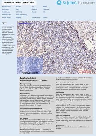
  • 1. Figure: Immunohistochemical analysis of paraffin embedded Human Tonsil tissue. 1: c-Src Polyclonal Antibody was diluted at 1:200 (4 degree Celsius,overnight). 2: Sodium citrate pH 6.0 was used for antibody retrieval (>98 degree Celsius,20min). 3: Secondary antibody was diluted at 1:200 (room temperature, 30min). Negative control was used by secondary antibody only. Report Number 92501-a Host Rabbit Application IHC-P Clonality Polyclonal Model Number STJ92501 Clone ID NA Antibody Name Anti-c-Src antibody Testing Species HUMAN Testing Tissue TONSIL ANTIBODY VALIDATION REPORT a. (A small amount of distilled water was added into the incubation box to prevent evaporation of antibody). 129. Secondary antibody incubation a. Slides were washed 3 times, with PBS on a shaker for 5min. Shortly after the slides were dried the corresponding secondary antibody solution was added (HRP labelled), covering the tissues, and incubated at room temperature for 30min. b. 130. DAB staining a. Slides were washed 3 times, with PBS on a shaker for 5min. b. Shortly after, the slides were dried and fresh DAB staining buffer was added inside the circles. The staining time was adjusted under a microscope. Yellow-brown colour represented a positive result. Slides were washed with water to stop the staining. c. 131. Haematoxylin staining a. Haematoxylin was used to counter-staining for 1min, and then the slides were washed with water. 1% Hydrochloric acid and alcohol was added for several seconds and then washed with water. Ammonia was used to reveal blue colour, and then flushed with water. b. 132. Desolation and Clearing i. Slides were incubated sequentially into: 75% alcohol 5min, 85% alcohol 5min, Anhydrous ethanol - 5min, Anhydrous ethanol - 5min & Xylene - 5min. Shortly after slides were dried and neutral gum was used to seal the slides. ii. 133. Visualization a. Results were validated with microscope, and the slides were scanned. Paraffin-Embedded Immunohistochemistry Protocol 123. 124. Tissue processing a. Slides were incubated sequentially into Xylene; 15min – Xylene, 15min - Anhydrous ethanol, 5min - Anhydrous ethanol, 5min - 85% alcohol, 5min - 75% alcohol & 5min – wash in distilled water. b. 125. Antigen retrieval a. Tissue slides were incubated with citric acid (PH6.0) antigen retrieval buffer and microwaved for antigen retrieval (heated until boiled and then stopped heating) for 8min. Slides were then heated with medium power for 7min. During this process slides were kept from drying out. After cooling down at room temperature, slides were washed with PBS on shaker for 5min, repeated for 3 times. b. 126. Inhibition of endogenous peroxidase a. Slides were placed in 3% Hydrogen peroxide solution, and incubated for 10 min at room temperature without light exposure. Slides were then washed 3 times with PBS on a shaker for 5mins. b. 127. BSA Blocking a. Shortly after slides were dried, a PAP pen was used to draw circles around the tissue sections (and to prevent draining of the antibody solution). Inside the circles, BSA was used to cover the tissue evenly, blocking for 30min. b. 128. Primary antibody incubation After blocking solution was removed a 1:200 solution of primary antibody/PBS was added on the slide, and incubated overnight at 4°C. St John's Laboratory Ltd. www.stjohnslabs.com
  • 2. Figure: Immunohistochemical analysis of paraffin embedded Human liver cancer tissue. 1: c-Src Polyclonal Antibody was diluted at 1:200 (4 degree Celsius,overnight). 2: Sodium citrate pH 6.0 was used for antibody retrieval (>98 degree Celsius,20min). 3: Secondary antibody was diluted at 1:200 (room temperature, 30min). Negative control was used by secondary antibody only. Report Number 92501-b Host Rabbit Application IHC-P Clonality Polyclonal Model Number STJ92501 Clone ID NA Antibody Name Anti-c-Src antibody Testing Species HUMAN Testing Tissue LIVER CANCER ANTIBODY VALIDATION REPORT a. (A small amount of distilled water was added into the incubation box to prevent evaporation of antibody). 118. Secondary antibody incubation a. Slides were washed 3 times, with PBS on a shaker for 5min. Shortly after the slides were dried the corresponding secondary antibody solution was added (HRP labelled), covering the tissues, and incubated at room temperature for 30min. b. 119. DAB staining a. Slides were washed 3 times, with PBS on a shaker for 5min. b. Shortly after, the slides were dried and fresh DAB staining buffer was added inside the circles. The staining time was adjusted under a microscope. Yellow-brown colour represented a positive result. Slides were washed with water to stop the staining. c. 120. Haematoxylin staining a. Haematoxylin was used to counter-staining for 1min, and then the slides were washed with water. 1% Hydrochloric acid and alcohol was added for several seconds and then washed with water. Ammonia was used to reveal blue colour, and then flushed with water. b. 121. Desolation and Clearing i. Slides were incubated sequentially into: 75% alcohol 5min, 85% alcohol 5min, Anhydrous ethanol - 5min, Anhydrous ethanol - 5min & Xylene - 5min. Shortly after slides were dried and neutral gum was used to seal the slides. ii. 122. Visualization a. Results were validated with microscope, and the slides were scanned. Paraffin-Embedded Immunohistochemistry Protocol 112. 113. Tissue processing a. Slides were incubated sequentially into Xylene; 15min – Xylene, 15min - Anhydrous ethanol, 5min - Anhydrous ethanol, 5min - 85% alcohol, 5min - 75% alcohol & 5min – wash in distilled water. b. 114. Antigen retrieval a. Tissue slides were incubated with citric acid (PH6.0) antigen retrieval buffer and microwaved for antigen retrieval (heated until boiled and then stopped heating) for 8min. Slides were then heated with medium power for 7min. During this process slides were kept from drying out. After cooling down at room temperature, slides were washed with PBS on shaker for 5min, repeated for 3 times. b. 115. Inhibition of endogenous peroxidase a. Slides were placed in 3% Hydrogen peroxide solution, and incubated for 10 min at room temperature without light exposure. Slides were then washed 3 times with PBS on a shaker for 5mins. b. 116. BSA Blocking a. Shortly after slides were dried, a PAP pen was used to draw circles around the tissue sections (and to prevent draining of the antibody solution). Inside the circles, BSA was used to cover the tissue evenly, blocking for 30min. b. 117. Primary antibody incubation After blocking solution was removed a 1:200 solution of primary antibody/PBS was added on the slide, and incubated overnight at 4°C. St John's Laboratory Ltd. www.stjohnslabs.com
  • 3. Figure: Immunohistochemical analysis of paraffin embedded Human stomach tissue. 1: c-Src Polyclonal Antibody was diluted at 1:200 (4 degree Celsius,overnight). 2: Sodium citrate pH 6.0 was used for antibody retrieval (>98 degree Celsius,20min). 3: Secondary antibody was diluted at 1:200 (room temperature, 30min). Negative control was used by secondary antibody only. Report Number 92501-c Host Rabbit Application IHC-P Clonality Polyclonal Model Number STJ92501 Clone ID NA Antibody Name Anti-c-Src antibody Testing Species HUMAN Testing Tissue STOMACH ANTIBODY VALIDATION REPORT a. (A small amount of distilled water was added into the incubation box to prevent evaporation of antibody). 107. Secondary antibody incubation a. Slides were washed 3 times, with PBS on a shaker for 5min. Shortly after the slides were dried the corresponding secondary antibody solution was added (HRP labelled), covering the tissues, and incubated at room temperature for 30min. b. 108. DAB staining a. Slides were washed 3 times, with PBS on a shaker for 5min. b. Shortly after, the slides were dried and fresh DAB staining buffer was added inside the circles. The staining time was adjusted under a microscope. Yellow-brown colour represented a positive result. Slides were washed with water to stop the staining. c. 109. Haematoxylin staining a. Haematoxylin was used to counter-staining for 1min, and then the slides were washed with water. 1% Hydrochloric acid and alcohol was added for several seconds and then washed with water. Ammonia was used to reveal blue colour, and then flushed with water. b. 110. Desolation and Clearing i. Slides were incubated sequentially into: 75% alcohol 5min, 85% alcohol 5min, Anhydrous ethanol - 5min, Anhydrous ethanol - 5min & Xylene - 5min. Shortly after slides were dried and neutral gum was used to seal the slides. ii. 111. Visualization a. Results were validated with microscope, and the slides were scanned. Paraffin-Embedded Immunohistochemistry Protocol 101. 102. Tissue processing a. Slides were incubated sequentially into Xylene; 15min – Xylene, 15min - Anhydrous ethanol, 5min - Anhydrous ethanol, 5min - 85% alcohol, 5min - 75% alcohol & 5min – wash in distilled water. b. 103. Antigen retrieval a. Tissue slides were incubated with citric acid (PH6.0) antigen retrieval buffer and microwaved for antigen retrieval (heated until boiled and then stopped heating) for 8min. Slides were then heated with medium power for 7min. During this process slides were kept from drying out. After cooling down at room temperature, slides were washed with PBS on shaker for 5min, repeated for 3 times. b. 104. Inhibition of endogenous peroxidase a. Slides were placed in 3% Hydrogen peroxide solution, and incubated for 10 min at room temperature without light exposure. Slides were then washed 3 times with PBS on a shaker for 5mins. b. 105. BSA Blocking a. Shortly after slides were dried, a PAP pen was used to draw circles around the tissue sections (and to prevent draining of the antibody solution). Inside the circles, BSA was used to cover the tissue evenly, blocking for 30min. b. 106. Primary antibody incubation After blocking solution was removed a 1:200 solution of primary antibody/PBS was added on the slide, and incubated overnight at 4°C. St John's Laboratory Ltd. www.stjohnslabs.com
  • 4. Figure: Immunohistochemical analysis of paraffin embedded Human stomach cancer tissue. 1: c-Src Polyclonal Antibody was diluted at 1:200 (4 degree Celsius,overnight). 2: Sodium citrate pH 6.0 was used for antibody retrieval (>98 degree Celsius,20min). 3: Secondary antibody was diluted at 1:200 (room temperature, 30min). Negative control was used by secondary antibody only. Report Number 92501-d Host Rabbit Application IHC-P Clonality Polyclonal Model Number STJ92501 Clone ID NA Antibody Name Anti-c-Src antibody Testing Species HUMAN Testing Tissue STOMACH CANCER ANTIBODY VALIDATION REPORT a. (A small amount of distilled water was added into the incubation box to prevent evaporation of antibody). 96. Secondary antibody incubation a. Slides were washed 3 times, with PBS on a shaker for 5min. Shortly after the slides were dried the corresponding secondary antibody solution was added (HRP labelled), covering the tissues, and incubated at room temperature for 30min. b. 97. DAB staining a. Slides were washed 3 times, with PBS on a shaker for 5min. b. Shortly after, the slides were dried and fresh DAB staining buffer was added inside the circles. The staining time was adjusted under a microscope. Yellow-brown colour represented a positive result. Slides were washed with water to stop the staining. c. 98. Haematoxylin staining a. Haematoxylin was used to counter-staining for 1min, and then the slides were washed with water. 1% Hydrochloric acid and alcohol was added for several seconds and then washed with water. Ammonia was used to reveal blue colour, and then flushed with water. b. 99. Desolation and Clearing i. Slides were incubated sequentially into: 75% alcohol 5min, 85% alcohol 5min, Anhydrous ethanol - 5min, Anhydrous ethanol - 5min & Xylene - 5min. Shortly after slides were dried and neutral gum was used to seal the slides. ii. 100. Visualization a. Results were validated with microscope, and the slides were scanned. Paraffin-Embedded Immunohistochemistry Protocol 90. 91. Tissue processing a. Slides were incubated sequentially into Xylene; 15min – Xylene, 15min - Anhydrous ethanol, 5min - Anhydrous ethanol, 5min - 85% alcohol, 5min - 75% alcohol & 5min – wash in distilled water. b. 92. Antigen retrieval a. Tissue slides were incubated with citric acid (PH6.0) antigen retrieval buffer and microwaved for antigen retrieval (heated until boiled and then stopped heating) for 8min. Slides were then heated with medium power for 7min. During this process slides were kept from drying out. After cooling down at room temperature, slides were washed with PBS on shaker for 5min, repeated for 3 times. b. 93. Inhibition of endogenous peroxidase a. Slides were placed in 3% Hydrogen peroxide solution, and incubated for 10 min at room temperature without light exposure. Slides were then washed 3 times with PBS on a shaker for 5mins. b. 94. BSA Blocking a. Shortly after slides were dried, a PAP pen was used to draw circles around the tissue sections (and to prevent draining of the antibody solution). Inside the circles, BSA was used to cover the tissue evenly, blocking for 30min. b. 95. Primary antibody incubation After blocking solution was removed a 1:200 solution of primary antibody/PBS was added on the slide, and incubated overnight at 4°C. St John's Laboratory Ltd. www.stjohnslabs.com
  • 5. Figure: Immunohistochemical analysis of paraffin embedded Human appendix tissue. 1: c-Src Polyclonal Antibody was diluted at 1:200 (4 degree Celsius,overnight). 2: Sodium citrate pH 6.0 was used for antibody retrieval (>98 degree Celsius,20min). 3: Secondary antibody was diluted at 1:200 (room temperature, 30min). Negative control was used by secondary antibody only. Report Number 92501-e Host Rabbit Application IHC-P Clonality Polyclonal Model Number STJ92501 Clone ID NA Antibody Name Anti-c-Src antibody Testing Species HUMAN Testing Tissue APPENDIX ANTIBODY VALIDATION REPORT a. (A small amount of distilled water was added into the incubation box to prevent evaporation of antibody). 85. Secondary antibody incubation a. Slides were washed 3 times, with PBS on a shaker for 5min. Shortly after the slides were dried the corresponding secondary antibody solution was added (HRP labelled), covering the tissues, and incubated at room temperature for 30min. b. 86. DAB staining a. Slides were washed 3 times, with PBS on a shaker for 5min. b. Shortly after, the slides were dried and fresh DAB staining buffer was added inside the circles. The staining time was adjusted under a microscope. Yellow-brown colour represented a positive result. Slides were washed with water to stop the staining. c. 87. Haematoxylin staining a. Haematoxylin was used to counter-staining for 1min, and then the slides were washed with water. 1% Hydrochloric acid and alcohol was added for several seconds and then washed with water. Ammonia was used to reveal blue colour, and then flushed with water. b. 88. Desolation and Clearing i. Slides were incubated sequentially into: 75% alcohol 5min, 85% alcohol 5min, Anhydrous ethanol - 5min, Anhydrous ethanol - 5min & Xylene - 5min. Shortly after slides were dried and neutral gum was used to seal the slides. ii. 89. Visualization a. Results were validated with microscope, and the slides were scanned. Paraffin-Embedded Immunohistochemistry Protocol 79. 80. Tissue processing a. Slides were incubated sequentially into Xylene; 15min – Xylene, 15min - Anhydrous ethanol, 5min - Anhydrous ethanol, 5min - 85% alcohol, 5min - 75% alcohol & 5min – wash in distilled water. b. 81. Antigen retrieval a. Tissue slides were incubated with citric acid (PH6.0) antigen retrieval buffer and microwaved for antigen retrieval (heated until boiled and then stopped heating) for 8min. Slides were then heated with medium power for 7min. During this process slides were kept from drying out. After cooling down at room temperature, slides were washed with PBS on shaker for 5min, repeated for 3 times. b. 82. Inhibition of endogenous peroxidase a. Slides were placed in 3% Hydrogen peroxide solution, and incubated for 10 min at room temperature without light exposure. Slides were then washed 3 times with PBS on a shaker for 5mins. b. 83. BSA Blocking a. Shortly after slides were dried, a PAP pen was used to draw circles around the tissue sections (and to prevent draining of the antibody solution). Inside the circles, BSA was used to cover the tissue evenly, blocking for 30min. b. 84. Primary antibody incubation After blocking solution was removed a 1:200 solution of primary antibody/PBS was added on the slide, and incubated overnight at 4°C. St John's Laboratory Ltd. www.stjohnslabs.com
  • 6. Figure: Immunohistochemical analysis of paraffin embedded Rat testis tissue. 1: c-Src Polyclonal Antibody was diluted at 1:200 (4 degree Celsius,overnight). 2: Sodium citrate pH 6.0 was used for antibody retrieval (>98 degree Celsius,20min). 3: Secondary antibody was diluted at 1:200 (room temperature, 30min). Negative control was used by secondary antibody only. Report Number 92501-f Host Rabbit Application IHC-P Clonality Polyclonal Model Number STJ92501 Clone ID NA Antibody Name Anti-c-Src antibody Testing Species RAT Testing Tissue TESTIS ANTIBODY VALIDATION REPORT a. (A small amount of distilled water was added into the incubation box to prevent evaporation of antibody). 74. Secondary antibody incubation a. Slides were washed 3 times, with PBS on a shaker for 5min. Shortly after the slides were dried the corresponding secondary antibody solution was added (HRP labelled), covering the tissues, and incubated at room temperature for 30min. b. 75. DAB staining a. Slides were washed 3 times, with PBS on a shaker for 5min. b. Shortly after, the slides were dried and fresh DAB staining buffer was added inside the circles. The staining time was adjusted under a microscope. Yellow-brown colour represented a positive result. Slides were washed with water to stop the staining. c. 76. Haematoxylin staining a. Haematoxylin was used to counter-staining for 1min, and then the slides were washed with water. 1% Hydrochloric acid and alcohol was added for several seconds and then washed with water. Ammonia was used to reveal blue colour, and then flushed with water. b. 77. Desolation and Clearing i. Slides were incubated sequentially into: 75% alcohol 5min, 85% alcohol 5min, Anhydrous ethanol - 5min, Anhydrous ethanol - 5min & Xylene - 5min. Shortly after slides were dried and neutral gum was used to seal the slides. ii. 78. Visualization a. Results were validated with microscope, and the slides were scanned. Paraffin-Embedded Immunohistochemistry Protocol 68. 69. Tissue processing a. Slides were incubated sequentially into Xylene; 15min – Xylene, 15min - Anhydrous ethanol, 5min - Anhydrous ethanol, 5min - 85% alcohol, 5min - 75% alcohol & 5min – wash in distilled water. b. 70. Antigen retrieval a. Tissue slides were incubated with citric acid (PH6.0) antigen retrieval buffer and microwaved for antigen retrieval (heated until boiled and then stopped heating) for 8min. Slides were then heated with medium power for 7min. During this process slides were kept from drying out. After cooling down at room temperature, slides were washed with PBS on shaker for 5min, repeated for 3 times. b. 71. Inhibition of endogenous peroxidase a. Slides were placed in 3% Hydrogen peroxide solution, and incubated for 10 min at room temperature without light exposure. Slides were then washed 3 times with PBS on a shaker for 5mins. b. 72. BSA Blocking a. Shortly after slides were dried, a PAP pen was used to draw circles around the tissue sections (and to prevent draining of the antibody solution). Inside the circles, BSA was used to cover the tissue evenly, blocking for 30min. b. 73. Primary antibody incubation After blocking solution was removed a 1:200 solution of primary antibody/PBS was added on the slide, and incubated overnight at 4°C. St John's Laboratory Ltd. www.stjohnslabs.com
  • 7. Figure: Immunohistochemical analysis of paraffin embedded Rat kidney tissue. 1: c-Src Polyclonal Antibody was diluted at 1:200 (4 degree Celsius,overnight). 2: Sodium citrate pH 6.0 was used for antibody retrieval (>98 degree Celsius,20min). 3: Secondary antibody was diluted at 1:200 (room temperature, 30min). Negative control was used by secondary antibody only. Report Number 92501-g Host Rabbit Application IHC-P Clonality Polyclonal Model Number STJ92501 Clone ID NA Antibody Name Anti-c-Src antibody Testing Species RAT Testing Tissue KIDNEY ANTIBODY VALIDATION REPORT a. (A small amount of distilled water was added into the incubation box to prevent evaporation of antibody). 63. Secondary antibody incubation a. Slides were washed 3 times, with PBS on a shaker for 5min. Shortly after the slides were dried the corresponding secondary antibody solution was added (HRP labelled), covering the tissues, and incubated at room temperature for 30min. b. 64. DAB staining a. Slides were washed 3 times, with PBS on a shaker for 5min. b. Shortly after, the slides were dried and fresh DAB staining buffer was added inside the circles. The staining time was adjusted under a microscope. Yellow-brown colour represented a positive result. Slides were washed with water to stop the staining. c. 65. Haematoxylin staining a. Haematoxylin was used to counter-staining for 1min, and then the slides were washed with water. 1% Hydrochloric acid and alcohol was added for several seconds and then washed with water. Ammonia was used to reveal blue colour, and then flushed with water. b. 66. Desolation and Clearing i. Slides were incubated sequentially into: 75% alcohol 5min, 85% alcohol 5min, Anhydrous ethanol - 5min, Anhydrous ethanol - 5min & Xylene - 5min. Shortly after slides were dried and neutral gum was used to seal the slides. ii. 67. Visualization a. Results were validated with microscope, and the slides were scanned. Paraffin-Embedded Immunohistochemistry Protocol 57. 58. Tissue processing a. Slides were incubated sequentially into Xylene; 15min – Xylene, 15min - Anhydrous ethanol, 5min - Anhydrous ethanol, 5min - 85% alcohol, 5min - 75% alcohol & 5min – wash in distilled water. b. 59. Antigen retrieval a. Tissue slides were incubated with citric acid (PH6.0) antigen retrieval buffer and microwaved for antigen retrieval (heated until boiled and then stopped heating) for 8min. Slides were then heated with medium power for 7min. During this process slides were kept from drying out. After cooling down at room temperature, slides were washed with PBS on shaker for 5min, repeated for 3 times. b. 60. Inhibition of endogenous peroxidase a. Slides were placed in 3% Hydrogen peroxide solution, and incubated for 10 min at room temperature without light exposure. Slides were then washed 3 times with PBS on a shaker for 5mins. b. 61. BSA Blocking a. Shortly after slides were dried, a PAP pen was used to draw circles around the tissue sections (and to prevent draining of the antibody solution). Inside the circles, BSA was used to cover the tissue evenly, blocking for 30min. b. 62. Primary antibody incubation After blocking solution was removed a 1:200 solution of primary antibody/PBS was added on the slide, and incubated overnight at 4°C. St John's Laboratory Ltd. www.stjohnslabs.com
  • 8. Figure: Immunohistochemical analysis of paraffin embedded Mouse heart tissue. 1: c-Src Polyclonal Antibody was diluted at 1:200 (4 degree Celsius,overnight). 2: Sodium citrate pH 6.0 was used for antibody retrieval (>98 degree Celsius,20min). 3: Secondary antibody was diluted at 1:200 (room temperature, 30min). Negative control was used by secondary antibody only. Report Number 92501-h Host Rabbit Application IHC-P Clonality Polyclonal Model Number STJ92501 Clone ID NA Antibody Name Anti-c-Src antibody Testing Species MOUSE Testing Tissue HEART ANTIBODY VALIDATION REPORT a. (A small amount of distilled water was added into the incubation box to prevent evaporation of antibody). 52. Secondary antibody incubation a. Slides were washed 3 times, with PBS on a shaker for 5min. Shortly after the slides were dried the corresponding secondary antibody solution was added (HRP labelled), covering the tissues, and incubated at room temperature for 30min. b. 53. DAB staining a. Slides were washed 3 times, with PBS on a shaker for 5min. b. Shortly after, the slides were dried and fresh DAB staining buffer was added inside the circles. The staining time was adjusted under a microscope. Yellow-brown colour represented a positive result. Slides were washed with water to stop the staining. c. 54. Haematoxylin staining a. Haematoxylin was used to counter-staining for 1min, and then the slides were washed with water. 1% Hydrochloric acid and alcohol was added for several seconds and then washed with water. Ammonia was used to reveal blue colour, and then flushed with water. b. 55. Desolation and Clearing i. Slides were incubated sequentially into: 75% alcohol 5min, 85% alcohol 5min, Anhydrous ethanol - 5min, Anhydrous ethanol - 5min & Xylene - 5min. Shortly after slides were dried and neutral gum was used to seal the slides. ii. 56. Visualization a. Results were validated with microscope, and the slides were scanned. Paraffin-Embedded Immunohistochemistry Protocol 46. 47. Tissue processing a. Slides were incubated sequentially into Xylene; 15min – Xylene, 15min - Anhydrous ethanol, 5min - Anhydrous ethanol, 5min - 85% alcohol, 5min - 75% alcohol & 5min – wash in distilled water. b. 48. Antigen retrieval a. Tissue slides were incubated with citric acid (PH6.0) antigen retrieval buffer and microwaved for antigen retrieval (heated until boiled and then stopped heating) for 8min. Slides were then heated with medium power for 7min. During this process slides were kept from drying out. After cooling down at room temperature, slides were washed with PBS on shaker for 5min, repeated for 3 times. b. 49. Inhibition of endogenous peroxidase a. Slides were placed in 3% Hydrogen peroxide solution, and incubated for 10 min at room temperature without light exposure. Slides were then washed 3 times with PBS on a shaker for 5mins. b. 50. BSA Blocking a. Shortly after slides were dried, a PAP pen was used to draw circles around the tissue sections (and to prevent draining of the antibody solution). Inside the circles, BSA was used to cover the tissue evenly, blocking for 30min. b. 51. Primary antibody incubation After blocking solution was removed a 1:200 solution of primary antibody/PBS was added on the slide, and incubated overnight at 4°C. St John's Laboratory Ltd. www.stjohnslabs.com
  • 9. Figure: Immunohistochemical analysis of paraffin embedded Mouse testis tissue. 1: c-Src Polyclonal Antibody was diluted at 1:200 (4 degree Celsius,overnight). 2: Sodium citrate pH 6.0 was used for antibody retrieval (>98 degree Celsius,20min). 3: Secondary antibody was diluted at 1:200 (room temperature, 30min). Negative control was used by secondary antibody only. Report Number 92501-i Host Rabbit Application IHC-P Clonality Polyclonal Model Number STJ92501 Clone ID NA Antibody Name Anti-c-Src antibody Testing Species MOUSE Testing Tissue TESTIS ANTIBODY VALIDATION REPORT a. (A small amount of distilled water was added into the incubation box to prevent evaporation of antibody). 41. Secondary antibody incubation a. Slides were washed 3 times, with PBS on a shaker for 5min. Shortly after the slides were dried the corresponding secondary antibody solution was added (HRP labelled), covering the tissues, and incubated at room temperature for 30min. b. 42. DAB staining a. Slides were washed 3 times, with PBS on a shaker for 5min. b. Shortly after, the slides were dried and fresh DAB staining buffer was added inside the circles. The staining time was adjusted under a microscope. Yellow-brown colour represented a positive result. Slides were washed with water to stop the staining. c. 43. Haematoxylin staining a. Haematoxylin was used to counter-staining for 1min, and then the slides were washed with water. 1% Hydrochloric acid and alcohol was added for several seconds and then washed with water. Ammonia was used to reveal blue colour, and then flushed with water. b. 44. Desolation and Clearing i. Slides were incubated sequentially into: 75% alcohol 5min, 85% alcohol 5min, Anhydrous ethanol - 5min, Anhydrous ethanol - 5min & Xylene - 5min. Shortly after slides were dried and neutral gum was used to seal the slides. ii. 45. Visualization a. Results were validated with microscope, and the slides were scanned. Paraffin-Embedded Immunohistochemistry Protocol 35. 36. Tissue processing a. Slides were incubated sequentially into Xylene; 15min – Xylene, 15min - Anhydrous ethanol, 5min - Anhydrous ethanol, 5min - 85% alcohol, 5min - 75% alcohol & 5min – wash in distilled water. b. 37. Antigen retrieval a. Tissue slides were incubated with citric acid (PH6.0) antigen retrieval buffer and microwaved for antigen retrieval (heated until boiled and then stopped heating) for 8min. Slides were then heated with medium power for 7min. During this process slides were kept from drying out. After cooling down at room temperature, slides were washed with PBS on shaker for 5min, repeated for 3 times. b. 38. Inhibition of endogenous peroxidase a. Slides were placed in 3% Hydrogen peroxide solution, and incubated for 10 min at room temperature without light exposure. Slides were then washed 3 times with PBS on a shaker for 5mins. b. 39. BSA Blocking a. Shortly after slides were dried, a PAP pen was used to draw circles around the tissue sections (and to prevent draining of the antibody solution). Inside the circles, BSA was used to cover the tissue evenly, blocking for 30min. b. 40. Primary antibody incubation After blocking solution was removed a 1:200 solution of primary antibody/PBS was added on the slide, and incubated overnight at 4°C. St John's Laboratory Ltd. www.stjohnslabs.com
  • 10. Figure: Immunohistochemical analysis of paraffin embedded Mouse liver tissue. 1: c-Src Polyclonal Antibody was diluted at 1:200 (4 degree Celsius,overnight). 2: Sodium citrate pH 6.0 was used for antibody retrieval (>98 degree Celsius,20min). 3: Secondary antibody was diluted at 1:200 (room temperature, 30min). Negative control was used by secondary antibody only. Report Number 92501-j Host Rabbit Application IHC-P Clonality Polyclonal Model Number STJ92501 Clone ID NA Antibody Name Anti-c-Src antibody Testing Species MOUSE Testing Tissue LIVER ANTIBODY VALIDATION REPORT b. (A small amount of distilled water was added into the incubation box to prevent evaporation of antibody). 30. Secondary antibody incubation a. Slides were washed 3 times, with PBS on a shaker for 5min. Shortly after the slides were dried the corresponding secondary antibody solution was added (HRP labelled), covering the tissues, and incubated at room temperature for 30min. b. 31. DAB staining a. Slides were washed 3 times, with PBS on a shaker for 5min. b. Shortly after, the slides were dried and fresh DAB staining buffer was added inside the circles. The staining time was adjusted under a microscope. Yellow-brown colour represented a positive result. Slides were washed with water to stop the staining. c. 32. Haematoxylin staining a. Haematoxylin was used to counter-staining for 1min, and then the slides were washed with water. 1% Hydrochloric acid and alcohol was added for several seconds and then washed with water. Ammonia was used to reveal blue colour, and then flushed with water. b. 33. Desolation and Clearing i. Slides were incubated sequentially into: 75% alcohol 5min, 85% alcohol 5min, Anhydrous ethanol - 5min, Anhydrous ethanol - 5min & Xylene - 5min. Shortly after slides were dried and neutral gum was used to seal the slides. ii. 34. Visualization a. Results were validated with microscope, and the slides were scanned. Paraffin-Embedded Immunohistochemistry Protocol 24. 25. Tissue processing a. Slides were incubated sequentially into Xylene; 15min – Xylene, 15min - Anhydrous ethanol, 5min - Anhydrous ethanol, 5min - 85% alcohol, 5min - 75% alcohol & 5min – wash in distilled water. b. 26. Antigen retrieval a. Tissue slides were incubated with citric acid (PH6.0) antigen retrieval buffer and microwaved for antigen retrieval (heated until boiled and then stopped heating) for 8min. Slides were then heated with medium power for 7min. During this process slides were kept from drying out. After cooling down at room temperature, slides were washed with PBS on shaker for 5min, repeated for 3 times. b. 27. Inhibition of endogenous peroxidase a. Slides were placed in 3% Hydrogen peroxide solution, and incubated for 10 min at room temperature without light exposure. Slides were then washed 3 times with PBS on a shaker for 5mins. b. 28. BSA Blocking a. Shortly after slides were dried, a PAP pen was used to draw circles around the tissue sections (and to prevent draining of the antibody solution). Inside the circles, BSA was used to cover the tissue evenly, blocking for 30min. b. 29. Primary antibody incubation After blocking solution was removed a 1:200 solution of primary antibody/PBS was added on the slide, and incubated overnight at 4°C. St John's Laboratory Ltd. www.stjohnslabs.com
  • 11. Figure: Immunohistochemical analysis of paraffin embedded Mouse lung tissue. 1: c-Src Polyclonal Antibody was diluted at 1:200 (4 degree Celsius,overnight). 2: Sodium citrate pH 6.0 was used for antibody retrieval (>98 degree Celsius,20min). 3: Secondary antibody was diluted at 1:200 (room temperature, 30min). Negative control was used by secondary antibody only. Report Number 92501-k Host Rabbit Application IHC-P Clonality Polyclonal Model Number STJ92501 Clone ID NA Antibody Name Anti-c-Src antibody Testing Species MOUSE Testing Tissue LUNG ANTIBODY VALIDATION REPORT b. (A small amount of distilled water was added into the incubation box to prevent evaporation of antibody). 19. Secondary antibody incubation a. Slides were washed 3 times, with PBS on a shaker for 5min. Shortly after the slides were dried the corresponding secondary antibody solution was added (HRP labelled), covering the tissues, and incubated at room temperature for 30min. b. 20. DAB staining a. Slides were washed 3 times, with PBS on a shaker for 5min. b. Shortly after, the slides were dried and fresh DAB staining buffer was added inside the circles. The staining time was adjusted under a microscope. Yellow-brown colour represented a positive result. Slides were washed with water to stop the staining. c. 21. Haematoxylin staining a. Haematoxylin was used to counter-staining for 1min, and then the slides were washed with water. 1% Hydrochloric acid and alcohol was added for several seconds and then washed with water. Ammonia was used to reveal blue colour, and then flushed with water. b. 22. Desolation and Clearing i. Slides were incubated sequentially into: 75% alcohol 5min, 85% alcohol 5min, Anhydrous ethanol - 5min, Anhydrous ethanol - 5min & Xylene - 5min. Shortly after slides were dried and neutral gum was used to seal the slides. ii. 23. Visualization a. Results were validated with microscope, and the slides were scanned. Paraffin-Embedded Immunohistochemistry Protocol 13. 14. Tissue processing a. Slides were incubated sequentially into Xylene; 15min – Xylene, 15min - Anhydrous ethanol, 5min - Anhydrous ethanol, 5min - 85% alcohol, 5min - 75% alcohol & 5min – wash in distilled water. b. 15. Antigen retrieval a. Tissue slides were incubated with citric acid (PH6.0) antigen retrieval buffer and microwaved for antigen retrieval (heated until boiled and then stopped heating) for 8min. Slides were then heated with medium power for 7min. During this process slides were kept from drying out. After cooling down at room temperature, slides were washed with PBS on shaker for 5min, repeated for 3 times. b. 16. Inhibition of endogenous peroxidase a. Slides were placed in 3% Hydrogen peroxide solution, and incubated for 10 min at room temperature without light exposure. Slides were then washed 3 times with PBS on a shaker for 5mins. b. 17. BSA Blocking a. Shortly after slides were dried, a PAP pen was used to draw circles around the tissue sections (and to prevent draining of the antibody solution). Inside the circles, BSA was used to cover the tissue evenly, blocking for 30min. b. 18. Primary antibody incubation After blocking solution was removed a 1:200 solution of primary antibody/PBS was added on the slide, and incubated overnight at 4°C. St John's Laboratory Ltd. www.stjohnslabs.com
  • 12. Figure: Immunohistochemical analysis of paraffin embedded Mouse kidney tissue. 1: c-Src Polyclonal Antibody was diluted at 1:200 (4 degree Celsius,overnight). 2: Sodium citrate pH 6.0 was used for antibody retrieval (>98 degree Celsius,20min). 3: Secondary antibody was diluted at 1:200 (room temperature, 30min). Negative control was used by secondary antibody only. Report Number 92501-l Host Rabbit Application IHC-P Clonality Polyclonal Model Number STJ92501 Clone ID NA Antibody Name Anti-c-Src antibody Testing Species MOUSE Testing Tissue KIDNEY ANTIBODY VALIDATION REPORT b. (A small amount of distilled water was added into the incubation box to prevent evaporation of antibody). 8. Secondary antibody incubation a. Slides were washed 3 times, with PBS on a shaker for 5min. Shortly after the slides were dried the corresponding secondary antibody solution was added (HRP labelled), covering the tissues, and incubated at room temperature for 30min. b. 9. DAB staining a. Slides were washed 3 times, with PBS on a shaker for 5min. b. Shortly after, the slides were dried and fresh DAB staining buffer was added inside the circles. The staining time was adjusted under a microscope. Yellow-brown colour represented a positive result. Slides were washed with water to stop the staining. c. 10. Haematoxylin staining a. Haematoxylin was used to counter-staining for 1min, and then the slides were washed with water. 1% Hydrochloric acid and alcohol was added for several seconds and then washed with water. Ammonia was used to reveal blue colour, and then flushed with water. b. 11. Desolation and Clearing i. Slides were incubated sequentially into: 75% alcohol 5min, 85% alcohol 5min, Anhydrous ethanol - 5min, Anhydrous ethanol - 5min & Xylene - 5min. Shortly after slides were dried and neutral gum was used to seal the slides. ii. 12. Visualization a. Results were validated with microscope, and the slides were scanned. Paraffin-Embedded Immunohistochemistry Protocol 2. 3. Tissue processing a. Slides were incubated sequentially into Xylene; 15min – Xylene, 15min - Anhydrous ethanol, 5min - Anhydrous ethanol, 5min - 85% alcohol, 5min - 75% alcohol & 5min – wash in distilled water. b. 4. Antigen retrieval a. Tissue slides were incubated with citric acid (PH6.0) antigen retrieval buffer and microwaved for antigen retrieval (heated until boiled and then stopped heating) for 8min. Slides were then heated with medium power for 7min. During this process slides were kept from drying out. After cooling down at room temperature, slides were washed with PBS on shaker for 5min, repeated for 3 times. b. 5. Inhibition of endogenous peroxidase a. Slides were placed in 3% Hydrogen peroxide solution, and incubated for 10 min at room temperature without light exposure. Slides were then washed 3 times with PBS on a shaker for 5mins. b. 6. BSA Blocking a. Shortly after slides were dried, a PAP pen was used to draw circles around the tissue sections (and to prevent draining of the antibody solution). Inside the circles, BSA was used to cover the tissue evenly, blocking for 30min. b. 7. Primary antibody incubation After blocking solution was removed a 1:200 solution of primary antibody/PBS was added on the slide, and incubated overnight at 4°C. St John's Laboratory Ltd. www.stjohnslabs.com