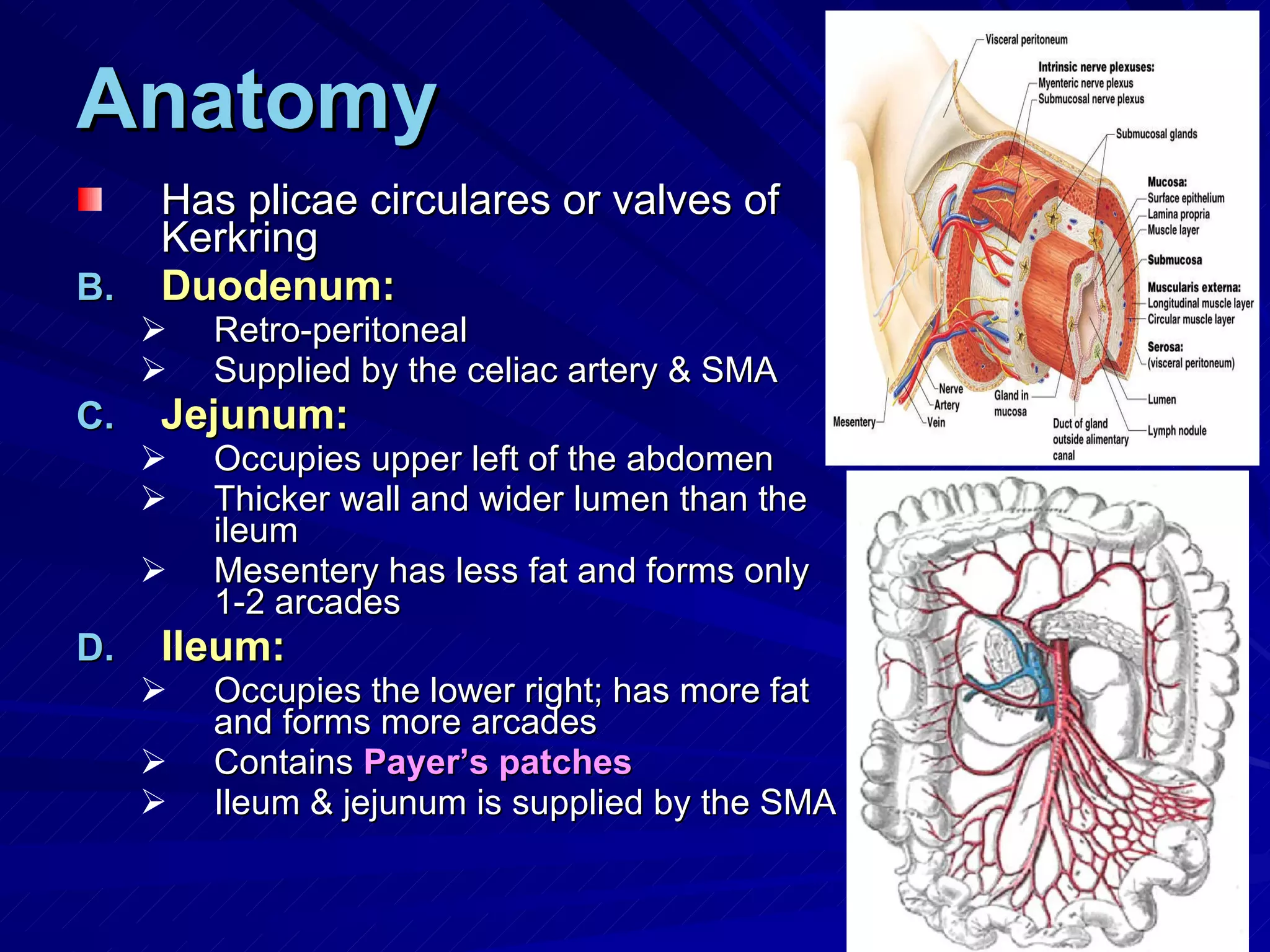
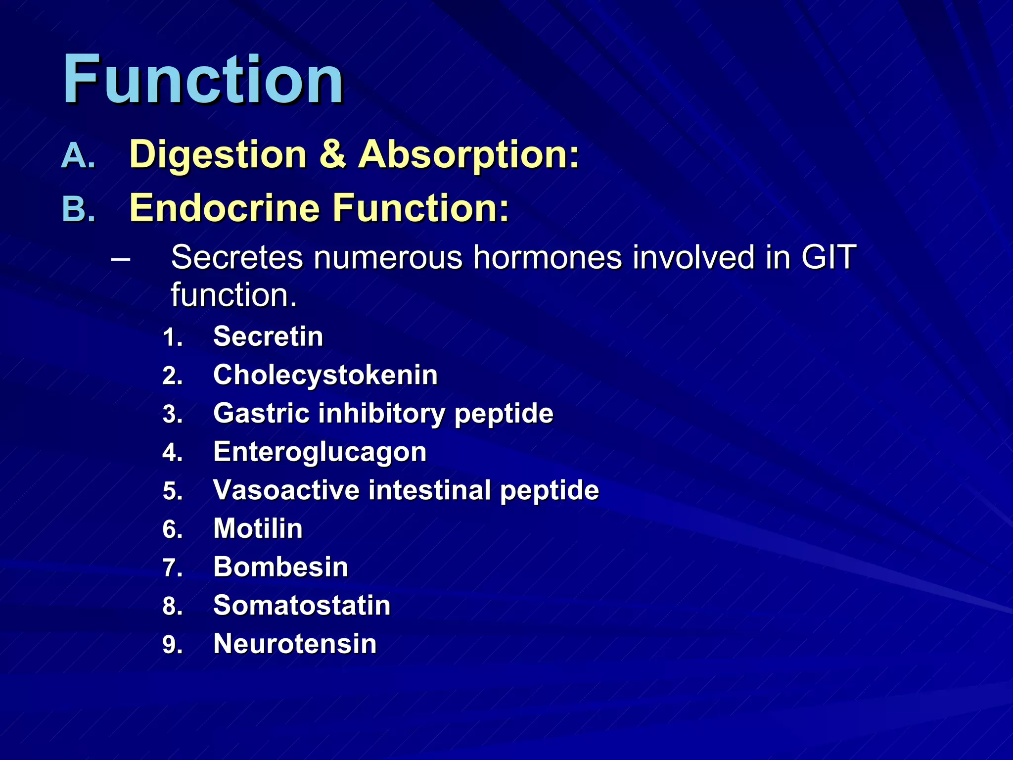
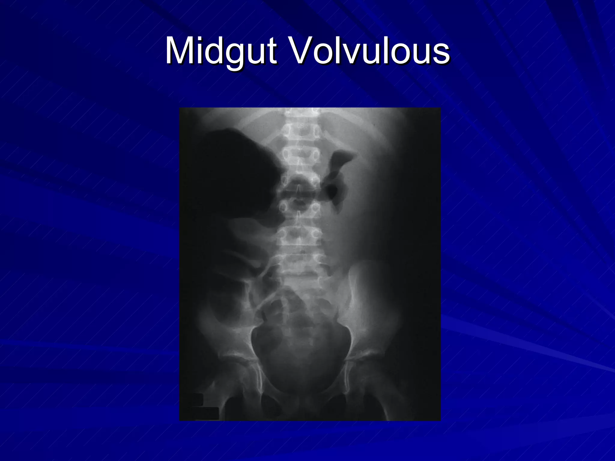
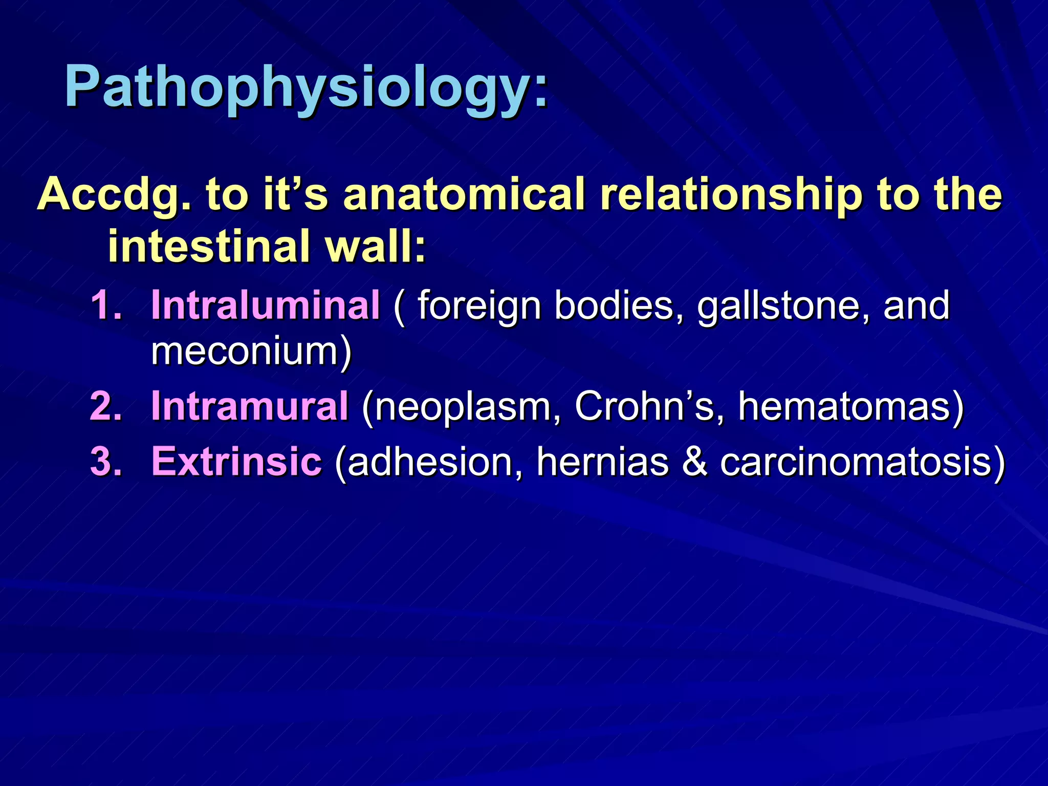
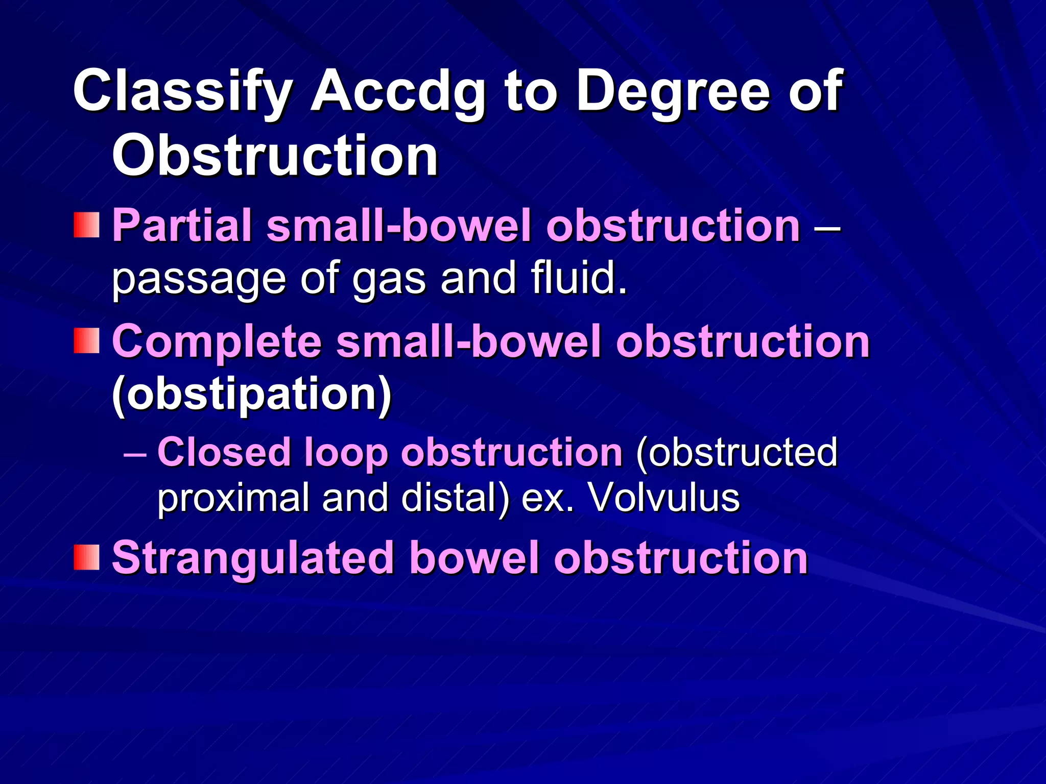
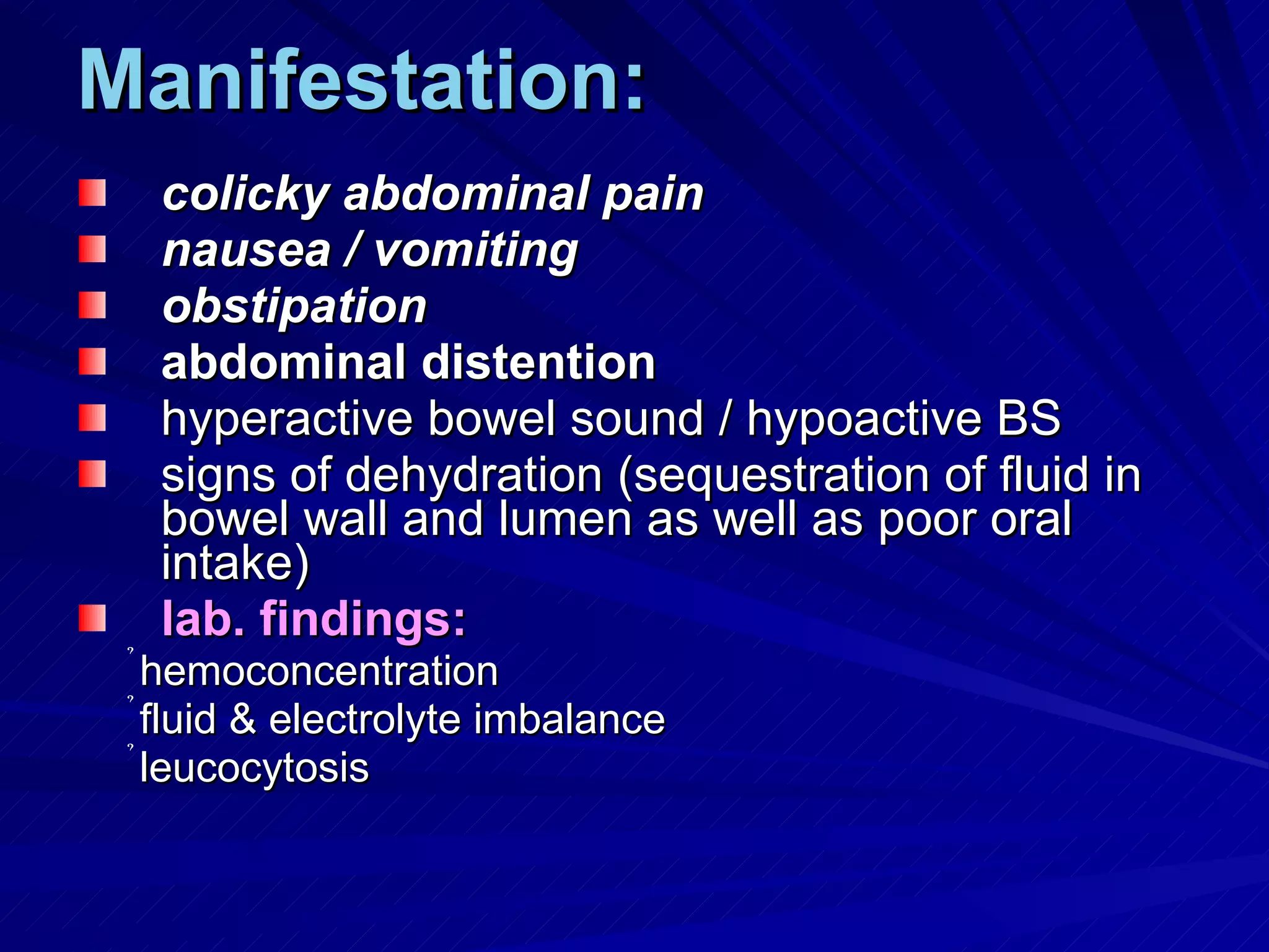
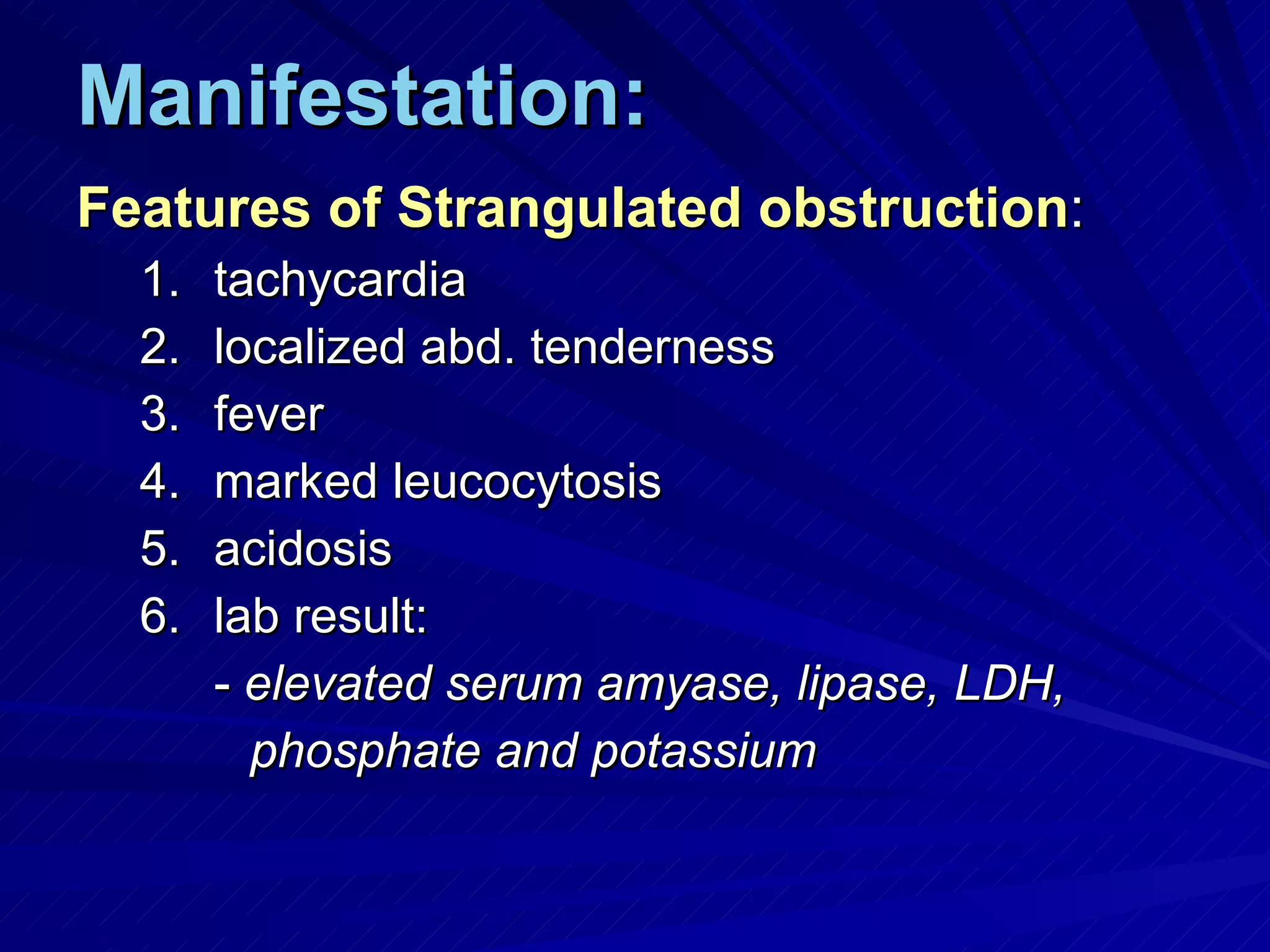
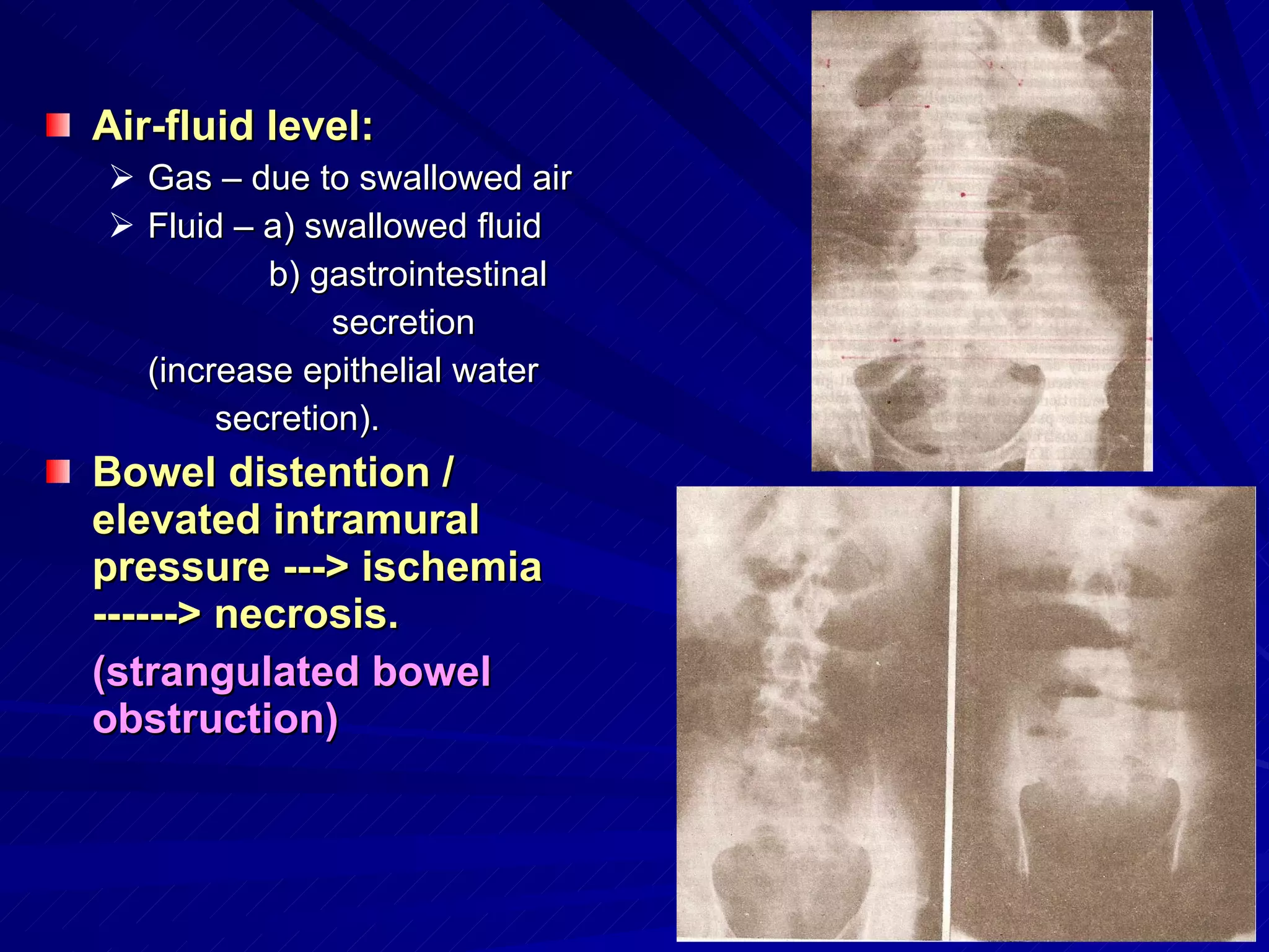
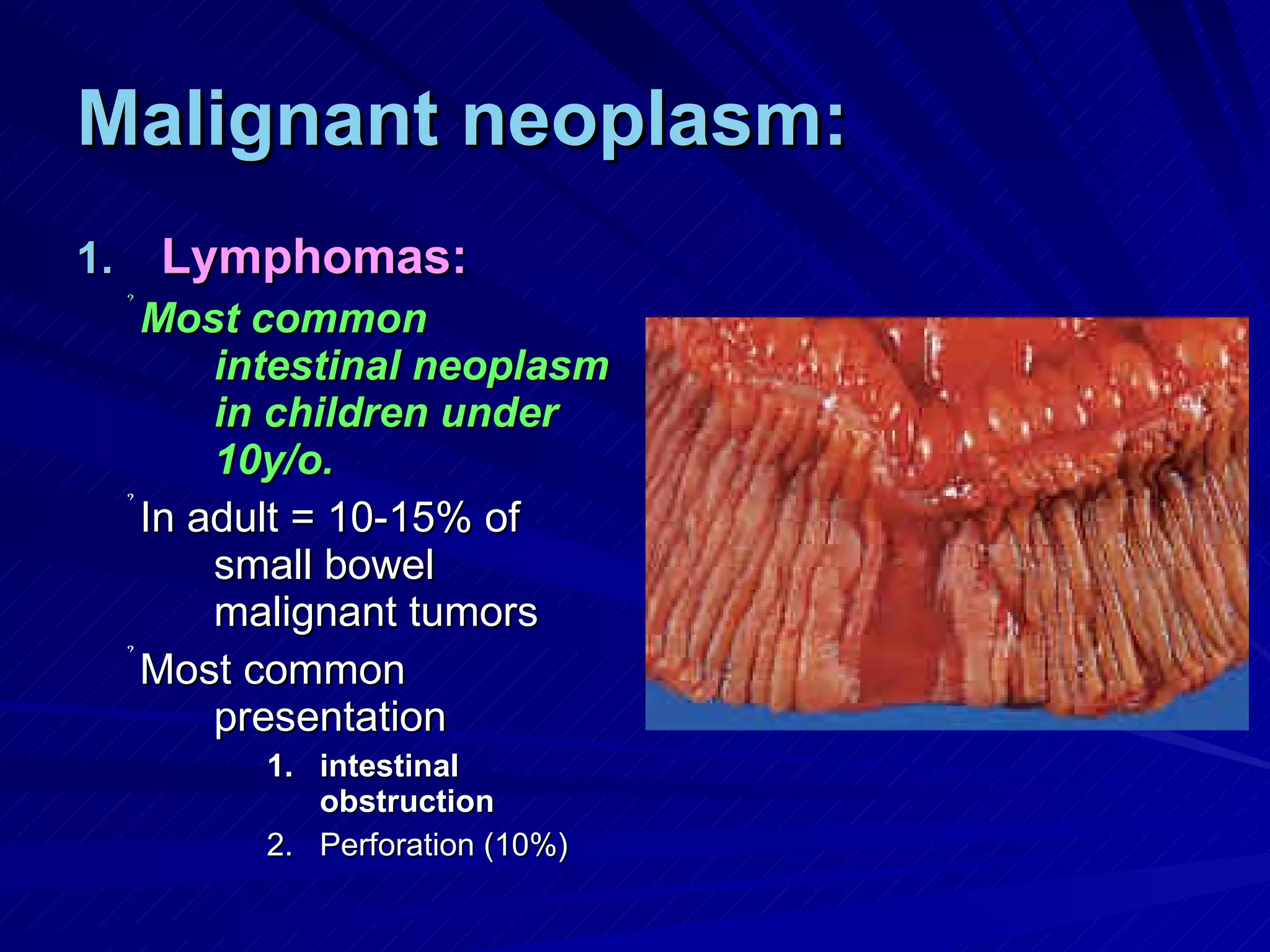
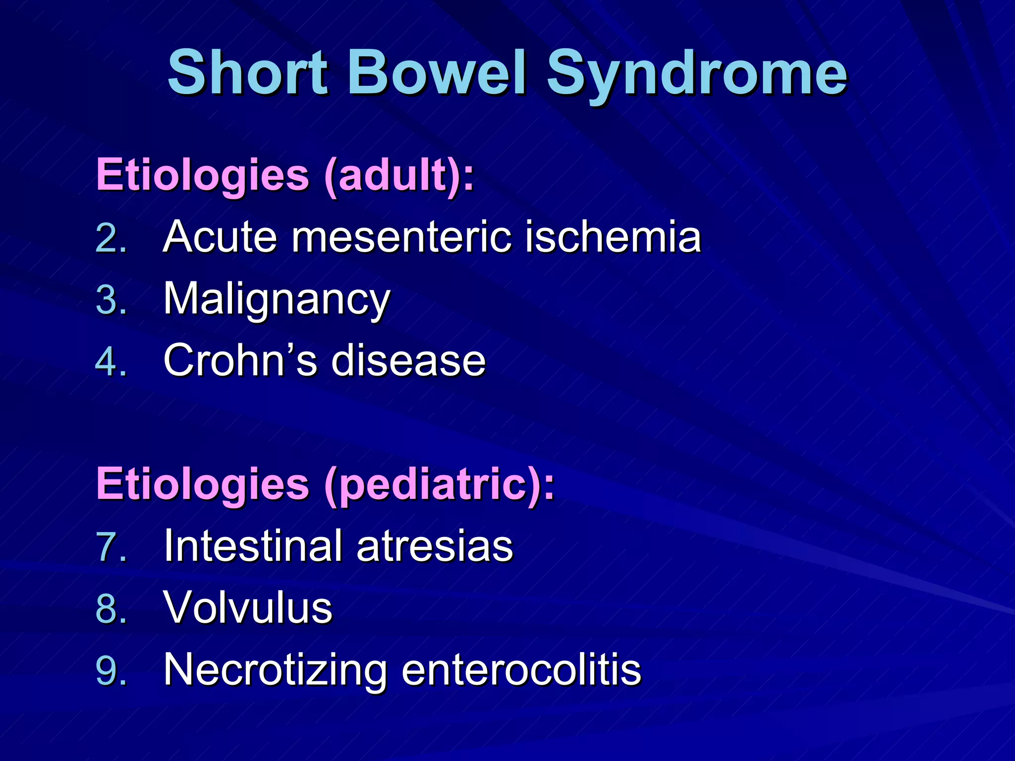
Small Intestine James Taclin C. Banez,  MD, FPCS, FPSGS, DPBS,DPSA
Small Intestine one of the most important organs for immune defense largest endocrine organ of the body Starts from the pylorus and ends at the cecum 3 parts: Duodenum  (20cm) Jejunum  (100 to 110cm) Ileum  (150 to 160 cm)
Anatomy Has plicae circulares or valves of Kerkring Duodenum: Retro-peritoneal Supplied by the celiac artery & SMA Jejunum: Occupies upper left of the abdomen Thicker wall and wider lumen than the ileum Mesentery has less fat and forms only 1-2 arcades Ileum: Occupies the lower right; has more fat and forms more arcades Contains  Payer’s patches Ileum & jejunum is supplied by the SMA
Function Digestion & Absorption: Endocrine Function: Secretes numerous hormones involved in GIT function. Secretin Cholecystokenin Gastric inhibitory peptide Enteroglucagon Vasoactive intestinal peptide Motilin Bombesin Somatostatin Neurotensin
Function Immune function: Major source of   IgA Integrity of the GUT wall  prevents bacterial translocation into the wall of the intestine and abdominal cavity which can lead to sepsis Gut associated lymphoid tissue  – part of the immune defense system which clears the abdominal cavity of pathogenic bacteria found in  Peyer’s patches
Small Bowel Surgical Lesions Small bowel obstruction: Mechanical Ileus Small bowel infection Chronic inflammation Neoplasm Diverticula Ischemic enteritis Short bowel syndrome
SMALL BOWEL OBSTRUCTION
Small Bowel Obstruction Causes of  Mechanical Obstruction : Post-operative adhesion  (75%) Midgut volvulous Hernias Crohn’s disease Neoplasm (primary or extrinsic compression or invasion) Superior mesenteric artery syndrome (compression of transverse duodenum)
Midgut Volvulous
Pathophysiology: Accdg. to it’s anatomical relationship to the intestinal wall: Intraluminal  ( foreign bodies, gallstone, and meconium) Intramural  (neoplasm, Crohn’s, hematomas) Extrinsic  (adhesion, hernias & carcinomatosis)
Classify Accdg to Degree of Obstruction Partial small-bowel obstruction  – passage of gas and fluid. Complete small-bowel obstruction  (obstipation) Closed loop obstruction  (obstructed proximal and distal) ex. Volvulus Strangulated bowel obstruction
Manifestation: colicky abdominal pain nausea / vomiting obstipation abdominal distention hyperactive bowel sound / hypoactive BS signs of dehydration (sequestration of fluid in bowel wall and lumen as well as poor oral intake) lab. findings: hemoconcentration fluid & electrolyte imbalance leucocytosis
Manifestation: Features of Strangulated obstruction : tachycardia localized abd. tenderness fever marked leucocytosis acidosis lab result: -  elevated serum amyase, lipase, LDH,  phosphate and potassium
Goals in its diagnosis: distinguish between mechanical obstruction from ileus whether it is partial or complete obstruction differentiate between simple and strangulating obstruction determine the etiology
Diagnosis: Clinical history & PE Radiological examination: FPA (supine and upright) Triad:   dilated small bowel (>3cm ) air-fluid levels seen in upright paucity of air in the colon
SMALL BOWEL OBSTRUCTION (Air Fluid Level)
Air-fluid level: Gas – due to swallowed air Fluid – a) swallowed fluid   b) gastrointestinal  secretion  (increase epithelial water  secretion). Bowel distention / elevated intramural pressure ---> ischemia ------> necrosis. (strangulated bowel obstruction)
Diagnosis: CT scan  (90% sensitive / 90% specific) Findings of small bowel obstruction: Discrete  transition zone Intra-luminal contrast unable to passed  beyond the  transition zone Colon containing little gas or fluid Strangulation is suggested: Thickening of the bowel wall Pneumatosis intestinalis Portal venous gas Mesentery haziness Poor uptake of intravenous contrast into the wall of the affected bowel Limitation:   unable to detect partial intestinal obstruction  (<50% sensitivity)
Treatment: Correct fluid & electrolyte imbalance : Isotonic fluid Monitor resuscitation (foley catheter/CVP) NPO / TPN Broad spectrum antibiotic  (due to bacterial translocation) Placed NGT  to decompress the stomach and decrease nausea, distention and risk of aspiration Expeditious celiotomy  (to minimize risk of strangulation). Type of operation based on operative finding causing intestinal obstruction
Ileus / Pseudo-Obstruction Impaired intestinal motility Most common cause of delayed discharge following abdominal operations Temporary and reversible
Ileus / Pseudo-Obstruction Etiologies: Abdominal surgery Infection & inflammation (sepsis/peritonitis) Electrolyte imbalance (Hypo K, Mg & Na) Drugs (anticholinergic, opiates) Visceral myopathies (degeneration/fibrosis of smooth muscle) Visceral neuropathies (degenerative disorders of myenteric & submucosal plexuses)
ILEUS
Symptoms: Inability to tolerate solid & liquid by mouth Nausea/vomiting Lack of flatus & bowel movements Diminished or absent bowel sound Abdominal pain and distention
Diagnosis: History of recent abdominal surgery Discontinue opiates  Serum electrolyte determination CT scan better than FPA in postoperative setting to exclude presence of abscess or mechanical obstruction
Therapy: NPO, if prolong TPN is required NGT to decompress the stomach Correct fluid & electrolyte imbalance Give  ketorolac  and reduce the dose of opioids
CHRONIC IDIOPATHIC INFLAMMATORY DISEASE OF THE BOWEL
CROHN’S DISEASE Regional, transmural, granulomatous enteritis.  Chronic, idiopathic inflammatory dse Ethnic groups ---> East Europe (Ashkenazi Jewish)  Female predominance, 2x higher smokers Familial association (30x in siblings / 13 x in 1 st  degree relatives). Higher socioeconomic status Breast feeding  is protective
Etiology: Unknown Hypothesis: Infectious:   - Chlamydia / Pseudomonas / Mycobacterium paratuberculosis / Listeria monocytogenesis / Measles / Yersinia enterocolitica Immunologic abnormalities: Humeral & cell-mediated immune reactions against gut cells. Genetic factors: Chromosome 16  (IBD1 --> NOD2)
Pathology: Affect any portion of GIT: Small bowel alone (30%) Ileocolitis (55%) Colon alone (15%) Hallmark  – focal, transmural inflammation of the intestine Earliest sign  -->  aphthous ulcers  surrounded by halo erythema over a non-caseating granuloma.
CHRON’S DISEASE
Pathology: As the aphthous ulcer enlarge and coalesce transversely forming  cobblestone appearance. Advanced dse  ---> transmural inflammation. This results to  COMPLICATIONS adhesions to adjacent bowel, stricture formation (fibrosis),  intra-abdominal abscesses,  fistula or free perforation (peritonitis) Skip lesions and w/ fat   wrapping  (encroachment of mesenteric fat onto the serosal surface) -->  pathognomonic for Crohn’s.
ADVANCED CHRON’S DSE
CHRON’S DSE. ANAL FISTULA
Clinical Manifestation: Most common symptom: Abdominal pain Diarrhea Weight loss Other symptoms depends on type of complications: obstruction (fibrosis) perforation (peritonitis, fistula, intraabdominal abscess) toxic megacolon (marked colonic dilatation, adb. tenderness, fever & leukocytosis) cancer (6x greater/more advanced---> poor prognosis) perianal dse (fissure, fistula, stricture or abscess) Extra-intestinal manifestation: erythema nodosum & peripheral arthritis are correlated w/ severity of intestinal inflammation.
Diagnosis: Endoscopy (esophagogastroduodenoscopy (EGD) /colonoscopy) w/ biopsy. Barium enema / intestinal series  Enteroclysis  (small bowel) more accurate CT scan – to reveal intra-abd. abscesses
Treatment: Medical: Intravenous fluids NGT to rest GIT (elemental diet/TPN) Medications:   to relieve diarrhea relieve pain control infection (antibiotic) Anti-inflammatory ( aminosalicylates, corticosteroid, immunomodulators – azathioprime 6-mercaptopurine and cyclosporine)
Surgical: Indicated if:  with complications Types: Segmental resection w/ primary anastomosis: Microscopic evidence of the dse at the resection margin does not compromise a safe anastomosis, hence, a frozen section is unnecessary. Stricturoplasty Bypass procedures (gastrojejunostomy)
Tuberculous Enteritis: In developing and under develop countries Resurgence in develop countries due to: AIDS epidemic Influx of Asian migrants Use of immunosuppressive agents Forms: Primary infection  (caused by M. tuberculosis bovine from ingested milk) Secondary infection  (swallowing bacilli from active pulmonary TB)
Tuberculous Enteritis
Tuberculous Enteritis: Patterns: Hypertrophic – causes stenosis or obstruction Ulcerative – diarrhea and bleeding Ulcero-hypertrophic Treatment: Chemotherapy (given 2 wks prior to surgery up to 1 yr). Rifampicin Isoniazid Ethambutol Surgery (perforation, obstruction, hemorrhage).
Typhoid enteritis: Caused by Salmonella typhi Diagnosis: Culture from blood or feces Agglutinins against O and H antigen Treatment: Medical: Chloramphenicol / trimethropin-sulfamethoxazole / amoxycillin / quinolones Surgical: perforations / hemorrhage Segmental resection (w/ primary anastomosis or ileostomy)
DIVERTICULAR DISEASE OF THE SMALL BOWEL
Meckels Diverticulum Most prevalent congenital anomaly of GIT True diverticula 60% contains heterotopic mucosa: Gastric mucosa  (60%) Pancreatic acini Brunner’s gland Pancreatic islets Colonic mucosa Endometriosis Hepatobiliary tissues
Meckels Diverticulum Rules of Twos: 2% prevalence 2:1 female predominance Location 2 feet proximal to the ileocecal valve in adults. Half of those are asymptomatic are younger than 2 years of age.
Meckels Diverticulum Complications: Bleeding (most common )  – due to ileal mucosal ulceration. Obstruction: Volvulus of the intestine Entrapment of intestine by the mesodiverticular band Intussuception Stricture due to diverticulitis As Littre’s hernia – found in inguinal or femoral hernia sac.
Meckels Diverticulum Clinical manifestation: Asymptomatic 4% symptomatic due to complication 50% are younger than 10y/o Symptomatic (Bleeding > obstruction > diverticulitis) bleeding is 50% in children and pt younger 18y/o bleeding is rare in pt older than 30y/o intestinal obstruction most common in adult diverticulitis  is indistinguishable to appendicitis Neoplasm seen: --- > Carcinoid
Meckels Diverticulum Diagnosis: For asymptomatic usually discovered as an incidental findings in radiographic imaging, endoscopy, or intraoperatively. Enteroclysis  has 75% accuracy but not applicable during acute cases. Radionuclide scans  (99m Tc-pertechnate) for ectopic gastric mucosa or in active bleeding Angiography  to localize site of bleeder
Meckels Diverticulum Management: Diverticulectomy: diverticulitis obstruction (w/ removal of associated band) Segmental resection for: Bleeding If with tumor
Acquired Small Bowel Diverticula Epidemiology: False diverticula Increases w/ age; seldom seen < 40y/o (50-70y/o) Duodenum:   Most common; usually adjacent to ampulla Called  periampullary, juxtapapillary, or peri-Vaterian diverticula 75% arise in the medial wall
Acquired Small Bowel Diverticula Jejunoileal: 80% - jejunum (tends to be large and multiple) 15% - ileum (tends to be small and solitary) 5% - both ileum and jejunum
Acquired Small Bowel Diverticula Pathophysiology: Abnormalities of intestinal smooth muscle  or dysregulated motility leading to herniation. Associated w/: Bacterial overgrowth  – vit B12 deficiency, megaloblastic anemia, malabsorption & steatorrhea Periampullary duodenal diverticula : Obstructive jaundice Pancreatitis Intestinal obstruction  due to compression of adjacent bowel
Acquired Small Bowel Diverticula Diagnosis: Best diagnosed w/  enteroclysis Treatment: Asymptomatic ---> left alone Bacterial overgrowth --> antibiotics Bleeding and obstruction ---> segmental resection for jejunoileal diverticula.
Acquired Small Bowel Diverticula Treatment: Diverticulectomy  if located  in the duodenum For medial duodenal diverticula ---> do lateral duodenotomy and oversewing of the bleeder May invaginate the diverticula into the duodenal lumen then excised If related to the ampulla ---> extended sphincterotoplasty If perforated ----> excised and closed w/ omental patch; if inflammed ---> placed gastrojejunostomy
MESENTERIC ISCHEMIA
Mesenteric Ischemia Clinical Syndrome: Acute mesenteric ischemia Pathophysiology Arterial embolus : (most common-50%; heart; usually lodge distal to origin of the middle colic Arterial thrombosis : occlusion occurs at proximal near it’s origin. Vasospasm  (nonocclusive mesenteric ischemia – NOMI): usually in critically-ill pt. receiving vasopressors. Venous thrombosis : (5-15%) and 95% SMV Primary – no etiologic factor identified Secondary – heritable or acquired coagulation disorder
Mesenteric Ischemia Clinical Syndrome: Chronic Mesenteric Ischemia: Develops insidiously allows for collateral circulation to develop Rarely leads to infarction. Usually due to  arteriosclerosis Usually two mesenteric arteries are involved
Mesenteric Ischemia Manifestation: Acute mesenteric ischemia: Severe abdominal pain out of proportion to the degree of abd. tenderness (hallmark) Colicky at the mid-abdomen. Nausea / vomiting, diarrhea  abd. distention,peritonitis, passage bloody stool Chronic mesenteric ischemia: Postprandial abd. pain “food-fear”, (most common)
Mesenteric Ischemia No laboratory test sensitive for the detection of acute mesenteric ischemia prior to the onset of intestinal infarction. The presence of it’s hallmark sign, is an indication for immediate celiotomy.
Mesenteric Ischemia Angiography   – most reliable; 74 – 100% sensitivity and 100% specificity;  It is  gold standard for the diagnosis of  arterial mesenteric ischemia.
Mesenteric Ischemia CT scanning  is used to: Disorder other abd. condition causing abd. pain Evidence of occlusion or stenosis of mesenteric vasculature. Evidence of ischemia in the intestine & mesentery Test of choice for acute mesenteric venous thrombosis
Mesenteric Ischemia Treatment: w/ signs of peritonitis --> celiotomy check for viability of the bowel: Necrotic ----> segmental resection Questionable viability ----> second look laparotomie s
Mesenteric Ischemia Surgical revascularization  (embolectomy / thrombectomy / mesenteric bypass). Not done if:  segment is necrotic is too unstable patient Done pt diagnosed w/ emboli or thrombus-induced acute mesenteric ischemia w/o signs of peritonitis. May give thrombolysis  (streptokinase, urokinase ,  recombinant tissue plasminogen activator).  Useful only in partially occluded vessels and has given w/in 12 hrs. after onset of symptoms.
NEOPLASM OF THE SMALL BOWEL
Neoplasm Rare: Rapid transit time Local immune system of the small bowel mucosa (IgA) Alkaline pH Relatively low concentration of bacteria; low concentration of carcinogenic products of bacterial metabolism. Presence of mucosal enzymes (hydrolases) that destroy certain carcinogens Efficient epithelial cellular apoptotic mechanisms  that serve to eliminate clones harboring genetic mutation
Neoplasm 50 – 60 y/o Risk factors: Red meat Ingestion of smoked or cured foods Crohn’s dse Celiac sprue Hereditary nonpolyposis colorectal cancer  (HNPCC) Familial adenomatous polyposis  (FAD) – 100% to develop  duodenal CA Peutz-Jeghers syndrome
Neoplasm Symptoms: Most are asymptomatic   Symptoms: Vague abdominal pain  (epigastric discomfort, N/V, abd. pain, diarrhea). Bleeding  (hematochezia or hematemesis) Obstruction  (intussuception, circumferencial growth, kinking of the bowel, intramural growth). Most common mode of presentation is  --->  crampy abd. pain, distention, nausea / vomiting Hemorrhage  usually indolent 2 nd  common mode of presentation
Neoplasm Diagnosis: For most are asymptomatic it is rarely diagnosed preoperatively Serological examination Serum 5-hydroxyindole acetic acid (HIAA)  for carcinoid. CEA  associated w/ small intestinal adenocarcinoma but only if w/ liver metastasis.
Neoplasm Diagnosis: Radiological examination: Enteroclysis  (test of choice – 90% sensitivity) UGIS w/ intestinal follow through CT scan Angiography / RBC scan --> bleeding lesions Endoscopy: EGD (esophagus, gastric, and duodenum) Colonoscopy
Benign tumors: Adenomas:  (most common benign neoplasm): True adenomas: Associated w/ bleeding and obstruction Usually seen in the  ileum Majority are asymptomatic Villous adenoma: Most common in the  duodenum “ soap bubble” appearance on contrast radiography No report of secretory diarrhea Brunner’s gland adenoma In the  duodenum No malignant potential Mimic PUD
Benign tumors: Leiomyoma: Most common symptomatic benign lesion Associated w/  bleeding Diagnosed by  angiography  and commonly located in the  jejunum 2 growth pattern: Intramurally ----> obstruction Both intramural and extramural  (Dumbbell shaped)
Benign tumors: Lipoma: Most common in the  ileum Causes obstruction (lead point of an intussusception) Bleeding due to ulcer formation No malignant degeneration
Benign tumors: Peutz-Jeghers Syndrome: Inherited syndrome of: Mucocutaneous melatonic pigmentation  (face, buccal mucosa, palm, sole, peri-anal area) Gastrointestinal polyp   (enteric  jejunum and ileum  are most frequent part of GIT followed by colon, rectum and stomach).
Benign tumors: Peutz-Jeghers Syndrome: Symptoms: colicky abd. pain (due to intermittent intussuception) Hemorrhage Treatment: Segmental resection of the bowel causing obstruction or bleeding. Cure impossible due to widespread intestinal involvement
Malignant neoplasm: Histologic types: Tumor type Cell of origin Frequency Predominant Site adenocarcinoma Epithelial cell 35 – 50% Duodenum carcinoid Enterochromaffin cell 20 – 40% Ileum lymphoma lymphocyte 10 – 15% Ileum GIST  (gastrointestinal stromal tumors) ? Interstitial cell of Cajal 10 – 15% -
Malignant neoplasm: Adenocarcinoma: Most common CA  of small bowel Most common in duodenum and proximal jejunum Half involve the ampulla of Vater.
Malignant neoplasm: Carcinoid: From  Enterochromaffin cells  or  Kultchitsky cells Arise from foregut, midgut & hindgut Appendix (46%) > Ileum (28%) > Rectum (17%)
Malignant neoplasm: Carcinoid: Aggressive behavior than the appendiceal carcinoid.  appendix – 3% metastasize; Ileum – 35% metastasize Appendix – solitary; Ileum – 30% multiple 25-50% w/ carcinoid tumor with liver metastasis develops  carcinoid syndrome . Secretes  serotonin, bradykinin and substance P Diarrhea Flushing Hypotension tachycardia  fibrosis of endocardium and valves of the right heart .
Malignant neoplasm: Lymphomas: Most common intestinal neoplasm in children under 10y/o. In adult = 10-15% of small bowel malignant tumors Most common presentation intestinal obstruction Perforation (10%)
Malignant neoplasm: Lymphomas: Criteria of primary lymphomas of the small bowel: Absence of peripheral lymphadenopathy Normal chest x-ray w/o evidence of mediastinal LN enlargement. Normal WBC count and differential At operation, the bowel lesion must predominate and the only nodes are associated w/ the bowel lesion Absence of disease in the liver and spleen
Treatment: For Benign lesions: All symptomatic benign tumors should be surgically resected or removed endoscopically (EGD / colonoscopy). Duodenal tumors: 1 cm. ----> endoscopic polypectomy 2cm. ----> surgically resected (Whipples – located near the ampulla of Vater). Duodenal adenomas  w/ FAP shd undergo Whipples for it is usually multiple and sessile and has  100% degenerate to CA.
Treatment: Malignant lesions: Adenocarcinoma: Wide local resection w/ it’s mesentery to achieve regional lymphadenectomy Chemotherapy has no proven efficacy in the adjuvant or palliative treatment of small-intestinal adenoCA. Small intestinal lymphoma: For localized: segmental resection w/ adjacent mesentery If w/ diffused involvement: -->chemotherapy rather than surgery, is primary therapy
Treatment: Carcinoid: Segmental intestinal resection & regional lymphadenectomy. <  1cm rarely has LN metastases > 3cm 75 to 90% LN metastases 30% are multiple, hence entire small bowel shd be examined prior to surgery .
Treatment: Carcinoid: If w/ metastatic lesions---> debulking, associated w/ long-term survival  & amelioration of symptoms of carcinoid syndrome Chemotherapy: ---> 30 -50% response Doxorubicin 5-fluorouracil Streptozocin Octreotide : - most effective for management of symptoms of carcinoid syndrome
Treatment: Metastatic cancers: Melanoma  associated w/ propensity for metastasis to the small bowel. Palliative resection / bypass procedure Systemic therapy depends on the responds of the primary site.
SHORT BOWEL SYNDROME
Short Bowel Syndrome Presence of  less than 200cm  of residual small bowel in adult pts. Functional definition: - insufficient intestinal absorptive capacity results in the clinical manifestations of: Diarrhea Dehydration malnutrition
Short Bowel Syndrome Etiologies (adult): Acute mesenteric ischemia Malignancy Crohn’s disease Etiologies (pediatric): Intestinal atresias Volvulus Necrotizing enterocolitis
Short Bowel Syndrome Medical therapy: Mx of primary condition causing intestinal resection Correct fluid & electrolyte imbalance due to severe diarrhea TPN, enteral nutrition is gradually introduced, once ileus is resolved
Short Bowel Syndrome Medical therapy: H2 receptor antagonist --> to reduce gastric acid secretion Antimotility agents (loperamide HCL or diphenoxylate)  Octreotide – to reduce volume of gastrointestinal secretion TPN complication: Catheter sepsis Venous thrombosis Liver and kidney failure osteoporosis
Short Bowel Syndrome Surgical Therapy : Non-transplant: Goal is to increase nutrient and fluid absorption by either slowing intestinal transit or increasing intestinal length Slow intestinal transit: Segmental reversal of the small bowel  Interposition of a segment of colon Construction of small intestinal valves Electrical pacing of the small bowel Limited case report Frequently associated w/ intestinal obstruction
GOD BLESS SALAMAT PO
THANK  YOU
Diagnosis: Enteroclysis   200 to 250 ml of barium followed by 1 to 2 L of methylcellulose in water is instilled into the proximal jejunum via a long naso-enteric tube
Short Bowel Syndrome Factors predictive of achieving independence from TPN: Presence or absence of an intact colon (capacity to absorb fluid & electrolytes and absorb short-chain FA). Intact ileocecal valve A healthy, rather disease, residual small intestine is associated w/ decreased severity of malabsorption Resection of jejunum is better tolerated than the ileum, due to bile salt and vit B12 absorption capacity of the ileum.
Short Bowel Syndrome Surgical Therapy: Non-transplant: Intestinal lengthening operation: Longitudinal Intestinal lengthening and tailoring (LILT) Serial transverse enteroplasty procedure (STEP) Intestinal transplant
Prognosis (CHRON’S DSE) High recurrence rate (most common proximal to the site of previous resection). 70% recur w/in 1 yr and 85% w/in 3 yrs. Most common complication: Wound infection Postoperative intra-abdominal abscess Anastomotic leaks 60-300 x more frequent to develop CA
Mesenteric Ischemia NOMI  – std tx. Is infusion of vasodilator  (papavarine hydrochloride)  into the SMA. If w/ signs of peritonitis --> immediate celiotomy and resect necrotic segment. Acute mesenteric venous thrombosis Std tx. anticoagulant (heparin / warfarin). Signs of peritonitis --> explore and resects if needed For chronic arterial mesenteric ischemia: Surgical revascularization  Aortomesenteric bypass grafting Mesenteric endarterectomy Percutaneous transluminal mesenteric angioplasty alone or w/ stent.
Malignant neoplasm: GISTs: (gastrointestinal stromal tumors) Most common  mesenchymal tumors  arising in the small bowel 70% arises from the  stomach  followed by the  small bowel 15% of small bowel malignancies Formerly classified as:  Leiomyomas Leiomyosarcomas Smooth muscle tumors of small bowel Associated w/  overt hemorrhage Has its expression of the receptor  tyrosine kinase   KIT (CD117).  There is pathological KIT signal transduction
Treatment: Small-intestine GISTs: Segmental resection If was preoperatively diagnosed, lymphadenectomy shd not be done, for rarely associated w/ LN metastases. Resistant to conventional chemotherapy IMATINIB (Gleevec): Formerly known as ST1571 80% of pt w/ unresectable lesions showed clinical benefits 50 – 60% showed evidence of reduction in tumor volume Role as neoadjuvant and adjuvant tx under investigation

Small Intestine Ii

  • 1.
    Small Intestine JamesTaclin C. Banez, MD, FPCS, FPSGS, DPBS,DPSA
  • 2.
    Small Intestine oneof the most important organs for immune defense largest endocrine organ of the body Starts from the pylorus and ends at the cecum 3 parts: Duodenum (20cm) Jejunum (100 to 110cm) Ileum (150 to 160 cm)
  • 3.
    Anatomy Has plicaecirculares or valves of Kerkring Duodenum: Retro-peritoneal Supplied by the celiac artery & SMA Jejunum: Occupies upper left of the abdomen Thicker wall and wider lumen than the ileum Mesentery has less fat and forms only 1-2 arcades Ileum: Occupies the lower right; has more fat and forms more arcades Contains Payer’s patches Ileum & jejunum is supplied by the SMA
  • 4.
    Function Digestion &Absorption: Endocrine Function: Secretes numerous hormones involved in GIT function. Secretin Cholecystokenin Gastric inhibitory peptide Enteroglucagon Vasoactive intestinal peptide Motilin Bombesin Somatostatin Neurotensin
  • 5.
    Function Immune function:Major source of IgA Integrity of the GUT wall prevents bacterial translocation into the wall of the intestine and abdominal cavity which can lead to sepsis Gut associated lymphoid tissue – part of the immune defense system which clears the abdominal cavity of pathogenic bacteria found in Peyer’s patches
  • 6.
    Small Bowel SurgicalLesions Small bowel obstruction: Mechanical Ileus Small bowel infection Chronic inflammation Neoplasm Diverticula Ischemic enteritis Short bowel syndrome
  • 7.
  • 8.
    Small Bowel ObstructionCauses of Mechanical Obstruction : Post-operative adhesion (75%) Midgut volvulous Hernias Crohn’s disease Neoplasm (primary or extrinsic compression or invasion) Superior mesenteric artery syndrome (compression of transverse duodenum)
  • 9.
  • 10.
    Pathophysiology: Accdg. toit’s anatomical relationship to the intestinal wall: Intraluminal ( foreign bodies, gallstone, and meconium) Intramural (neoplasm, Crohn’s, hematomas) Extrinsic (adhesion, hernias & carcinomatosis)
  • 11.
    Classify Accdg toDegree of Obstruction Partial small-bowel obstruction – passage of gas and fluid. Complete small-bowel obstruction (obstipation) Closed loop obstruction (obstructed proximal and distal) ex. Volvulus Strangulated bowel obstruction
  • 12.
    Manifestation: colicky abdominalpain nausea / vomiting obstipation abdominal distention hyperactive bowel sound / hypoactive BS signs of dehydration (sequestration of fluid in bowel wall and lumen as well as poor oral intake) lab. findings: hemoconcentration fluid & electrolyte imbalance leucocytosis
  • 13.
    Manifestation: Features ofStrangulated obstruction : tachycardia localized abd. tenderness fever marked leucocytosis acidosis lab result: - elevated serum amyase, lipase, LDH, phosphate and potassium
  • 14.
    Goals in itsdiagnosis: distinguish between mechanical obstruction from ileus whether it is partial or complete obstruction differentiate between simple and strangulating obstruction determine the etiology
  • 15.
    Diagnosis: Clinical history& PE Radiological examination: FPA (supine and upright) Triad: dilated small bowel (>3cm ) air-fluid levels seen in upright paucity of air in the colon
  • 16.
    SMALL BOWEL OBSTRUCTION(Air Fluid Level)
  • 17.
    Air-fluid level: Gas– due to swallowed air Fluid – a) swallowed fluid b) gastrointestinal secretion (increase epithelial water secretion). Bowel distention / elevated intramural pressure ---> ischemia ------> necrosis. (strangulated bowel obstruction)
  • 18.
    Diagnosis: CT scan (90% sensitive / 90% specific) Findings of small bowel obstruction: Discrete transition zone Intra-luminal contrast unable to passed beyond the transition zone Colon containing little gas or fluid Strangulation is suggested: Thickening of the bowel wall Pneumatosis intestinalis Portal venous gas Mesentery haziness Poor uptake of intravenous contrast into the wall of the affected bowel Limitation: unable to detect partial intestinal obstruction (<50% sensitivity)
  • 19.
    Treatment: Correct fluid& electrolyte imbalance : Isotonic fluid Monitor resuscitation (foley catheter/CVP) NPO / TPN Broad spectrum antibiotic (due to bacterial translocation) Placed NGT to decompress the stomach and decrease nausea, distention and risk of aspiration Expeditious celiotomy (to minimize risk of strangulation). Type of operation based on operative finding causing intestinal obstruction
  • 20.
    Ileus / Pseudo-ObstructionImpaired intestinal motility Most common cause of delayed discharge following abdominal operations Temporary and reversible
  • 21.
    Ileus / Pseudo-ObstructionEtiologies: Abdominal surgery Infection & inflammation (sepsis/peritonitis) Electrolyte imbalance (Hypo K, Mg & Na) Drugs (anticholinergic, opiates) Visceral myopathies (degeneration/fibrosis of smooth muscle) Visceral neuropathies (degenerative disorders of myenteric & submucosal plexuses)
  • 22.
  • 23.
    Symptoms: Inability totolerate solid & liquid by mouth Nausea/vomiting Lack of flatus & bowel movements Diminished or absent bowel sound Abdominal pain and distention
  • 24.
    Diagnosis: History ofrecent abdominal surgery Discontinue opiates Serum electrolyte determination CT scan better than FPA in postoperative setting to exclude presence of abscess or mechanical obstruction
  • 25.
    Therapy: NPO, ifprolong TPN is required NGT to decompress the stomach Correct fluid & electrolyte imbalance Give ketorolac and reduce the dose of opioids
  • 26.
    CHRONIC IDIOPATHIC INFLAMMATORYDISEASE OF THE BOWEL
  • 27.
    CROHN’S DISEASE Regional,transmural, granulomatous enteritis. Chronic, idiopathic inflammatory dse Ethnic groups ---> East Europe (Ashkenazi Jewish) Female predominance, 2x higher smokers Familial association (30x in siblings / 13 x in 1 st degree relatives). Higher socioeconomic status Breast feeding is protective
  • 28.
    Etiology: Unknown Hypothesis:Infectious: - Chlamydia / Pseudomonas / Mycobacterium paratuberculosis / Listeria monocytogenesis / Measles / Yersinia enterocolitica Immunologic abnormalities: Humeral & cell-mediated immune reactions against gut cells. Genetic factors: Chromosome 16 (IBD1 --> NOD2)
  • 29.
    Pathology: Affect anyportion of GIT: Small bowel alone (30%) Ileocolitis (55%) Colon alone (15%) Hallmark – focal, transmural inflammation of the intestine Earliest sign --> aphthous ulcers surrounded by halo erythema over a non-caseating granuloma.
  • 30.
  • 31.
    Pathology: As theaphthous ulcer enlarge and coalesce transversely forming cobblestone appearance. Advanced dse ---> transmural inflammation. This results to COMPLICATIONS adhesions to adjacent bowel, stricture formation (fibrosis), intra-abdominal abscesses, fistula or free perforation (peritonitis) Skip lesions and w/ fat wrapping (encroachment of mesenteric fat onto the serosal surface) --> pathognomonic for Crohn’s.
  • 32.
  • 33.
  • 34.
    Clinical Manifestation: Mostcommon symptom: Abdominal pain Diarrhea Weight loss Other symptoms depends on type of complications: obstruction (fibrosis) perforation (peritonitis, fistula, intraabdominal abscess) toxic megacolon (marked colonic dilatation, adb. tenderness, fever & leukocytosis) cancer (6x greater/more advanced---> poor prognosis) perianal dse (fissure, fistula, stricture or abscess) Extra-intestinal manifestation: erythema nodosum & peripheral arthritis are correlated w/ severity of intestinal inflammation.
  • 35.
    Diagnosis: Endoscopy (esophagogastroduodenoscopy(EGD) /colonoscopy) w/ biopsy. Barium enema / intestinal series Enteroclysis (small bowel) more accurate CT scan – to reveal intra-abd. abscesses
  • 36.
    Treatment: Medical: Intravenousfluids NGT to rest GIT (elemental diet/TPN) Medications: to relieve diarrhea relieve pain control infection (antibiotic) Anti-inflammatory ( aminosalicylates, corticosteroid, immunomodulators – azathioprime 6-mercaptopurine and cyclosporine)
  • 37.
    Surgical: Indicated if: with complications Types: Segmental resection w/ primary anastomosis: Microscopic evidence of the dse at the resection margin does not compromise a safe anastomosis, hence, a frozen section is unnecessary. Stricturoplasty Bypass procedures (gastrojejunostomy)
  • 38.
    Tuberculous Enteritis: Indeveloping and under develop countries Resurgence in develop countries due to: AIDS epidemic Influx of Asian migrants Use of immunosuppressive agents Forms: Primary infection (caused by M. tuberculosis bovine from ingested milk) Secondary infection (swallowing bacilli from active pulmonary TB)
  • 39.
  • 40.
    Tuberculous Enteritis: Patterns:Hypertrophic – causes stenosis or obstruction Ulcerative – diarrhea and bleeding Ulcero-hypertrophic Treatment: Chemotherapy (given 2 wks prior to surgery up to 1 yr). Rifampicin Isoniazid Ethambutol Surgery (perforation, obstruction, hemorrhage).
  • 41.
    Typhoid enteritis: Causedby Salmonella typhi Diagnosis: Culture from blood or feces Agglutinins against O and H antigen Treatment: Medical: Chloramphenicol / trimethropin-sulfamethoxazole / amoxycillin / quinolones Surgical: perforations / hemorrhage Segmental resection (w/ primary anastomosis or ileostomy)
  • 42.
    DIVERTICULAR DISEASE OFTHE SMALL BOWEL
  • 43.
    Meckels Diverticulum Mostprevalent congenital anomaly of GIT True diverticula 60% contains heterotopic mucosa: Gastric mucosa (60%) Pancreatic acini Brunner’s gland Pancreatic islets Colonic mucosa Endometriosis Hepatobiliary tissues
  • 44.
    Meckels Diverticulum Rulesof Twos: 2% prevalence 2:1 female predominance Location 2 feet proximal to the ileocecal valve in adults. Half of those are asymptomatic are younger than 2 years of age.
  • 45.
    Meckels Diverticulum Complications:Bleeding (most common ) – due to ileal mucosal ulceration. Obstruction: Volvulus of the intestine Entrapment of intestine by the mesodiverticular band Intussuception Stricture due to diverticulitis As Littre’s hernia – found in inguinal or femoral hernia sac.
  • 46.
    Meckels Diverticulum Clinicalmanifestation: Asymptomatic 4% symptomatic due to complication 50% are younger than 10y/o Symptomatic (Bleeding > obstruction > diverticulitis) bleeding is 50% in children and pt younger 18y/o bleeding is rare in pt older than 30y/o intestinal obstruction most common in adult diverticulitis is indistinguishable to appendicitis Neoplasm seen: --- > Carcinoid
  • 47.
    Meckels Diverticulum Diagnosis:For asymptomatic usually discovered as an incidental findings in radiographic imaging, endoscopy, or intraoperatively. Enteroclysis has 75% accuracy but not applicable during acute cases. Radionuclide scans (99m Tc-pertechnate) for ectopic gastric mucosa or in active bleeding Angiography to localize site of bleeder
  • 48.
    Meckels Diverticulum Management:Diverticulectomy: diverticulitis obstruction (w/ removal of associated band) Segmental resection for: Bleeding If with tumor
  • 49.
    Acquired Small BowelDiverticula Epidemiology: False diverticula Increases w/ age; seldom seen < 40y/o (50-70y/o) Duodenum: Most common; usually adjacent to ampulla Called periampullary, juxtapapillary, or peri-Vaterian diverticula 75% arise in the medial wall
  • 50.
    Acquired Small BowelDiverticula Jejunoileal: 80% - jejunum (tends to be large and multiple) 15% - ileum (tends to be small and solitary) 5% - both ileum and jejunum
  • 51.
    Acquired Small BowelDiverticula Pathophysiology: Abnormalities of intestinal smooth muscle or dysregulated motility leading to herniation. Associated w/: Bacterial overgrowth – vit B12 deficiency, megaloblastic anemia, malabsorption & steatorrhea Periampullary duodenal diverticula : Obstructive jaundice Pancreatitis Intestinal obstruction due to compression of adjacent bowel
  • 52.
    Acquired Small BowelDiverticula Diagnosis: Best diagnosed w/ enteroclysis Treatment: Asymptomatic ---> left alone Bacterial overgrowth --> antibiotics Bleeding and obstruction ---> segmental resection for jejunoileal diverticula.
  • 53.
    Acquired Small BowelDiverticula Treatment: Diverticulectomy if located in the duodenum For medial duodenal diverticula ---> do lateral duodenotomy and oversewing of the bleeder May invaginate the diverticula into the duodenal lumen then excised If related to the ampulla ---> extended sphincterotoplasty If perforated ----> excised and closed w/ omental patch; if inflammed ---> placed gastrojejunostomy
  • 54.
  • 55.
    Mesenteric Ischemia ClinicalSyndrome: Acute mesenteric ischemia Pathophysiology Arterial embolus : (most common-50%; heart; usually lodge distal to origin of the middle colic Arterial thrombosis : occlusion occurs at proximal near it’s origin. Vasospasm (nonocclusive mesenteric ischemia – NOMI): usually in critically-ill pt. receiving vasopressors. Venous thrombosis : (5-15%) and 95% SMV Primary – no etiologic factor identified Secondary – heritable or acquired coagulation disorder
  • 56.
    Mesenteric Ischemia ClinicalSyndrome: Chronic Mesenteric Ischemia: Develops insidiously allows for collateral circulation to develop Rarely leads to infarction. Usually due to arteriosclerosis Usually two mesenteric arteries are involved
  • 57.
    Mesenteric Ischemia Manifestation:Acute mesenteric ischemia: Severe abdominal pain out of proportion to the degree of abd. tenderness (hallmark) Colicky at the mid-abdomen. Nausea / vomiting, diarrhea abd. distention,peritonitis, passage bloody stool Chronic mesenteric ischemia: Postprandial abd. pain “food-fear”, (most common)
  • 58.
    Mesenteric Ischemia Nolaboratory test sensitive for the detection of acute mesenteric ischemia prior to the onset of intestinal infarction. The presence of it’s hallmark sign, is an indication for immediate celiotomy.
  • 59.
    Mesenteric Ischemia Angiography – most reliable; 74 – 100% sensitivity and 100% specificity; It is gold standard for the diagnosis of arterial mesenteric ischemia.
  • 60.
    Mesenteric Ischemia CTscanning is used to: Disorder other abd. condition causing abd. pain Evidence of occlusion or stenosis of mesenteric vasculature. Evidence of ischemia in the intestine & mesentery Test of choice for acute mesenteric venous thrombosis
  • 61.
    Mesenteric Ischemia Treatment:w/ signs of peritonitis --> celiotomy check for viability of the bowel: Necrotic ----> segmental resection Questionable viability ----> second look laparotomie s
  • 62.
    Mesenteric Ischemia Surgicalrevascularization (embolectomy / thrombectomy / mesenteric bypass). Not done if: segment is necrotic is too unstable patient Done pt diagnosed w/ emboli or thrombus-induced acute mesenteric ischemia w/o signs of peritonitis. May give thrombolysis (streptokinase, urokinase , recombinant tissue plasminogen activator). Useful only in partially occluded vessels and has given w/in 12 hrs. after onset of symptoms.
  • 63.
    NEOPLASM OF THESMALL BOWEL
  • 64.
    Neoplasm Rare: Rapidtransit time Local immune system of the small bowel mucosa (IgA) Alkaline pH Relatively low concentration of bacteria; low concentration of carcinogenic products of bacterial metabolism. Presence of mucosal enzymes (hydrolases) that destroy certain carcinogens Efficient epithelial cellular apoptotic mechanisms that serve to eliminate clones harboring genetic mutation
  • 65.
    Neoplasm 50 –60 y/o Risk factors: Red meat Ingestion of smoked or cured foods Crohn’s dse Celiac sprue Hereditary nonpolyposis colorectal cancer (HNPCC) Familial adenomatous polyposis (FAD) – 100% to develop duodenal CA Peutz-Jeghers syndrome
  • 66.
    Neoplasm Symptoms: Mostare asymptomatic Symptoms: Vague abdominal pain (epigastric discomfort, N/V, abd. pain, diarrhea). Bleeding (hematochezia or hematemesis) Obstruction (intussuception, circumferencial growth, kinking of the bowel, intramural growth). Most common mode of presentation is ---> crampy abd. pain, distention, nausea / vomiting Hemorrhage usually indolent 2 nd common mode of presentation
  • 67.
    Neoplasm Diagnosis: Formost are asymptomatic it is rarely diagnosed preoperatively Serological examination Serum 5-hydroxyindole acetic acid (HIAA) for carcinoid. CEA associated w/ small intestinal adenocarcinoma but only if w/ liver metastasis.
  • 68.
    Neoplasm Diagnosis: Radiologicalexamination: Enteroclysis (test of choice – 90% sensitivity) UGIS w/ intestinal follow through CT scan Angiography / RBC scan --> bleeding lesions Endoscopy: EGD (esophagus, gastric, and duodenum) Colonoscopy
  • 69.
    Benign tumors: Adenomas: (most common benign neoplasm): True adenomas: Associated w/ bleeding and obstruction Usually seen in the ileum Majority are asymptomatic Villous adenoma: Most common in the duodenum “ soap bubble” appearance on contrast radiography No report of secretory diarrhea Brunner’s gland adenoma In the duodenum No malignant potential Mimic PUD
  • 70.
    Benign tumors: Leiomyoma:Most common symptomatic benign lesion Associated w/ bleeding Diagnosed by angiography and commonly located in the jejunum 2 growth pattern: Intramurally ----> obstruction Both intramural and extramural (Dumbbell shaped)
  • 71.
    Benign tumors: Lipoma:Most common in the ileum Causes obstruction (lead point of an intussusception) Bleeding due to ulcer formation No malignant degeneration
  • 72.
    Benign tumors: Peutz-JeghersSyndrome: Inherited syndrome of: Mucocutaneous melatonic pigmentation (face, buccal mucosa, palm, sole, peri-anal area) Gastrointestinal polyp (enteric jejunum and ileum are most frequent part of GIT followed by colon, rectum and stomach).
  • 73.
    Benign tumors: Peutz-JeghersSyndrome: Inherited syndrome of: Mucocutaneous melatonic pigmentation (face, buccal mucosa, palm, sole, peri-anal area) Gastrointestinal polyp (enteric jejunum and ileum are most frequent part of GIT followed by colon, rectum and stomach).
  • 74.
    Benign tumors: Peutz-JeghersSyndrome: Symptoms: colicky abd. pain (due to intermittent intussuception) Hemorrhage Treatment: Segmental resection of the bowel causing obstruction or bleeding. Cure impossible due to widespread intestinal involvement
  • 75.
    Malignant neoplasm: Histologictypes: Tumor type Cell of origin Frequency Predominant Site adenocarcinoma Epithelial cell 35 – 50% Duodenum carcinoid Enterochromaffin cell 20 – 40% Ileum lymphoma lymphocyte 10 – 15% Ileum GIST (gastrointestinal stromal tumors) ? Interstitial cell of Cajal 10 – 15% -
  • 76.
    Malignant neoplasm: Adenocarcinoma:Most common CA of small bowel Most common in duodenum and proximal jejunum Half involve the ampulla of Vater.
  • 77.
    Malignant neoplasm: Carcinoid:From Enterochromaffin cells or Kultchitsky cells Arise from foregut, midgut & hindgut Appendix (46%) > Ileum (28%) > Rectum (17%)
  • 78.
    Malignant neoplasm: Carcinoid:Aggressive behavior than the appendiceal carcinoid. appendix – 3% metastasize; Ileum – 35% metastasize Appendix – solitary; Ileum – 30% multiple 25-50% w/ carcinoid tumor with liver metastasis develops carcinoid syndrome . Secretes serotonin, bradykinin and substance P Diarrhea Flushing Hypotension tachycardia fibrosis of endocardium and valves of the right heart .
  • 79.
    Malignant neoplasm: Lymphomas:Most common intestinal neoplasm in children under 10y/o. In adult = 10-15% of small bowel malignant tumors Most common presentation intestinal obstruction Perforation (10%)
  • 80.
    Malignant neoplasm: Lymphomas:Criteria of primary lymphomas of the small bowel: Absence of peripheral lymphadenopathy Normal chest x-ray w/o evidence of mediastinal LN enlargement. Normal WBC count and differential At operation, the bowel lesion must predominate and the only nodes are associated w/ the bowel lesion Absence of disease in the liver and spleen
  • 81.
    Treatment: For Benignlesions: All symptomatic benign tumors should be surgically resected or removed endoscopically (EGD / colonoscopy). Duodenal tumors: 1 cm. ----> endoscopic polypectomy 2cm. ----> surgically resected (Whipples – located near the ampulla of Vater). Duodenal adenomas w/ FAP shd undergo Whipples for it is usually multiple and sessile and has 100% degenerate to CA.
  • 82.
    Treatment: Malignant lesions:Adenocarcinoma: Wide local resection w/ it’s mesentery to achieve regional lymphadenectomy Chemotherapy has no proven efficacy in the adjuvant or palliative treatment of small-intestinal adenoCA. Small intestinal lymphoma: For localized: segmental resection w/ adjacent mesentery If w/ diffused involvement: -->chemotherapy rather than surgery, is primary therapy
  • 83.
    Treatment: Carcinoid: Segmentalintestinal resection & regional lymphadenectomy. < 1cm rarely has LN metastases > 3cm 75 to 90% LN metastases 30% are multiple, hence entire small bowel shd be examined prior to surgery .
  • 84.
    Treatment: Carcinoid: Ifw/ metastatic lesions---> debulking, associated w/ long-term survival & amelioration of symptoms of carcinoid syndrome Chemotherapy: ---> 30 -50% response Doxorubicin 5-fluorouracil Streptozocin Octreotide : - most effective for management of symptoms of carcinoid syndrome
  • 85.
    Treatment: Metastatic cancers:Melanoma associated w/ propensity for metastasis to the small bowel. Palliative resection / bypass procedure Systemic therapy depends on the responds of the primary site.
  • 86.
  • 87.
    Short Bowel SyndromePresence of less than 200cm of residual small bowel in adult pts. Functional definition: - insufficient intestinal absorptive capacity results in the clinical manifestations of: Diarrhea Dehydration malnutrition
  • 88.
    Short Bowel SyndromeEtiologies (adult): Acute mesenteric ischemia Malignancy Crohn’s disease Etiologies (pediatric): Intestinal atresias Volvulus Necrotizing enterocolitis
  • 89.
    Short Bowel SyndromeMedical therapy: Mx of primary condition causing intestinal resection Correct fluid & electrolyte imbalance due to severe diarrhea TPN, enteral nutrition is gradually introduced, once ileus is resolved
  • 90.
    Short Bowel SyndromeMedical therapy: H2 receptor antagonist --> to reduce gastric acid secretion Antimotility agents (loperamide HCL or diphenoxylate) Octreotide – to reduce volume of gastrointestinal secretion TPN complication: Catheter sepsis Venous thrombosis Liver and kidney failure osteoporosis
  • 91.
    Short Bowel SyndromeSurgical Therapy : Non-transplant: Goal is to increase nutrient and fluid absorption by either slowing intestinal transit or increasing intestinal length Slow intestinal transit: Segmental reversal of the small bowel Interposition of a segment of colon Construction of small intestinal valves Electrical pacing of the small bowel Limited case report Frequently associated w/ intestinal obstruction
  • 92.
  • 93.
  • 94.
    Diagnosis: Enteroclysis 200 to 250 ml of barium followed by 1 to 2 L of methylcellulose in water is instilled into the proximal jejunum via a long naso-enteric tube
  • 95.
    Short Bowel SyndromeFactors predictive of achieving independence from TPN: Presence or absence of an intact colon (capacity to absorb fluid & electrolytes and absorb short-chain FA). Intact ileocecal valve A healthy, rather disease, residual small intestine is associated w/ decreased severity of malabsorption Resection of jejunum is better tolerated than the ileum, due to bile salt and vit B12 absorption capacity of the ileum.
  • 96.
    Short Bowel SyndromeSurgical Therapy: Non-transplant: Intestinal lengthening operation: Longitudinal Intestinal lengthening and tailoring (LILT) Serial transverse enteroplasty procedure (STEP) Intestinal transplant
  • 97.
    Prognosis (CHRON’S DSE)High recurrence rate (most common proximal to the site of previous resection). 70% recur w/in 1 yr and 85% w/in 3 yrs. Most common complication: Wound infection Postoperative intra-abdominal abscess Anastomotic leaks 60-300 x more frequent to develop CA
  • 98.
    Mesenteric Ischemia NOMI – std tx. Is infusion of vasodilator (papavarine hydrochloride) into the SMA. If w/ signs of peritonitis --> immediate celiotomy and resect necrotic segment. Acute mesenteric venous thrombosis Std tx. anticoagulant (heparin / warfarin). Signs of peritonitis --> explore and resects if needed For chronic arterial mesenteric ischemia: Surgical revascularization Aortomesenteric bypass grafting Mesenteric endarterectomy Percutaneous transluminal mesenteric angioplasty alone or w/ stent.
  • 99.
    Malignant neoplasm: GISTs:(gastrointestinal stromal tumors) Most common mesenchymal tumors arising in the small bowel 70% arises from the stomach followed by the small bowel 15% of small bowel malignancies Formerly classified as: Leiomyomas Leiomyosarcomas Smooth muscle tumors of small bowel Associated w/ overt hemorrhage Has its expression of the receptor tyrosine kinase KIT (CD117). There is pathological KIT signal transduction
  • 100.
    Treatment: Small-intestine GISTs:Segmental resection If was preoperatively diagnosed, lymphadenectomy shd not be done, for rarely associated w/ LN metastases. Resistant to conventional chemotherapy IMATINIB (Gleevec): Formerly known as ST1571 80% of pt w/ unresectable lesions showed clinical benefits 50 – 60% showed evidence of reduction in tumor volume Role as neoadjuvant and adjuvant tx under investigation