SlideShare a Scribd company logo
1 of 31
Download to read offline
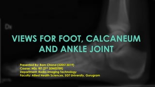
VIEWS FOR FOOT, CALCANEUM
AND ANKLE JOINT
Presented By: Ram Chand (220513019)
Course: MSc RIT (2nd SEMESTER)
Department: Radio-Imaging Technology
Faculty: Allied Health Sciences, SGT University, Gurugram
INTRODUCTION
A foot, ankle, and calcaneum X-ray is a diagnostic tool used
to assess the bones and joints in the lower leg and foot. It is a
safe and effective way to identify conditions or injuries that
may require treatment.
ANATOMY
Image Sources: Research gate, Pinterest
INDICATIONS
• foot trauma
• bony tenderness at the base of the metatarsal
• bony tenderness at the navicular bone
• inability to weight-bear more than four steps
• non-traumatic foot pain
PREPARATION
• Patient history is collected
• Patient is asked to remove all radiopaque materials
• Wear hospital gown
• Chaperone is necessary for female patient
• Shielding is provided to patient
FOOT SERIES
• Dorsoplantar view
• Medial Oblique view
• Lateral view
FOOT (DORSOPLANTER VIEW) POSITIONING
• the patient may be supine or
upright depending on comfort
• the affected leg must be flexed
enough that the plantar aspect of
the foot is resting on the image
receptor
Image Sources: Department of Radiology, Apollo Hospital, Bhubaneswar
TECHNICAL FACTORS
• centring point
• x-ray beam centred to the base of the 3rd metatarsal
• Orientation: Portrait
• detector size: 18 cm x 24 cm
• exposure
• 50-55 kVp
• 3-4 mAs
• SID: 100 cm
• Grid: no
Image Sources: Bontrager’s
FOOT MEDIAL OBLIQUE VIEW POSITIONING
• the patient may be supine or upright
depending on comfort
• the affected leg must be flexed enough
that the plantar aspect of the foot is resting
on the image receptor
• the foot is medially rotated until the
plantar surface sits at a 45° angle to the
image receptor
Image Sources: Department of Radiology, Apollo Hospital, Bhubaneswar
TECHNICAL FACTORS
• centring point
• x-ray beam centred to the base of the 3rd metatarsal
• the beam will be perpendicular to the image receptor
• Orientation: Portrait
• detector size: 18 cm x 24 cm
• exposure
• 50-55 kVp
• 3-4 mAs
• SID: 100 cm
• Grid: no
Image Sources: Bontrager’s
FOOT LATERAL VIEW POSITIONING
• the patient may be supine or upright depending on
comfort
• the affected leg is externally rotated until the distal limb is
parallel to the table, in many cases, the patient will have
to half roll onto the affected side
• the lateral aspect of the foot will be in contact with the
image receptor
• the non-affected side is kept posterior to prevent over
rotation
• the foot is in slight dorsiflexion
• the planter surface should be perpendicular to the image
receptor
Image Sources: Department of Radiology, Apollo Hospital, Bhubaneswar
TECHNICAL FACTORS
• centring point
• base of metatarsals or midfoot
• Orientation: Landscape
• detector size: 18 cm x 24 cm
• exposure
• 50-60 kVp
• 4-6 mAs
• SID: 100 cm
• Grid: no
Image Sources: Bontrager’s
CALCANEUM
SERIES
• Axial view
• Lateral view
AXIAL VIEW OF CALCANIUM POSITIONING
• The patient is seated on x ray table
with legs extended.
• Affected side ankle is dorsiflexed
placing the heel on the cassette.
• The patient is asked to hold the
ankle by the help of a bandage
cloth sling over the foot.
• Patient is immobilized side is
marked and beam is collimated.
TECHNICAL FACTORS
• centring point
• Middle sole of the foot
• Orientation: Portrait
• detector size: 18 cm x 24 cm
• exposure
• 50-60 kVp
• 3-5 mAs
• SID: 100 cm
• Grid: no
Image Sources: Bontrager’s
LATERAL VIEW OF CALCANIUM POSITIONING
• The patient is seated on x ray table
with legs extended.
• Recumbent, on affected side, knee
flexed with unaffected limb behind,
to prevent over-rotation
• Place support under knee and leg
as needed for a true lateral
• Dorsiflex foot so the plantar surface
is near 90° to leg if possible.
TECHNICAL FACTORS
• centring point
• Mid calcaneus, inferior to medial malleolus
• Orientation: Portrait
• detector size: 18 cm x 24 cm
• exposure
• 50-60 kVp
• 3-5 mAs
• SID: 100 cm
• Grid: no
Image Sources: Bontrager’s
ANKLE SERIES
• AP view
• Lateral view
•Special projections
• Mortise view
• Stress view
ANKLE AP VIEW POSITIONG
• the patient may be supine or sitting
upright with their leg straighten on the
table
• the foot is in dorsiflexion
• the toes will be pointing directly
toward the ceiling
Image Sources: Department of Radiology, Apollo Hospital, Bhubaneswar
TECHNICAL FACTORS
• centring point
• the midpoint of the lateral and medial malleoli
• Orientation: Portrait
• detector size: 24 cm x 30 cm
• exposure
• 50-60 kVp
• 3-5 mAs
• SID: 100 cm
• Grid: no
Image Sources: Bontrager’s
ANKLE LATERAL VIEW POSITIONG
• patient is in a lateral recumbent position on the
table
• the lateral aspect of the knee and ankle joint
should be in contact with the table resulting in the
tibia lying parallel to the table
• the leg can be bent or straight
• foot in dorsiflexion
• place the opposite leg behind the injured limb to
avoid over-rotation
Image Sources: Department of Radiology, Apollo Hospital, Bhubaneswar
TECHNICAL FACTORS
• centring point
• the bony prominence of the medial malleolus of the distal tibia
• Orientation: Portrait
• detector size: 18 cm x 24 cm
• exposure
• 50-60 kVp
• 3-5 mAs
• SID: 100 cm
• Grid: no
Image Sources: Bontrager’s
SPECIAL PROJECTIONS
MORTISE VIEW STRESS VIEW
This projection is use to assess the
articulation of the Tibial Plafond and tow
Malleoli with the Talar Dome
Stress view is important in the evaluation of
the Ligamentous Tears, joint stability and
fracture unions
Image Sources: Bontrager’s
ANKLE MORTISE VIEW POSITIONG
• the patient may be supine or sitting upright with
the leg straightened on the table
• the leg must be rotated internally 15° to 20°, thus
aligning the intermalleolar line parallel to the
detector. This usually results in the 5th toe being
directly in line with the centre of the calcaneum
• internal rotation must be from the hip; isolated
rotation of the ankle will result in a non-diagnostic
image
• foot should be in slight dorsiflexion
Image Sources: Bontrager’s
TECHNICAL FACTORS
• centring point
• the midpoint of the lateral and medial malleoli
• Orientation: Portrait
• detector size: 24 cm x 30 cm
• exposure
• 50-60 kVp
• 3-5 mAs
• SID: 100 cm
• Grid: no
Image Sources: Bontrager’s
ANKLE STRESS VIEW POSITIONG
• the patient may be supine or sitting
upright with the leg straightened on the
table
• the leg must be rotated internally 15° to
20°
• the second person (often requesting
physician) will then place the ankle into
supination and external rotation
Image Sources: Bontrager’s
TECHNICAL FACTORS
• centring point
• the midpoint of the lateral and medial malleoli
• Orientation: Landscape
• detector size: 24 cm x 30 cm
• exposure
• 50-60 kVp
• 3-5 mAs
• SID: 100 cm
• Grid: no
Image Sources: Bontrager’s
BIBLIOGRAPHY
https://courseware.cutm.ac.in/teachers/sambangi-
satyananda-shiva-sagar/
https://radiopaedia.org/articles/
Bontrager’s HANDBOOK OF RADIOGRAPHIC POSITIONING AND
TECHNIQUES
FOOTNOTES
Disclosure. This topic is more often based on the practical
aspects under authorized personnel.
QUESTIONS
Can you identify the following figure positionings?
A B
Image Sources: Department of Radiology, Apollo Hospital, Bhubaneswar
QUESTIONS
What is Tibial Plafond?
Image Sources: Bontrager’s
A tibial plafond fracture (also known as a pilon fracture) is a fracture of the
distal end of the tibia
THANK
YOU

More Related Content

Similar to X-RAY views for foot, calcaneum, and ankle joint.

Portals in a'scopy
Portals in a'scopyPortals in a'scopy
Portals in a'scopyRaj Kishore
 
General Principles of Arthroscopy Pramesh.pptx
General Principles of Arthroscopy Pramesh.pptxGeneral Principles of Arthroscopy Pramesh.pptx
General Principles of Arthroscopy Pramesh.pptxBirajkc5
 
Radiography Positioning Spine
Radiography Positioning SpineRadiography Positioning Spine
Radiography Positioning SpineDeepak Prasath
 
Hip and sacroiliac joints_compressed.pdf
Hip and sacroiliac joints_compressed.pdfHip and sacroiliac joints_compressed.pdf
Hip and sacroiliac joints_compressed.pdfchristapherjohn1433
 
Radiographic views of proximal femur and pelvis
Radiographic views of proximal femur and pelvisRadiographic views of proximal femur and pelvis
Radiographic views of proximal femur and pelvisChandan Prasad
 
X-RAY PELVIS PROJECTIONS
X-RAY PELVIS PROJECTIONSX-RAY PELVIS PROJECTIONS
X-RAY PELVIS PROJECTIONSJai Kumar
 
RADIOGRAPHIC VIEWS OF ANKLE JOINT
RADIOGRAPHIC VIEWS OF ANKLE JOINTRADIOGRAPHIC VIEWS OF ANKLE JOINT
RADIOGRAPHIC VIEWS OF ANKLE JOINTGanesan Yogananthem
 
Radiographic evaluation of shoulder
Radiographic evaluation of shoulderRadiographic evaluation of shoulder
Radiographic evaluation of shouldermrinal joshi
 
Technique 1 Lower limbs 2
Technique 1 Lower limbs 2Technique 1 Lower limbs 2
Technique 1 Lower limbs 2Behzad Ommani
 
Knee joint (Radiographic projection's)
Knee joint  (Radiographic  projection's)Knee joint  (Radiographic  projection's)
Knee joint (Radiographic projection's)harshit chauhan
 
Final Image Evaluation: Left Lateral Lumbar Spine
Final Image Evaluation: Left Lateral Lumbar SpineFinal Image Evaluation: Left Lateral Lumbar Spine
Final Image Evaluation: Left Lateral Lumbar SpineNBlankz
 
Technique 1 Lower limbs 3
Technique 1 Lower limbs 3Technique 1 Lower limbs 3
Technique 1 Lower limbs 3Behzad Ommani
 
Extraoral radiograph lecture
Extraoral radiograph lectureExtraoral radiograph lecture
Extraoral radiograph lectureLama K Banna
 
dental operating microscope.ppt
dental operating microscope.pptdental operating microscope.ppt
dental operating microscope.pptRoopaBabannavar1
 
Extraoral Imaging Techniques
Extraoral Imaging TechniquesExtraoral Imaging Techniques
Extraoral Imaging TechniquesSarbajit Halder
 

Similar to X-RAY views for foot, calcaneum, and ankle joint. (20)

Portals in a'scopy
Portals in a'scopyPortals in a'scopy
Portals in a'scopy
 
General Principles of Arthroscopy Pramesh.pptx
General Principles of Arthroscopy Pramesh.pptxGeneral Principles of Arthroscopy Pramesh.pptx
General Principles of Arthroscopy Pramesh.pptx
 
Radiography Positioning Spine
Radiography Positioning SpineRadiography Positioning Spine
Radiography Positioning Spine
 
Hip and sacroiliac joints_compressed.pdf
Hip and sacroiliac joints_compressed.pdfHip and sacroiliac joints_compressed.pdf
Hip and sacroiliac joints_compressed.pdf
 
Radiographic views of proximal femur and pelvis
Radiographic views of proximal femur and pelvisRadiographic views of proximal femur and pelvis
Radiographic views of proximal femur and pelvis
 
X-RAY PELVIS PROJECTIONS
X-RAY PELVIS PROJECTIONSX-RAY PELVIS PROJECTIONS
X-RAY PELVIS PROJECTIONS
 
RADIOGRAPHIC VIEWS OF ANKLE JOINT
RADIOGRAPHIC VIEWS OF ANKLE JOINTRADIOGRAPHIC VIEWS OF ANKLE JOINT
RADIOGRAPHIC VIEWS OF ANKLE JOINT
 
Seminar new
Seminar newSeminar new
Seminar new
 
Various X-ray views of Knee Joint
Various X-ray views of Knee JointVarious X-ray views of Knee Joint
Various X-ray views of Knee Joint
 
Radiographic evaluation of shoulder
Radiographic evaluation of shoulderRadiographic evaluation of shoulder
Radiographic evaluation of shoulder
 
Technique 1 Lower limbs 2
Technique 1 Lower limbs 2Technique 1 Lower limbs 2
Technique 1 Lower limbs 2
 
Knee joint (Radiographic projection's)
Knee joint  (Radiographic  projection's)Knee joint  (Radiographic  projection's)
Knee joint (Radiographic projection's)
 
Final Image Evaluation: Left Lateral Lumbar Spine
Final Image Evaluation: Left Lateral Lumbar SpineFinal Image Evaluation: Left Lateral Lumbar Spine
Final Image Evaluation: Left Lateral Lumbar Spine
 
Technique 1 Lower limbs 3
Technique 1 Lower limbs 3Technique 1 Lower limbs 3
Technique 1 Lower limbs 3
 
Ortho Journal Club 2 by Dr Saumya Agarwal
Ortho Journal Club 2 by Dr Saumya AgarwalOrtho Journal Club 2 by Dr Saumya Agarwal
Ortho Journal Club 2 by Dr Saumya Agarwal
 
Extraoral radiograph lecture
Extraoral radiograph lectureExtraoral radiograph lecture
Extraoral radiograph lecture
 
Extraoral radiography
Extraoral radiographyExtraoral radiography
Extraoral radiography
 
dental operating microscope.ppt
dental operating microscope.pptdental operating microscope.ppt
dental operating microscope.ppt
 
Fracture Neck Of Femur
Fracture Neck Of FemurFracture Neck Of Femur
Fracture Neck Of Femur
 
Extraoral Imaging Techniques
Extraoral Imaging TechniquesExtraoral Imaging Techniques
Extraoral Imaging Techniques
 

Recently uploaded

Presentation by Andreas Schleicher Tackling the School Absenteeism Crisis 30 ...
Presentation by Andreas Schleicher Tackling the School Absenteeism Crisis 30 ...Presentation by Andreas Schleicher Tackling the School Absenteeism Crisis 30 ...
Presentation by Andreas Schleicher Tackling the School Absenteeism Crisis 30 ...EduSkills OECD
 
SOCIAL AND HISTORICAL CONTEXT - LFTVD.pptx
SOCIAL AND HISTORICAL CONTEXT - LFTVD.pptxSOCIAL AND HISTORICAL CONTEXT - LFTVD.pptx
SOCIAL AND HISTORICAL CONTEXT - LFTVD.pptxiammrhaywood
 
Hybridoma Technology ( Production , Purification , and Application )
Hybridoma Technology  ( Production , Purification , and Application  ) Hybridoma Technology  ( Production , Purification , and Application  )
Hybridoma Technology ( Production , Purification , and Application ) Sakshi Ghasle
 
Privatization and Disinvestment - Meaning, Objectives, Advantages and Disadva...
Privatization and Disinvestment - Meaning, Objectives, Advantages and Disadva...Privatization and Disinvestment - Meaning, Objectives, Advantages and Disadva...
Privatization and Disinvestment - Meaning, Objectives, Advantages and Disadva...RKavithamani
 
Z Score,T Score, Percential Rank and Box Plot Graph
Z Score,T Score, Percential Rank and Box Plot GraphZ Score,T Score, Percential Rank and Box Plot Graph
Z Score,T Score, Percential Rank and Box Plot GraphThiyagu K
 
Accessible design: Minimum effort, maximum impact
Accessible design: Minimum effort, maximum impactAccessible design: Minimum effort, maximum impact
Accessible design: Minimum effort, maximum impactdawncurless
 
How to Make a Pirate ship Primary Education.pptx
How to Make a Pirate ship Primary Education.pptxHow to Make a Pirate ship Primary Education.pptx
How to Make a Pirate ship Primary Education.pptxmanuelaromero2013
 
Kisan Call Centre - To harness potential of ICT in Agriculture by answer farm...
Kisan Call Centre - To harness potential of ICT in Agriculture by answer farm...Kisan Call Centre - To harness potential of ICT in Agriculture by answer farm...
Kisan Call Centre - To harness potential of ICT in Agriculture by answer farm...Krashi Coaching
 
Nutritional Needs Presentation - HLTH 104
Nutritional Needs Presentation - HLTH 104Nutritional Needs Presentation - HLTH 104
Nutritional Needs Presentation - HLTH 104misteraugie
 
“Oh GOSH! Reflecting on Hackteria's Collaborative Practices in a Global Do-It...
“Oh GOSH! Reflecting on Hackteria's Collaborative Practices in a Global Do-It...“Oh GOSH! Reflecting on Hackteria's Collaborative Practices in a Global Do-It...
“Oh GOSH! Reflecting on Hackteria's Collaborative Practices in a Global Do-It...Marc Dusseiller Dusjagr
 
microwave assisted reaction. General introduction
microwave assisted reaction. General introductionmicrowave assisted reaction. General introduction
microwave assisted reaction. General introductionMaksud Ahmed
 
Beyond the EU: DORA and NIS 2 Directive's Global Impact
Beyond the EU: DORA and NIS 2 Directive's Global ImpactBeyond the EU: DORA and NIS 2 Directive's Global Impact
Beyond the EU: DORA and NIS 2 Directive's Global ImpactPECB
 
BASLIQ CURRENT LOOKBOOK LOOKBOOK(1) (1).pdf
BASLIQ CURRENT LOOKBOOK  LOOKBOOK(1) (1).pdfBASLIQ CURRENT LOOKBOOK  LOOKBOOK(1) (1).pdf
BASLIQ CURRENT LOOKBOOK LOOKBOOK(1) (1).pdfSoniaTolstoy
 
Call Girls in Dwarka Mor Delhi Contact Us 9654467111
Call Girls in Dwarka Mor Delhi Contact Us 9654467111Call Girls in Dwarka Mor Delhi Contact Us 9654467111
Call Girls in Dwarka Mor Delhi Contact Us 9654467111Sapana Sha
 
Separation of Lanthanides/ Lanthanides and Actinides
Separation of Lanthanides/ Lanthanides and ActinidesSeparation of Lanthanides/ Lanthanides and Actinides
Separation of Lanthanides/ Lanthanides and ActinidesFatimaKhan178732
 
Web & Social Media Analytics Previous Year Question Paper.pdf
Web & Social Media Analytics Previous Year Question Paper.pdfWeb & Social Media Analytics Previous Year Question Paper.pdf
Web & Social Media Analytics Previous Year Question Paper.pdfJayanti Pande
 
Software Engineering Methodologies (overview)
Software Engineering Methodologies (overview)Software Engineering Methodologies (overview)
Software Engineering Methodologies (overview)eniolaolutunde
 
CARE OF CHILD IN INCUBATOR..........pptx
CARE OF CHILD IN INCUBATOR..........pptxCARE OF CHILD IN INCUBATOR..........pptx
CARE OF CHILD IN INCUBATOR..........pptxGaneshChakor2
 

Recently uploaded (20)

Presentation by Andreas Schleicher Tackling the School Absenteeism Crisis 30 ...
Presentation by Andreas Schleicher Tackling the School Absenteeism Crisis 30 ...Presentation by Andreas Schleicher Tackling the School Absenteeism Crisis 30 ...
Presentation by Andreas Schleicher Tackling the School Absenteeism Crisis 30 ...
 
SOCIAL AND HISTORICAL CONTEXT - LFTVD.pptx
SOCIAL AND HISTORICAL CONTEXT - LFTVD.pptxSOCIAL AND HISTORICAL CONTEXT - LFTVD.pptx
SOCIAL AND HISTORICAL CONTEXT - LFTVD.pptx
 
Hybridoma Technology ( Production , Purification , and Application )
Hybridoma Technology  ( Production , Purification , and Application  ) Hybridoma Technology  ( Production , Purification , and Application  )
Hybridoma Technology ( Production , Purification , and Application )
 
Privatization and Disinvestment - Meaning, Objectives, Advantages and Disadva...
Privatization and Disinvestment - Meaning, Objectives, Advantages and Disadva...Privatization and Disinvestment - Meaning, Objectives, Advantages and Disadva...
Privatization and Disinvestment - Meaning, Objectives, Advantages and Disadva...
 
Z Score,T Score, Percential Rank and Box Plot Graph
Z Score,T Score, Percential Rank and Box Plot GraphZ Score,T Score, Percential Rank and Box Plot Graph
Z Score,T Score, Percential Rank and Box Plot Graph
 
Accessible design: Minimum effort, maximum impact
Accessible design: Minimum effort, maximum impactAccessible design: Minimum effort, maximum impact
Accessible design: Minimum effort, maximum impact
 
Mattingly "AI & Prompt Design: The Basics of Prompt Design"
Mattingly "AI & Prompt Design: The Basics of Prompt Design"Mattingly "AI & Prompt Design: The Basics of Prompt Design"
Mattingly "AI & Prompt Design: The Basics of Prompt Design"
 
How to Make a Pirate ship Primary Education.pptx
How to Make a Pirate ship Primary Education.pptxHow to Make a Pirate ship Primary Education.pptx
How to Make a Pirate ship Primary Education.pptx
 
Kisan Call Centre - To harness potential of ICT in Agriculture by answer farm...
Kisan Call Centre - To harness potential of ICT in Agriculture by answer farm...Kisan Call Centre - To harness potential of ICT in Agriculture by answer farm...
Kisan Call Centre - To harness potential of ICT in Agriculture by answer farm...
 
Nutritional Needs Presentation - HLTH 104
Nutritional Needs Presentation - HLTH 104Nutritional Needs Presentation - HLTH 104
Nutritional Needs Presentation - HLTH 104
 
“Oh GOSH! Reflecting on Hackteria's Collaborative Practices in a Global Do-It...
“Oh GOSH! Reflecting on Hackteria's Collaborative Practices in a Global Do-It...“Oh GOSH! Reflecting on Hackteria's Collaborative Practices in a Global Do-It...
“Oh GOSH! Reflecting on Hackteria's Collaborative Practices in a Global Do-It...
 
microwave assisted reaction. General introduction
microwave assisted reaction. General introductionmicrowave assisted reaction. General introduction
microwave assisted reaction. General introduction
 
Código Creativo y Arte de Software | Unidad 1
Código Creativo y Arte de Software | Unidad 1Código Creativo y Arte de Software | Unidad 1
Código Creativo y Arte de Software | Unidad 1
 
Beyond the EU: DORA and NIS 2 Directive's Global Impact
Beyond the EU: DORA and NIS 2 Directive's Global ImpactBeyond the EU: DORA and NIS 2 Directive's Global Impact
Beyond the EU: DORA and NIS 2 Directive's Global Impact
 
BASLIQ CURRENT LOOKBOOK LOOKBOOK(1) (1).pdf
BASLIQ CURRENT LOOKBOOK  LOOKBOOK(1) (1).pdfBASLIQ CURRENT LOOKBOOK  LOOKBOOK(1) (1).pdf
BASLIQ CURRENT LOOKBOOK LOOKBOOK(1) (1).pdf
 
Call Girls in Dwarka Mor Delhi Contact Us 9654467111
Call Girls in Dwarka Mor Delhi Contact Us 9654467111Call Girls in Dwarka Mor Delhi Contact Us 9654467111
Call Girls in Dwarka Mor Delhi Contact Us 9654467111
 
Separation of Lanthanides/ Lanthanides and Actinides
Separation of Lanthanides/ Lanthanides and ActinidesSeparation of Lanthanides/ Lanthanides and Actinides
Separation of Lanthanides/ Lanthanides and Actinides
 
Web & Social Media Analytics Previous Year Question Paper.pdf
Web & Social Media Analytics Previous Year Question Paper.pdfWeb & Social Media Analytics Previous Year Question Paper.pdf
Web & Social Media Analytics Previous Year Question Paper.pdf
 
Software Engineering Methodologies (overview)
Software Engineering Methodologies (overview)Software Engineering Methodologies (overview)
Software Engineering Methodologies (overview)
 
CARE OF CHILD IN INCUBATOR..........pptx
CARE OF CHILD IN INCUBATOR..........pptxCARE OF CHILD IN INCUBATOR..........pptx
CARE OF CHILD IN INCUBATOR..........pptx
 

X-RAY views for foot, calcaneum, and ankle joint.

  • 1. VIEWS FOR FOOT, CALCANEUM AND ANKLE JOINT Presented By: Ram Chand (220513019) Course: MSc RIT (2nd SEMESTER) Department: Radio-Imaging Technology Faculty: Allied Health Sciences, SGT University, Gurugram
  • 2. INTRODUCTION A foot, ankle, and calcaneum X-ray is a diagnostic tool used to assess the bones and joints in the lower leg and foot. It is a safe and effective way to identify conditions or injuries that may require treatment.
  • 4. INDICATIONS • foot trauma • bony tenderness at the base of the metatarsal • bony tenderness at the navicular bone • inability to weight-bear more than four steps • non-traumatic foot pain
  • 5. PREPARATION • Patient history is collected • Patient is asked to remove all radiopaque materials • Wear hospital gown • Chaperone is necessary for female patient • Shielding is provided to patient
  • 6. FOOT SERIES • Dorsoplantar view • Medial Oblique view • Lateral view
  • 7. FOOT (DORSOPLANTER VIEW) POSITIONING • the patient may be supine or upright depending on comfort • the affected leg must be flexed enough that the plantar aspect of the foot is resting on the image receptor Image Sources: Department of Radiology, Apollo Hospital, Bhubaneswar
  • 8. TECHNICAL FACTORS • centring point • x-ray beam centred to the base of the 3rd metatarsal • Orientation: Portrait • detector size: 18 cm x 24 cm • exposure • 50-55 kVp • 3-4 mAs • SID: 100 cm • Grid: no Image Sources: Bontrager’s
  • 9. FOOT MEDIAL OBLIQUE VIEW POSITIONING • the patient may be supine or upright depending on comfort • the affected leg must be flexed enough that the plantar aspect of the foot is resting on the image receptor • the foot is medially rotated until the plantar surface sits at a 45° angle to the image receptor Image Sources: Department of Radiology, Apollo Hospital, Bhubaneswar
  • 10. TECHNICAL FACTORS • centring point • x-ray beam centred to the base of the 3rd metatarsal • the beam will be perpendicular to the image receptor • Orientation: Portrait • detector size: 18 cm x 24 cm • exposure • 50-55 kVp • 3-4 mAs • SID: 100 cm • Grid: no Image Sources: Bontrager’s
  • 11. FOOT LATERAL VIEW POSITIONING • the patient may be supine or upright depending on comfort • the affected leg is externally rotated until the distal limb is parallel to the table, in many cases, the patient will have to half roll onto the affected side • the lateral aspect of the foot will be in contact with the image receptor • the non-affected side is kept posterior to prevent over rotation • the foot is in slight dorsiflexion • the planter surface should be perpendicular to the image receptor Image Sources: Department of Radiology, Apollo Hospital, Bhubaneswar
  • 12. TECHNICAL FACTORS • centring point • base of metatarsals or midfoot • Orientation: Landscape • detector size: 18 cm x 24 cm • exposure • 50-60 kVp • 4-6 mAs • SID: 100 cm • Grid: no Image Sources: Bontrager’s
  • 14. AXIAL VIEW OF CALCANIUM POSITIONING • The patient is seated on x ray table with legs extended. • Affected side ankle is dorsiflexed placing the heel on the cassette. • The patient is asked to hold the ankle by the help of a bandage cloth sling over the foot. • Patient is immobilized side is marked and beam is collimated.
  • 15. TECHNICAL FACTORS • centring point • Middle sole of the foot • Orientation: Portrait • detector size: 18 cm x 24 cm • exposure • 50-60 kVp • 3-5 mAs • SID: 100 cm • Grid: no Image Sources: Bontrager’s
  • 16. LATERAL VIEW OF CALCANIUM POSITIONING • The patient is seated on x ray table with legs extended. • Recumbent, on affected side, knee flexed with unaffected limb behind, to prevent over-rotation • Place support under knee and leg as needed for a true lateral • Dorsiflex foot so the plantar surface is near 90° to leg if possible.
  • 17. TECHNICAL FACTORS • centring point • Mid calcaneus, inferior to medial malleolus • Orientation: Portrait • detector size: 18 cm x 24 cm • exposure • 50-60 kVp • 3-5 mAs • SID: 100 cm • Grid: no Image Sources: Bontrager’s
  • 18. ANKLE SERIES • AP view • Lateral view •Special projections • Mortise view • Stress view
  • 19. ANKLE AP VIEW POSITIONG • the patient may be supine or sitting upright with their leg straighten on the table • the foot is in dorsiflexion • the toes will be pointing directly toward the ceiling Image Sources: Department of Radiology, Apollo Hospital, Bhubaneswar
  • 20. TECHNICAL FACTORS • centring point • the midpoint of the lateral and medial malleoli • Orientation: Portrait • detector size: 24 cm x 30 cm • exposure • 50-60 kVp • 3-5 mAs • SID: 100 cm • Grid: no Image Sources: Bontrager’s
  • 21. ANKLE LATERAL VIEW POSITIONG • patient is in a lateral recumbent position on the table • the lateral aspect of the knee and ankle joint should be in contact with the table resulting in the tibia lying parallel to the table • the leg can be bent or straight • foot in dorsiflexion • place the opposite leg behind the injured limb to avoid over-rotation Image Sources: Department of Radiology, Apollo Hospital, Bhubaneswar
  • 22. TECHNICAL FACTORS • centring point • the bony prominence of the medial malleolus of the distal tibia • Orientation: Portrait • detector size: 18 cm x 24 cm • exposure • 50-60 kVp • 3-5 mAs • SID: 100 cm • Grid: no Image Sources: Bontrager’s
  • 23. SPECIAL PROJECTIONS MORTISE VIEW STRESS VIEW This projection is use to assess the articulation of the Tibial Plafond and tow Malleoli with the Talar Dome Stress view is important in the evaluation of the Ligamentous Tears, joint stability and fracture unions Image Sources: Bontrager’s
  • 24. ANKLE MORTISE VIEW POSITIONG • the patient may be supine or sitting upright with the leg straightened on the table • the leg must be rotated internally 15° to 20°, thus aligning the intermalleolar line parallel to the detector. This usually results in the 5th toe being directly in line with the centre of the calcaneum • internal rotation must be from the hip; isolated rotation of the ankle will result in a non-diagnostic image • foot should be in slight dorsiflexion Image Sources: Bontrager’s
  • 25. TECHNICAL FACTORS • centring point • the midpoint of the lateral and medial malleoli • Orientation: Portrait • detector size: 24 cm x 30 cm • exposure • 50-60 kVp • 3-5 mAs • SID: 100 cm • Grid: no Image Sources: Bontrager’s
  • 26. ANKLE STRESS VIEW POSITIONG • the patient may be supine or sitting upright with the leg straightened on the table • the leg must be rotated internally 15° to 20° • the second person (often requesting physician) will then place the ankle into supination and external rotation Image Sources: Bontrager’s
  • 27. TECHNICAL FACTORS • centring point • the midpoint of the lateral and medial malleoli • Orientation: Landscape • detector size: 24 cm x 30 cm • exposure • 50-60 kVp • 3-5 mAs • SID: 100 cm • Grid: no Image Sources: Bontrager’s
  • 28. BIBLIOGRAPHY https://courseware.cutm.ac.in/teachers/sambangi- satyananda-shiva-sagar/ https://radiopaedia.org/articles/ Bontrager’s HANDBOOK OF RADIOGRAPHIC POSITIONING AND TECHNIQUES FOOTNOTES Disclosure. This topic is more often based on the practical aspects under authorized personnel.
  • 29. QUESTIONS Can you identify the following figure positionings? A B Image Sources: Department of Radiology, Apollo Hospital, Bhubaneswar
  • 30. QUESTIONS What is Tibial Plafond? Image Sources: Bontrager’s A tibial plafond fracture (also known as a pilon fracture) is a fracture of the distal end of the tibia