SlideShare a Scribd company logo
1 of 57
Ms. M. Arthi, M.Sc., M.Phil., NET., SET.,
Assistant Professor of Microbiology
The process by which a cell
makes proteins using the
genetic information carried
in messenger RNA (mRNA).
INTRODUCTION
• Proteins are the end products of most information pathways.
• A typical cell requires thousands of different proteins at any given moment.
• It is synthesized in response to the cell’s current needs, transported (targeted) to
their appropriate cellular locations, and degraded when no longer needed.
• This biosynthetic machinery is well conserved in all life-forms from bacteria to
higher eukaryotes - present in the last universal common ancestor (LUCA) of all
extant organisms.
• It’s a most complex biosynthetic process.
• Eukaryotic protein synthesis involves –
• Ribosomal proteins - more than 70 types.
• For the activation of amino acid precursors - 20 or more enzymes
• For initiation, elongation, and termination of polypeptides - dozen or more
auxiliary enzymes and other protein factors.
• For the final processing of different proteins - 100 additional enzymes
• Transfer and ribosomal RNAs - 40 or more kinds.
• Overall - almost 300 different macromolecules cooperate to synthesize
polypeptides.
• Many of these macromolecules are organized into the complex 3D structure of the
ribosome.
• Typical bacterial cell
– Ribosomes - 15,000
– protein synthesis–related protein factors and enzymes - 100,000 molecules
– tRNA molecules - 200,000
• All this can account for more than 35% of the cell’s dry weight.
• A polypeptide of 100 residues is synthesized in an Escherichia coli cell (at 370C) in about 5
seconds.
• Elucidation of 3D structures of ribosomes - began in
the year 2000
• Two decades earlier to the discovery of 3D structure
of ribosomes - Proteins are synthesized by a gigantic
RNA enzyme! - Hypothesis was first put forward by
Harry Noller.
• 1950s, Paul Zamecnik - labeled a.as appeared in a fraction containing small
ribonucleoprotein particles (visible in animal tissues by electron microscopy),
identified as the site of protein synthesis - later named as ribosomes.
• Mahlon Hoagland and Zamecnik - amino acids + ATP + cytosolic fraction of
liver cells  “activated” A.as.
• Robert Holley - The amino acids attaches to heat-stable soluble RNA - later
called transfer RNA (tRNA), to form aminoacyl-tRNAs. The enzymes that
catalyze this process are the aminoacyl-tRNA synthetases.
• Francis Crick’s – Crick’s adaptor hypothesis. The tRNA adaptor “translates” the
nucleotide sequence of an mRNA into the amino acid sequence of a
polypeptide. The overall process of mRNA-guided protein synthesis is often
referred to simply as translation.
• Masayasu Nomura – discovered 30S or 50S subunits.
• A codon is a triplet of nucleotides
that codes for a specific amino acid.
• nucleotide triplets are read in a
successive, nonoverlapping fashion.
• A specific first codon in the sequence
establishes the reading frame, in
which a new codon begins every
three nucleotide residues.
• There is no punctuation between
codons for successive amino acid
residues
• mRNA sequence has three possible reading frames. Each reading frame gives a
different sequence of codons, but only one is likely to encode a given protein.
• The initiation codon AUG is the most common signal for the beginning of a
polypeptide in all cells, in addition to coding for Met residues in internal positions of
polypeptides.
• The termination codons (UAA, UAG, and UGA), also called stop codons or nonsense
codons, normally signal the end of polypeptide synthesis and do not code for any
known amino acids.
• a reading frame without a termination codon among 50 or more codons is referred
to as an open reading frame (ORF).
Phe
Ser
Leu
Protein Biosynthesis Takes Place in Five Stages
• Activation of amino acids
• Initiation
• Elongation
• Termination & release
• Folding and Posttranslational Processing
Before looking at these five stages in detail, we must examine
two key components in protein biosynthesis: the ribosome and
tRNAs.
• E. coli cell contains 15,000 or more ribosomes, which comprise
nearly a quarter of the dry weight of the cell.
• Bacterial ribosomes contain about 65% rRNA and 35% protein;
they have a diameter of about 18 nm.
• composed of two unequal subunits with sedimentation
coefficients of 30S and 50S and a combined sedimentation
coefficient of 70S.
• Both subunits contain dozens of ribosomal proteins and at least
one large rRNA.
• The ribosomal subunits are huge RNA molecules. In the 50S
subunit, the 5S and 23S rRNAs form the structural core. The
proteins are secondary elements in the complex, decorating the
surface.
• there is no protein within 18 Å of the active site for peptide
bond formation.
• the ribosome is a ribozyme.
Structure of Ribosomes
• The bacterial ribosome is complex, with a combined
molecular weight of ,2.7 million.
• The two irregularly shaped ribosomal subunits fit
together to form a cleft through which the mRNA
passes as the ribosome moves along it during
translation.
• The 57 proteins in bacterial ribosomes vary enormously
in size and structure. Molecular weights range from
about 6,000 to 75,000.
• Most of the proteins have globular domains arranged
on the ribosome surface. Some also have snakelike
extensions that protrude into the rRNA core of the
ribosome, stabilizing its structure.
• Each of the three single-stranded rRNAs of E. coli has a
specific three-dimensional conformation featuring
extensive intrachain base pairing.
• The folding patterns of the rRNAs are highly conserved
in all organisms, particularly the regions implicated in
key functions.
Structure of tRNA
• Transfer RNAs are small and consist of a single strand of RNA folded into a
precise 3D structure.
• The tRNAs in bacteria and in the cytosol of eukaryotes - have b/w 73 and 93 nts
• Molecular weights of 24,000 to 31,000.
• Mitochondria and chloroplasts contain - smaller tRNAs.
• Cells have at least one kind of tRNA for each amino acid; at least 32 tRNAs are
required to recognize all the amino acid codons, but some cells use more than
32.
• Yeast alanine tRNA (tRNAAla) - the first nucleic acid to be completely sequenced
• It contains 76 nucleotide residues, of which 10 are modified bases.
This structure was deduced in 1965 by Robert
W. Holley and his colleagues
D - 5,6-dihydrouridine
m1G - 1-methylguanosine;
m2G - N2-dimethylguanosine.
I- inosine; ; m1I - 1-methylinosine;
ψ - pseudouridine;
T- ribothymidine;
• tRNAs have a guanylate (pG) residue at the 5’ end
• Have the trinucleotide sequence CCA at the 3’ end.
• D and TC arms helps in the overall folding of tRNA
molecules,
• TC arm interacts with the large-subunit rRNA
Protein Biosynthesis Takes Place in Five Stages
• Activation of amino acids
• Initiation
• Elongation
• Termination & release
• Folding and Posttranslational Processing
Components Required for the Five Major Stages of Protein Synthesis in E. coli
1. Activation of amino acids
Aminoacyl-tRNA Synthetases Attach the Correct Amino Acids to Their tRNAs
• This reaction takes place in the cytosol, not on the ribosome.
• 20 amino acids are covalently attached to a specific tRNA at the expense of ATP energy, using Mg2+ dependent
activating enzymes known as aminoacyl-tRNA synthetases.
• When attached to their amino acid (aminoacylated) the tRNAs are said to be “charged.”
• Specificity – each a.a has a specific activating enzyme – tRNA amino acyl synthetase (20 diff for 20 a.as). Some can
activate more than 1 a.a
– Eg: Isoleucine tRNA synthetase – activates Isoleucine & Valine
– Valine tRNA synthetase – activates only valine
• Based on differences in 10 and 30 structure and in reaction mechanism, aminoacyl-tRNA synthetases of E. coli is
divided into two classes.
Reaction Mechanism
• This reaction occurs in two steps in the enzyme’s active site.
• In step 1 – coo- group of a.a reacts with the α-phosphoryl group of ATP to form an
anhydride linkage, with displacement of pyrophosphate. Forms an enzyme-bound
intermediate - aminoacyl adenylate (aminoacyl-AMP).
• In step 2 - aminoacyl group is transferred from enzyme-bound aminoacyl-AMP to its
corresponding specific tRNA. The pyrophosphate formed in the activation reaction
undergoes hydrolysis to phosphate by inorganic pyrophosphatase.
Amino acid + tRNA + ATP --------------------------------------- aminoacyl-tRNA + AMP + PPi
aminoacyl-tRNA synthetase
Mg2+
Proofreading by Aminoacyl-tRNA Synthetases
• The amino acid attached to a tRNA is not checked on the ribosome, so attachment of
the correct amino acid to the tRNA is essential to the fidelity of protein synthesis.
• Hence some amino acyl tRNA synthetases enzyme has proof reading function.
• Ile-tRNA synthetase, which distinguishes valine and isoleucine (single methylene group -
CH2-).
• If available binding interactions do not provide sufficient discrimination between two
substrates, the necessary specificity can be achieved by substrate-specific binding.
• Eg : Methylene group (in Ile) could enhance substrate binding
Enzyme has 3 filters:
• 1st filter - initial binding of the amino acid to the enzyme and its activation to
aminoacyl-AMP.
• 2nd filter - binding of any incorrect aminoacyl-AMP products to a separate ( second)
active site on the enzyme is hydrolyzed.
– Eg. The R group of valine is slightly smaller than that of isoleucine, so Val-AMP fits
the hydrolytic (proofreading) site of the Ile-tRNA synthetase but Ile-AMP does not.
Thus Val-AMP is hydrolyzed to valine and AMP in the proofreading active site.
• 3rd filter - most aminoacyl-tRNA synthetases can also hydrolyze the ester linkage
between amino acids and tRNAs in the aminoacyl-tRNAs. This hydrolysis is greatly
accelerated for incorrectly charged tRNAs.
• All the 3 filters enhance the fidelity of the overall process.
• blue dots - same in all tRNAs
• Orange dots - recognition points for one aminoacyl-tRNA
synthetases.
• green dots - recognition points for more aminoacyl-tRNA
synthetases.
Interaction between an Aminoacyl-tRNA Synthetase and a tRNA: A
“Second Genetic Code”
• An individual aminoacyl-tRNA synthetase is
specific not only for a single amino acid but
for certain tRNAs as well.
• Eg. tRNA ala
• Ten or more specific nucleotides may be
involved in recognition of a tRNA by its
specific aminoacyl-tRNA synthetase.
Single G=U base pair in the amino acid arm of tRNAAla recognized by the
Ala-tRNA synthetases.
Fidelity of protein synthesis
• Discriminating dozens of tRNAs and amino acids is important for the overall fidelity of
protein biosynthesis.
• The overall error rate of protein synthesis (~1 mistake per 104 amino acids
incorporated) is not low as DNA replication.
• Proteins have less biological significance becoz not passed on to future generations.
• Incorrect proteins are eliminated by degradation.
• The degree of fidelity in protein synthesis is essential to save energy required to
synthesize a protein.
• One defective protein molecule is usually unimportant when many correct copies of the
same protein are present.
2. Initiation
• Howard Dintzis in 1961 proved that Protein synthesis begins at the amino-terminal end and proceeds by
the stepwise addition of amino acids to the carboxyl-terminal end of the growing polypeptide using
Reticulocytes (immature erythrocytes) that actively synthesizing hemoglobin were incubated with
radioactive leucine.
• The AUG initiation codon specifies methionine residue (amino-terminal).
• Methionine has only one codon, (5’)AUG but all organisms have two tRNAs for methionine.
– One is for initiation codon, which was designated as tRNAfMet .
– The other is for the addition of Met residue in an internal position in a polypeptide - tRNAMet.
• The amino acid incorporated in response to the (5’)AUG initiation codon is N-formylmethionine (fMet).
• It arrives at the ribosome as N-formylmethionyl-tRNAfMet (fMet-tRNAfMet)
Formed in two successive reactions.
• Step 1 - methionine is attached to tRNAfMet by the Met-tRNA synthetase (which in E. coli aminoacylates
both tRNAfMet and tRNAMet)
Methionine + tRNAfMet+ ATP ----------- Met-tRNAfMet + AMP + Ppi
• Step 2 - Transformylase transfers a formyl group from N10-formyltetrahydrofolate to the amino group of
the Met residue
N10-Formyltetrahydrofolate + Met-tRNAfMet ------- tetrahydrofolate + fMet-tRNAfMet
Met-tRNA synthetase
Transformylase
• In eukaryotic cells polypeptides synthesized by
• cytosolic ribosomes - begin with a Met residue (rather than fMet)
• mitochondrial and chloroplast ribosomes - begin with N-formylmethionine.
Requirements for the initiation in bacteria
(1) the 30S ribosomal subunit,
(2) the mRNA coding for the polypeptide to be made,
(3) the initiating fMet-tRNAfMet,
(4) a set of three proteins called initiation factors (IF-1, IF-2, and IF-3),
(5) GTP,
(6) the 50S ribosomal subunit, and
(7) Mg2.
The Three Steps of Initiation
• Step 1 - the 30S ribosomal subunit binds two
initiation factors, IF-1 (binds at the A site and
prevents the tRNA binding at this site during
initiation) and IF-3 (prevents the reassociation of 30S
and 50S subunits prematurely).
• The mRNA (Shine dalgarno sequence) then binds to
the 30S subunit (16S rRNA).
• This mRNA-rRNA interaction positions the initiating
(5’)AUG sequence of the mRNA at the P site (fMet-
tRNAfMet binds) on the 30S subunit.
ShineDalgarno sequence
• Named for Australian researchers John Shine and Lynn Dalgarno, who identified it
in the mRNA.
• This consensus sequence is an initiation signal of four to nine purine residues,
located 8 to 13 bp upstream to the 5’ side of the initiation codon.
• This sequence base-pairs with a complementary pyrimidine-rich sequence near
the 3’ end of the 16S rRNA of the 30S ribosomal subunit.
Bacterial ribosomes have three sites
• Peptidyl (P) site - The initiating (5)AUG is positioned at
the P site, the only site to which fMettRNAfMet can
bind.
• Aminoacyl (A) site - during the elongation stage, all
other incoming aminoacyl-tRNAs bind first to the A site
• Exit (E) site - from which the “uncharged” tRNAs leave
during elongation
confined to the 50S subunit
confined to both 50S & 30S subunit
• Step 2 - The anticodon of initiating fMet-
tRNAfMet pairs correctly with the
initiation codon of mRNA. mRNA is joined
by both GTP-bound IF-2 and the initiating
fMet-tRNAfMet.
The correct binding of the complex is assured by at least three points of
recognition and attachment:
–Codon-anticodon interaction
–Shine-Dalgarno sequence and the 16S rRNA interactions
–Ribosomal P site and the fMet-tRNAfMet interaction.
• The initiation complex is now ready for elongation.
• Step 3 - this large complex combines with the 50S
ribosomal subunit; simultaneously, the GTP
bound to IF-2 is hydrolyzed to GDP and Pi
• Subsequently all the 3 initiation factors are
departed from the ribosome.
• Completion of the steps produces a functional
70S ribosome called the initiation complex.
Initiation in Eukaryotic Cells
• Eukaryotic mRNAs are bound to the ribosome with the
help of a specific binding proteins.
• They tie the 5’ and 3’ ends of the message together.
• The 3’ end of the mRNA is bound to the poly(A)
binding protein (PAB).
• The 5’ end of mRNA bind with eIF4E.
• Eukaryotic cells have at least nine initiation factors.
• The initiating (5’)AUG is detected within the mRNA not
by its proximity to a Shine-Dalgarno-like sequence but
by a scanning process (facilitated by eIF4F & eIF4B).
3. Elongation
• Requirements for elongation:
(1)the initiation complex,
(2)aminoacyl-tRNAs,
(3)a set of three soluble cytosolic proteins called elongation
factors (EF-Tu, EF-Ts, and EF-G in bacteria), and
(4)GTP.
The Three Steps of Elongation
• Cells use three steps to add each amino acid residue, and the steps are repeated
as many times as there are residues to be added.
»Step 1: Binding of an Incoming Aminoacyl-tRNA
» Step 2: Peptide Bond Formation
»Step 3: Translocation
• The appropriate incoming aminoacyl-tRNA binds to a
complex of GTP-bound EF-Tu.
• The resulting aminoacyltRNA–EF-Tu–GTP complex binds
to the A site of the 70S initiation complex.
• The GTP is hydrolyzed and an EF-Tu–GDP complex is
released from the 70S ribosome.
• The EF-Tu–GTP complex is regenerated in a process
involving EF-Ts and GTP.
Step 1: Binding of an Incoming Aminoacyl-tRNA
Step 2: Peptide Bond Formation
• A peptide bond is formed between the two amino acids bound by
their tRNAs to the A and P sites on the ribosome.
• This occurs by the transfer of the initiating N-formylmethionyl group
from its tRNA to the amino group of the second amino acid, in the A
site.
• The α-amino group of the amino acid in the A site acts as a
nucleophile, displacing the tRNA in the P site to form the peptide
bond.
• This reaction produces a dipeptidyltRNA in the A site, and the now
“uncharged” (deacylated) tRNAfMet remains bound to the P site.
• The enzymatic activity that catalyzes peptide bond formation has
historically been referred to as peptidyl transferase, now know that
this reaction is catalyzed by the 23S rRNA (ribozymes).
Step 3: Translocation
• The ribosome moves one codon toward the 3’ end of the
mRNA.
• This movement shifts the anticodon of the dipeptidyltRNA
(attached to the second codon of the mRNA) from the A site to
the P site.
• Shifts the deacylated tRNA from the P site to the E site, from
where the tRNA is released into the cytosol.
• The third codon of the mRNA now lies in the A site and the
second codon in the P site.
• Movement of the ribosome along the mRNA requires EF-G
(also known as translocase) and the energy provided by
hydrolysis of another molecule of GTP.
• EF-G binds to the A site and displace the peptidyl-tRNA.
Elongation in Eukaryotic Cells
• The elongation cycle in eukaryotes is quite similar to that in prokaryotes.
• Three eukaryotic elongation factors (eEF1α, eEF1βγ, and eEF2) have functions
analogous to bacterial elongation factors (EF-Tu, EF-Ts, and EF-G, respectively).
• Eukaryotic ribosomes do not have an E site; uncharged tRNAs are expelled
directly from the P site.
4. Termination
• Elongation continues until the ribosome adds the last amino acid coded by the mRNA.
• Termination, is signalled by the presence of one of three termination codons in the
mRNA (UAA, UAG, UGA), immediately following the final coded amino acid.
• In bacteria, once a termination codon occupies the ribosomal A site, three termination
factors, or release factors—
»RF-1 - recognizes the termination codons UAG and UAA
»RF-2 - recognizes UGA and UAA
»RF-3 – function is unknown
• In eukaryotes, a single release factor, eRF, recognizes all three termination codons.
Three termination factors, or release factors contribute to
(1) hydrolysis of the terminal peptidyltRNA bond (peptidyl
transferase to transfer the growing polypeptide to a water
molecule rather than to another amino acid).
(2) release of the free polypeptide and the last tRNA, now
uncharged, from the P site.
(3) dissociation of the 70S ribosome into its 30S and 50S
subunits, ready to start a new cycle of polypeptide synthesis.
• In bacteria, transcription and translation are tightly coupled.
• mRNAs are synthesized and translated in the same 5’- 3’ direction.
• Bacterial mRNAs generally exist for just a few minutes before they are degraded by nucleases.
• In order to maintain high rates of protein synthesis, the mRNA for a given protein or set of
proteins must be made continuously and translated with maximum efficiency.
• In eukaryotic cells, newly transcribed mRNAs must leave the nucleus before they can be
translated.
4. Newly Synthesized Polypeptide Chains Undergo Folding
and Processing
• The nascent polypeptide chain is folded and processed into its biologically active form.
• During or after its synthesis, the polypeptide progressively assumes its native
conformation, with the formation of appropriate
»hydrogen bonds & van der Waals,
»Ionic & hydrophobic interactions.
• Linear, or 1D, genetic message in the mRNA is converted into the 3D structure of the
protein.
• Some newly made proteins, both prokaryotic and eukaryotic, do not attain their final
biologically active conformation until they have been altered by one or more processing
reactions called post-translational modifications.
a. Amino-Terminal and Carboxyl-Terminal Modifications
• The first residue inserted in all polypeptides is N-formylmethionine (in bacteria)
or methionine (in eukaryotes).
• However, the formyl group, the amino-terminal Met residue, and additional
amino-terminal (and, in some cases, carboxyl-terminal) residues may be removed
enzymatically in formation of the final functional protein.
• In 50% of eukaryotic proteins, the amino group of the amino-terminal residue is
N-acetylated after translation. Carboxyl-terminal residues are also sometimes
modified.
b. Loss of Signal Sequences
• 15 to 30 residues at the amino-terminal end of some proteins directs the protein
to its ultimate destination in the cell.
• Such signal sequences are removed by specific peptidases.
c. Modification of Individual Amino Acids
Addition of phosphate groups
The phosphate groups add negative
charges to these polypeptides.
Serine/thr/tyr -> Phosphoserine
Eg – Milk protein casein
Binds calcium (Ca2+)
Addition of carboxyl groups
Glutamate -> γ carboxyglutamate
Eg – blood clotting protein
prothrombin
Binds Ca2+ required to initiate clotting
mechanism
Addition of Methyl groups
Monomethyl, dimethyl & trimethyl
added to lysine residues
occur in some muscle proteins,
cytochrome c & calmodulin
In other proteins, the carboxyl groups of
some Glu residues undergo methylation,
removing their negative charge.
d. Attachment of Carbohydrate Side Chain
• The carbohydrate side chains of glycoproteins are
attached covalently during or after synthesis of the
polypeptide.
• The carbohydrate side chain is attached enzymatically to
side chains of
–Asn residues (N-linked oligosaccharides),
–Ser or Thr residues (O-linked oligosaccharides).
• Many proteins that function extracellularly, as well as
the lubricating proteoglycans that coat mucous
membranes, contain oligosaccharide side chains.
e. Addition of Isoprenyl Groups
• Eukaryotic proteins are modified by the addition isoprenyl groups
derived from isoprene (The isoprenyl groups are derived from
pyrophosphorylated intermediates of the cholesterol biosynthetic
pathway such as farnesyl pyrophosphate).
• A thioether bond is formed between the isoprenyl group and a Cys
residue of the protein.
• Eg : Ras proteins, G proteins and lamin proteins found in the nuclear
matrix.
• The isoprenyl group helps to anchor the protein in a membrane.
• Ras oncogene ---> ras protein - isoprenylation responsible for cancer
- (carcinogenic) activity is lost when isoprenylation of the Ras protein
is blocked.
• identifying inhibitors for this posttranslational modification helps in
cancer chemotherapy.
isoprenoids of 15 and 20 carbons
f. Addition of Prosthetic Groups
• Many prokaryotic and eukaryotic proteins requires a covalently bound prosthetic
groups for their activity.
• Two examples are
»biotin molecule of acetyl-CoA carboxylase and
»the heme group of hemoglobin or cytochrome c.
g. Proteolytic Processing
• Many proteins are initially synthesized as large,
inactive precursor polypeptides that are
proteolytically trimmed to form their smaller,
active forms.
• Examples include proinsulin, some viral proteins,
and proteases such as chymotrypsinogen and
trypsinogen.
three polypeptide chains (A, B, and C) of chymotrypsin
are linked by disulfide bonds
h. Formation of Disulfide Cross-Links
• After folding into their native conformations, some proteins form intrachain or
interchain disulfide bridges between Cys residues.
• In eukaryotes, disulfide bonds are common in proteins to be exported from cells.
• The cross-links helps to protect the native conformation of the protein molecule
from denaturation in the extracellular environment.
Peptidyl puromycin
Inhibitors of protein synthesis
• Puromycin - Streptomyces alboniger
– Its structure is very similar to the 3’ end of an aminoacyl-tRNA,
– Participate in peptide bond formation, producing peptidyl-puromycin
– Prematurely terminates polypeptide synthesis.
• Tetracyclines
– Mode of action - In bacteria blocks the A site on the ribosome & prevents
the binding of aminoacyl-tRNAs.
• Chloramphenicol
– Mode of action – in bacterial, mitochondrial & chloroplast ribosomes -
blocks peptidyl transfer; it does not affect cytosolic protein synthesis in
eukaryotes.
• Cycloheximide
– Mode of action - blocks the peptidyl transferase of 80S eukaryotic
ribosomes but not that of 70S bacterial (and mitochondrial and chloroplast)
ribosomes.
• Streptomycin
– Mode of action - a basic trisaccharide, causes misreading of the genetic
code (in bacteria) at relatively low concentrations and inhibits initiation at
higher concentrations.
Toxin inhibitors
• other inhibitors of protein synthesis are notable because of their toxicity to humans and
other mammals.
• Diphtheria toxin (Mr 58,330) - catalyzes the ADP-ribosylation of a diphthamide (a
modified histidine) residue of eukaryotic elongation factor eEF2, thereby inactivating it.
• Ricin (Mr 29,895) - an extremely toxic protein of the castor bean, inactivates the 60S
subunit of eukaryotic ribosomes by depurinating a specific adenosine in 23S rRNA.
Translation                         .pptx

More Related Content

What's hot

Translation
TranslationTranslation
Translation
aljeirou
 

What's hot (20)

DNA Transcription (Pharmaceutical Biotechnology)
DNA Transcription (Pharmaceutical Biotechnology)DNA Transcription (Pharmaceutical Biotechnology)
DNA Transcription (Pharmaceutical Biotechnology)
 
Translation
TranslationTranslation
Translation
 
PROKARYOTIC DNA REPLICATION PRESENTATION
PROKARYOTIC DNA REPLICATION PRESENTATIONPROKARYOTIC DNA REPLICATION PRESENTATION
PROKARYOTIC DNA REPLICATION PRESENTATION
 
Translation
TranslationTranslation
Translation
 
DNA Damage, Repair and Recombination
DNA Damage, Repair and RecombinationDNA Damage, Repair and Recombination
DNA Damage, Repair and Recombination
 
Translation(molecular biology)
Translation(molecular biology)Translation(molecular biology)
Translation(molecular biology)
 
Post transcriptional modification
Post transcriptional modificationPost transcriptional modification
Post transcriptional modification
 
RNA Polymerase Slides
RNA Polymerase SlidesRNA Polymerase Slides
RNA Polymerase Slides
 
RNA Editing.
RNA Editing.RNA Editing.
RNA Editing.
 
Blotting techniques
Blotting techniquesBlotting techniques
Blotting techniques
 
Topoisomerases
TopoisomerasesTopoisomerases
Topoisomerases
 
Microphysio 5
Microphysio 5Microphysio 5
Microphysio 5
 
Translation
TranslationTranslation
Translation
 
Prokaryotic & Eukaryotic Replication.pptx
Prokaryotic & Eukaryotic Replication.pptxProkaryotic & Eukaryotic Replication.pptx
Prokaryotic & Eukaryotic Replication.pptx
 
Rna polymerase
Rna polymeraseRna polymerase
Rna polymerase
 
Transcription (Eukaryotic and prokaryotic )
Transcription (Eukaryotic and prokaryotic )Transcription (Eukaryotic and prokaryotic )
Transcription (Eukaryotic and prokaryotic )
 
Transcription and translation
Transcription   and   translationTranscription   and   translation
Transcription and translation
 
Charging of tRNA, Aminoacyl tRNA Synthetases
Charging of tRNA, Aminoacyl tRNA Synthetases Charging of tRNA, Aminoacyl tRNA Synthetases
Charging of tRNA, Aminoacyl tRNA Synthetases
 
Post transcriptional processing
Post transcriptional processingPost transcriptional processing
Post transcriptional processing
 
Transcription factors
Transcription factorsTranscription factors
Transcription factors
 

Similar to Translation .pptx

104 Genetics and cellular functionLearning Objective.docx
104 Genetics and cellular functionLearning Objective.docx104 Genetics and cellular functionLearning Objective.docx
104 Genetics and cellular functionLearning Objective.docx
aulasnilda
 
27 28 105 fa13 transcription and translation skel
27 28 105 fa13 transcription and translation skel27 28 105 fa13 transcription and translation skel
27 28 105 fa13 transcription and translation skel
Afton Chase
 

Similar to Translation .pptx (20)

Basics of Protein Translation
Basics of Protein TranslationBasics of Protein Translation
Basics of Protein Translation
 
Translation.pptx
Translation.pptxTranslation.pptx
Translation.pptx
 
Ch14-2translation.pptx
Ch14-2translation.pptxCh14-2translation.pptx
Ch14-2translation.pptx
 
Ch14-2translation.pdf
Ch14-2translation.pdfCh14-2translation.pdf
Ch14-2translation.pdf
 
281 lec17 tRNA and rRNA
281 lec17 tRNA and rRNA281 lec17 tRNA and rRNA
281 lec17 tRNA and rRNA
 
Campbell6e lecture ch12
Campbell6e lecture ch12Campbell6e lecture ch12
Campbell6e lecture ch12
 
Protein synathesis in eukariyotes
Protein synathesis in eukariyotesProtein synathesis in eukariyotes
Protein synathesis in eukariyotes
 
Gene expression & protein synthesis
Gene expression & protein synthesisGene expression & protein synthesis
Gene expression & protein synthesis
 
Translation & Post Translational Modifications
Translation & Post Translational ModificationsTranslation & Post Translational Modifications
Translation & Post Translational Modifications
 
104 Genetics and cellular functionLearning Objective.docx
104 Genetics and cellular functionLearning Objective.docx104 Genetics and cellular functionLearning Objective.docx
104 Genetics and cellular functionLearning Objective.docx
 
TRANSLATION (Protein synthesis) presentation.pdf
TRANSLATION (Protein synthesis) presentation.pdfTRANSLATION (Protein synthesis) presentation.pdf
TRANSLATION (Protein synthesis) presentation.pdf
 
transcription activators, repressors, & control RNA splicing, procesing and e...
transcription activators, repressors, & control RNA splicing, procesing and e...transcription activators, repressors, & control RNA splicing, procesing and e...
transcription activators, repressors, & control RNA splicing, procesing and e...
 
lec 4 bio اشعة.pptx
lec 4 bio اشعة.pptxlec 4 bio اشعة.pptx
lec 4 bio اشعة.pptx
 
Translation
TranslationTranslation
Translation
 
Translation.pptx
Translation.pptxTranslation.pptx
Translation.pptx
 
27 28 105 fa13 transcription and translation skel
27 28 105 fa13 transcription and translation skel27 28 105 fa13 transcription and translation skel
27 28 105 fa13 transcription and translation skel
 
Translation in Eukaryotes.
Translation in Eukaryotes.Translation in Eukaryotes.
Translation in Eukaryotes.
 
Molecular basis of Inheritance.
Molecular basis of Inheritance.Molecular basis of Inheritance.
Molecular basis of Inheritance.
 
translation-160204065032.pptx
translation-160204065032.pptxtranslation-160204065032.pptx
translation-160204065032.pptx
 
Lecture 6. Protein synthesis.ppt
Lecture 6. Protein synthesis.pptLecture 6. Protein synthesis.ppt
Lecture 6. Protein synthesis.ppt
 

More from MicrobiologyMicro

More from MicrobiologyMicro (17)

1. Immunology and its history.pptx
1.       Immunology and its history.pptx1.       Immunology and its history.pptx
1. Immunology and its history.pptx
 
5. ATOMIC FORCE MICROSCOPE (AFM).pdf
5.     ATOMIC FORCE MICROSCOPE (AFM).pdf5.     ATOMIC FORCE MICROSCOPE (AFM).pdf
5. ATOMIC FORCE MICROSCOPE (AFM).pdf
 
5. Fluorescence Microscope.pdf
5.            Fluorescence Microscope.pdf5.            Fluorescence Microscope.pdf
5. Fluorescence Microscope.pdf
 
4. Phase-contrast microscope.pdf
4.         Phase-contrast microscope.pdf4.         Phase-contrast microscope.pdf
4. Phase-contrast microscope.pdf
 
3. Dark-field microscopy.pdf
3.             Dark-field microscopy.pdf3.             Dark-field microscopy.pdf
3. Dark-field microscopy.pdf
 
2. Types and Bright field microscopy.pdf
2. Types and Bright field microscopy.pdf2. Types and Bright field microscopy.pdf
2. Types and Bright field microscopy.pdf
 
1. Introduction to Microscopy.pdf
1.        Introduction to Microscopy.pdf1.        Introduction to Microscopy.pdf
1. Introduction to Microscopy.pdf
 
3. Differences between Prokaryotes and Eukaryotes.pdf
3. Differences between Prokaryotes and Eukaryotes.pdf3. Differences between Prokaryotes and Eukaryotes.pdf
3. Differences between Prokaryotes and Eukaryotes.pdf
 
History of Microbiology - General microbiology.pdf
History of Microbiology - General microbiology.pdfHistory of Microbiology - General microbiology.pdf
History of Microbiology - General microbiology.pdf
 
Definition and scope of microbiology.pdf
Definition and scope of microbiology.pdfDefinition and scope of microbiology.pdf
Definition and scope of microbiology.pdf
 
Transposons, types and transposition.pptx
Transposons, types and transposition.pptxTransposons, types and transposition.pptx
Transposons, types and transposition.pptx
 
Microbial Assessment of Air Quality.pptx
Microbial Assessment of Air Quality.pptxMicrobial Assessment of Air Quality.pptx
Microbial Assessment of Air Quality.pptx
 
Air Sanitation.pptx
Air Sanitation.pptxAir Sanitation.pptx
Air Sanitation.pptx
 
Composition and Distribution of Air.pptx
Composition and Distribution of Air.pptxComposition and Distribution of Air.pptx
Composition and Distribution of Air.pptx
 
Interaction of microorganisms with Insects.pptx
Interaction of microorganisms with Insects.pptxInteraction of microorganisms with Insects.pptx
Interaction of microorganisms with Insects.pptx
 
Interaction of microorganisms with Animals.pptx
Interaction of microorganisms with Animals.pptxInteraction of microorganisms with Animals.pptx
Interaction of microorganisms with Animals.pptx
 
Interaction of microorganisms with vascular plants.pptx
Interaction of microorganisms with vascular plants.pptxInteraction of microorganisms with vascular plants.pptx
Interaction of microorganisms with vascular plants.pptx
 

Recently uploaded

Kisan Call Centre - To harness potential of ICT in Agriculture by answer farm...
Kisan Call Centre - To harness potential of ICT in Agriculture by answer farm...Kisan Call Centre - To harness potential of ICT in Agriculture by answer farm...
Kisan Call Centre - To harness potential of ICT in Agriculture by answer farm...
Krashi Coaching
 
The basics of sentences session 2pptx copy.pptx
The basics of sentences session 2pptx copy.pptxThe basics of sentences session 2pptx copy.pptx
The basics of sentences session 2pptx copy.pptx
heathfieldcps1
 
BASLIQ CURRENT LOOKBOOK LOOKBOOK(1) (1).pdf
BASLIQ CURRENT LOOKBOOK  LOOKBOOK(1) (1).pdfBASLIQ CURRENT LOOKBOOK  LOOKBOOK(1) (1).pdf
BASLIQ CURRENT LOOKBOOK LOOKBOOK(1) (1).pdf
SoniaTolstoy
 

Recently uploaded (20)

Kisan Call Centre - To harness potential of ICT in Agriculture by answer farm...
Kisan Call Centre - To harness potential of ICT in Agriculture by answer farm...Kisan Call Centre - To harness potential of ICT in Agriculture by answer farm...
Kisan Call Centre - To harness potential of ICT in Agriculture by answer farm...
 
Measures of Central Tendency: Mean, Median and Mode
Measures of Central Tendency: Mean, Median and ModeMeasures of Central Tendency: Mean, Median and Mode
Measures of Central Tendency: Mean, Median and Mode
 
Mattingly "AI & Prompt Design: Structured Data, Assistants, & RAG"
Mattingly "AI & Prompt Design: Structured Data, Assistants, & RAG"Mattingly "AI & Prompt Design: Structured Data, Assistants, & RAG"
Mattingly "AI & Prompt Design: Structured Data, Assistants, & RAG"
 
Interactive Powerpoint_How to Master effective communication
Interactive Powerpoint_How to Master effective communicationInteractive Powerpoint_How to Master effective communication
Interactive Powerpoint_How to Master effective communication
 
SOCIAL AND HISTORICAL CONTEXT - LFTVD.pptx
SOCIAL AND HISTORICAL CONTEXT - LFTVD.pptxSOCIAL AND HISTORICAL CONTEXT - LFTVD.pptx
SOCIAL AND HISTORICAL CONTEXT - LFTVD.pptx
 
Sports & Fitness Value Added Course FY..
Sports & Fitness Value Added Course FY..Sports & Fitness Value Added Course FY..
Sports & Fitness Value Added Course FY..
 
Holdier Curriculum Vitae (April 2024).pdf
Holdier Curriculum Vitae (April 2024).pdfHoldier Curriculum Vitae (April 2024).pdf
Holdier Curriculum Vitae (April 2024).pdf
 
The basics of sentences session 2pptx copy.pptx
The basics of sentences session 2pptx copy.pptxThe basics of sentences session 2pptx copy.pptx
The basics of sentences session 2pptx copy.pptx
 
Accessible design: Minimum effort, maximum impact
Accessible design: Minimum effort, maximum impactAccessible design: Minimum effort, maximum impact
Accessible design: Minimum effort, maximum impact
 
Nutritional Needs Presentation - HLTH 104
Nutritional Needs Presentation - HLTH 104Nutritional Needs Presentation - HLTH 104
Nutritional Needs Presentation - HLTH 104
 
Sanyam Choudhary Chemistry practical.pdf
Sanyam Choudhary Chemistry practical.pdfSanyam Choudhary Chemistry practical.pdf
Sanyam Choudhary Chemistry practical.pdf
 
Grant Readiness 101 TechSoup and Remy Consulting
Grant Readiness 101 TechSoup and Remy ConsultingGrant Readiness 101 TechSoup and Remy Consulting
Grant Readiness 101 TechSoup and Remy Consulting
 
Paris 2024 Olympic Geographies - an activity
Paris 2024 Olympic Geographies - an activityParis 2024 Olympic Geographies - an activity
Paris 2024 Olympic Geographies - an activity
 
IGNOU MSCCFT and PGDCFT Exam Question Pattern: MCFT003 Counselling and Family...
IGNOU MSCCFT and PGDCFT Exam Question Pattern: MCFT003 Counselling and Family...IGNOU MSCCFT and PGDCFT Exam Question Pattern: MCFT003 Counselling and Family...
IGNOU MSCCFT and PGDCFT Exam Question Pattern: MCFT003 Counselling and Family...
 
Unit-IV- Pharma. Marketing Channels.pptx
Unit-IV- Pharma. Marketing Channels.pptxUnit-IV- Pharma. Marketing Channels.pptx
Unit-IV- Pharma. Marketing Channels.pptx
 
Z Score,T Score, Percential Rank and Box Plot Graph
Z Score,T Score, Percential Rank and Box Plot GraphZ Score,T Score, Percential Rank and Box Plot Graph
Z Score,T Score, Percential Rank and Box Plot Graph
 
General AI for Medical Educators April 2024
General AI for Medical Educators April 2024General AI for Medical Educators April 2024
General AI for Medical Educators April 2024
 
Mattingly "AI & Prompt Design: The Basics of Prompt Design"
Mattingly "AI & Prompt Design: The Basics of Prompt Design"Mattingly "AI & Prompt Design: The Basics of Prompt Design"
Mattingly "AI & Prompt Design: The Basics of Prompt Design"
 
Advanced Views - Calendar View in Odoo 17
Advanced Views - Calendar View in Odoo 17Advanced Views - Calendar View in Odoo 17
Advanced Views - Calendar View in Odoo 17
 
BASLIQ CURRENT LOOKBOOK LOOKBOOK(1) (1).pdf
BASLIQ CURRENT LOOKBOOK  LOOKBOOK(1) (1).pdfBASLIQ CURRENT LOOKBOOK  LOOKBOOK(1) (1).pdf
BASLIQ CURRENT LOOKBOOK LOOKBOOK(1) (1).pdf
 

Translation .pptx

  • 1. Ms. M. Arthi, M.Sc., M.Phil., NET., SET., Assistant Professor of Microbiology
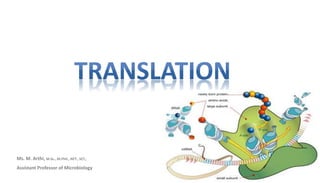
  • 2. The process by which a cell makes proteins using the genetic information carried in messenger RNA (mRNA).
  • 3. INTRODUCTION • Proteins are the end products of most information pathways. • A typical cell requires thousands of different proteins at any given moment. • It is synthesized in response to the cell’s current needs, transported (targeted) to their appropriate cellular locations, and degraded when no longer needed. • This biosynthetic machinery is well conserved in all life-forms from bacteria to higher eukaryotes - present in the last universal common ancestor (LUCA) of all extant organisms.
  • 4. • It’s a most complex biosynthetic process. • Eukaryotic protein synthesis involves – • Ribosomal proteins - more than 70 types. • For the activation of amino acid precursors - 20 or more enzymes • For initiation, elongation, and termination of polypeptides - dozen or more auxiliary enzymes and other protein factors. • For the final processing of different proteins - 100 additional enzymes • Transfer and ribosomal RNAs - 40 or more kinds. • Overall - almost 300 different macromolecules cooperate to synthesize polypeptides. • Many of these macromolecules are organized into the complex 3D structure of the ribosome.
  • 5. • Typical bacterial cell – Ribosomes - 15,000 – protein synthesis–related protein factors and enzymes - 100,000 molecules – tRNA molecules - 200,000 • All this can account for more than 35% of the cell’s dry weight. • A polypeptide of 100 residues is synthesized in an Escherichia coli cell (at 370C) in about 5 seconds. • Elucidation of 3D structures of ribosomes - began in the year 2000 • Two decades earlier to the discovery of 3D structure of ribosomes - Proteins are synthesized by a gigantic RNA enzyme! - Hypothesis was first put forward by Harry Noller.
  • 6. • 1950s, Paul Zamecnik - labeled a.as appeared in a fraction containing small ribonucleoprotein particles (visible in animal tissues by electron microscopy), identified as the site of protein synthesis - later named as ribosomes. • Mahlon Hoagland and Zamecnik - amino acids + ATP + cytosolic fraction of liver cells  “activated” A.as. • Robert Holley - The amino acids attaches to heat-stable soluble RNA - later called transfer RNA (tRNA), to form aminoacyl-tRNAs. The enzymes that catalyze this process are the aminoacyl-tRNA synthetases. • Francis Crick’s – Crick’s adaptor hypothesis. The tRNA adaptor “translates” the nucleotide sequence of an mRNA into the amino acid sequence of a polypeptide. The overall process of mRNA-guided protein synthesis is often referred to simply as translation. • Masayasu Nomura – discovered 30S or 50S subunits.
  • 7. • A codon is a triplet of nucleotides that codes for a specific amino acid. • nucleotide triplets are read in a successive, nonoverlapping fashion. • A specific first codon in the sequence establishes the reading frame, in which a new codon begins every three nucleotide residues. • There is no punctuation between codons for successive amino acid residues
  • 8. • mRNA sequence has three possible reading frames. Each reading frame gives a different sequence of codons, but only one is likely to encode a given protein. • The initiation codon AUG is the most common signal for the beginning of a polypeptide in all cells, in addition to coding for Met residues in internal positions of polypeptides. • The termination codons (UAA, UAG, and UGA), also called stop codons or nonsense codons, normally signal the end of polypeptide synthesis and do not code for any known amino acids. • a reading frame without a termination codon among 50 or more codons is referred to as an open reading frame (ORF). Phe Ser Leu
  • 9. Protein Biosynthesis Takes Place in Five Stages • Activation of amino acids • Initiation • Elongation • Termination & release • Folding and Posttranslational Processing
  • 10. Before looking at these five stages in detail, we must examine two key components in protein biosynthesis: the ribosome and tRNAs.
  • 11. • E. coli cell contains 15,000 or more ribosomes, which comprise nearly a quarter of the dry weight of the cell. • Bacterial ribosomes contain about 65% rRNA and 35% protein; they have a diameter of about 18 nm. • composed of two unequal subunits with sedimentation coefficients of 30S and 50S and a combined sedimentation coefficient of 70S. • Both subunits contain dozens of ribosomal proteins and at least one large rRNA. • The ribosomal subunits are huge RNA molecules. In the 50S subunit, the 5S and 23S rRNAs form the structural core. The proteins are secondary elements in the complex, decorating the surface. • there is no protein within 18 Å of the active site for peptide bond formation. • the ribosome is a ribozyme.
  • 12. Structure of Ribosomes • The bacterial ribosome is complex, with a combined molecular weight of ,2.7 million. • The two irregularly shaped ribosomal subunits fit together to form a cleft through which the mRNA passes as the ribosome moves along it during translation. • The 57 proteins in bacterial ribosomes vary enormously in size and structure. Molecular weights range from about 6,000 to 75,000. • Most of the proteins have globular domains arranged on the ribosome surface. Some also have snakelike extensions that protrude into the rRNA core of the ribosome, stabilizing its structure. • Each of the three single-stranded rRNAs of E. coli has a specific three-dimensional conformation featuring extensive intrachain base pairing. • The folding patterns of the rRNAs are highly conserved in all organisms, particularly the regions implicated in key functions.
  • 13. Structure of tRNA • Transfer RNAs are small and consist of a single strand of RNA folded into a precise 3D structure. • The tRNAs in bacteria and in the cytosol of eukaryotes - have b/w 73 and 93 nts • Molecular weights of 24,000 to 31,000. • Mitochondria and chloroplasts contain - smaller tRNAs. • Cells have at least one kind of tRNA for each amino acid; at least 32 tRNAs are required to recognize all the amino acid codons, but some cells use more than 32. • Yeast alanine tRNA (tRNAAla) - the first nucleic acid to be completely sequenced • It contains 76 nucleotide residues, of which 10 are modified bases.
  • 14. This structure was deduced in 1965 by Robert W. Holley and his colleagues D - 5,6-dihydrouridine m1G - 1-methylguanosine; m2G - N2-dimethylguanosine. I- inosine; ; m1I - 1-methylinosine; ψ - pseudouridine; T- ribothymidine; • tRNAs have a guanylate (pG) residue at the 5’ end • Have the trinucleotide sequence CCA at the 3’ end. • D and TC arms helps in the overall folding of tRNA molecules, • TC arm interacts with the large-subunit rRNA
  • 15. Protein Biosynthesis Takes Place in Five Stages • Activation of amino acids • Initiation • Elongation • Termination & release • Folding and Posttranslational Processing
  • 16.
  • 17. Components Required for the Five Major Stages of Protein Synthesis in E. coli
  • 18. 1. Activation of amino acids Aminoacyl-tRNA Synthetases Attach the Correct Amino Acids to Their tRNAs • This reaction takes place in the cytosol, not on the ribosome. • 20 amino acids are covalently attached to a specific tRNA at the expense of ATP energy, using Mg2+ dependent activating enzymes known as aminoacyl-tRNA synthetases. • When attached to their amino acid (aminoacylated) the tRNAs are said to be “charged.” • Specificity – each a.a has a specific activating enzyme – tRNA amino acyl synthetase (20 diff for 20 a.as). Some can activate more than 1 a.a – Eg: Isoleucine tRNA synthetase – activates Isoleucine & Valine – Valine tRNA synthetase – activates only valine • Based on differences in 10 and 30 structure and in reaction mechanism, aminoacyl-tRNA synthetases of E. coli is divided into two classes.
  • 19. Reaction Mechanism • This reaction occurs in two steps in the enzyme’s active site. • In step 1 – coo- group of a.a reacts with the α-phosphoryl group of ATP to form an anhydride linkage, with displacement of pyrophosphate. Forms an enzyme-bound intermediate - aminoacyl adenylate (aminoacyl-AMP). • In step 2 - aminoacyl group is transferred from enzyme-bound aminoacyl-AMP to its corresponding specific tRNA. The pyrophosphate formed in the activation reaction undergoes hydrolysis to phosphate by inorganic pyrophosphatase. Amino acid + tRNA + ATP --------------------------------------- aminoacyl-tRNA + AMP + PPi aminoacyl-tRNA synthetase Mg2+
  • 20.
  • 21. Proofreading by Aminoacyl-tRNA Synthetases • The amino acid attached to a tRNA is not checked on the ribosome, so attachment of the correct amino acid to the tRNA is essential to the fidelity of protein synthesis. • Hence some amino acyl tRNA synthetases enzyme has proof reading function. • Ile-tRNA synthetase, which distinguishes valine and isoleucine (single methylene group - CH2-). • If available binding interactions do not provide sufficient discrimination between two substrates, the necessary specificity can be achieved by substrate-specific binding. • Eg : Methylene group (in Ile) could enhance substrate binding
  • 22. Enzyme has 3 filters: • 1st filter - initial binding of the amino acid to the enzyme and its activation to aminoacyl-AMP. • 2nd filter - binding of any incorrect aminoacyl-AMP products to a separate ( second) active site on the enzyme is hydrolyzed. – Eg. The R group of valine is slightly smaller than that of isoleucine, so Val-AMP fits the hydrolytic (proofreading) site of the Ile-tRNA synthetase but Ile-AMP does not. Thus Val-AMP is hydrolyzed to valine and AMP in the proofreading active site. • 3rd filter - most aminoacyl-tRNA synthetases can also hydrolyze the ester linkage between amino acids and tRNAs in the aminoacyl-tRNAs. This hydrolysis is greatly accelerated for incorrectly charged tRNAs. • All the 3 filters enhance the fidelity of the overall process.
  • 23. • blue dots - same in all tRNAs • Orange dots - recognition points for one aminoacyl-tRNA synthetases. • green dots - recognition points for more aminoacyl-tRNA synthetases. Interaction between an Aminoacyl-tRNA Synthetase and a tRNA: A “Second Genetic Code” • An individual aminoacyl-tRNA synthetase is specific not only for a single amino acid but for certain tRNAs as well. • Eg. tRNA ala • Ten or more specific nucleotides may be involved in recognition of a tRNA by its specific aminoacyl-tRNA synthetase. Single G=U base pair in the amino acid arm of tRNAAla recognized by the Ala-tRNA synthetases.
  • 24. Fidelity of protein synthesis • Discriminating dozens of tRNAs and amino acids is important for the overall fidelity of protein biosynthesis. • The overall error rate of protein synthesis (~1 mistake per 104 amino acids incorporated) is not low as DNA replication. • Proteins have less biological significance becoz not passed on to future generations. • Incorrect proteins are eliminated by degradation. • The degree of fidelity in protein synthesis is essential to save energy required to synthesize a protein. • One defective protein molecule is usually unimportant when many correct copies of the same protein are present.
  • 25. 2. Initiation • Howard Dintzis in 1961 proved that Protein synthesis begins at the amino-terminal end and proceeds by the stepwise addition of amino acids to the carboxyl-terminal end of the growing polypeptide using Reticulocytes (immature erythrocytes) that actively synthesizing hemoglobin were incubated with radioactive leucine. • The AUG initiation codon specifies methionine residue (amino-terminal). • Methionine has only one codon, (5’)AUG but all organisms have two tRNAs for methionine. – One is for initiation codon, which was designated as tRNAfMet . – The other is for the addition of Met residue in an internal position in a polypeptide - tRNAMet.
  • 26. • The amino acid incorporated in response to the (5’)AUG initiation codon is N-formylmethionine (fMet). • It arrives at the ribosome as N-formylmethionyl-tRNAfMet (fMet-tRNAfMet) Formed in two successive reactions. • Step 1 - methionine is attached to tRNAfMet by the Met-tRNA synthetase (which in E. coli aminoacylates both tRNAfMet and tRNAMet) Methionine + tRNAfMet+ ATP ----------- Met-tRNAfMet + AMP + Ppi • Step 2 - Transformylase transfers a formyl group from N10-formyltetrahydrofolate to the amino group of the Met residue N10-Formyltetrahydrofolate + Met-tRNAfMet ------- tetrahydrofolate + fMet-tRNAfMet Met-tRNA synthetase Transformylase
  • 27. • In eukaryotic cells polypeptides synthesized by • cytosolic ribosomes - begin with a Met residue (rather than fMet) • mitochondrial and chloroplast ribosomes - begin with N-formylmethionine.
  • 28. Requirements for the initiation in bacteria (1) the 30S ribosomal subunit, (2) the mRNA coding for the polypeptide to be made, (3) the initiating fMet-tRNAfMet, (4) a set of three proteins called initiation factors (IF-1, IF-2, and IF-3), (5) GTP, (6) the 50S ribosomal subunit, and (7) Mg2.
  • 29. The Three Steps of Initiation • Step 1 - the 30S ribosomal subunit binds two initiation factors, IF-1 (binds at the A site and prevents the tRNA binding at this site during initiation) and IF-3 (prevents the reassociation of 30S and 50S subunits prematurely). • The mRNA (Shine dalgarno sequence) then binds to the 30S subunit (16S rRNA). • This mRNA-rRNA interaction positions the initiating (5’)AUG sequence of the mRNA at the P site (fMet- tRNAfMet binds) on the 30S subunit.
  • 30. ShineDalgarno sequence • Named for Australian researchers John Shine and Lynn Dalgarno, who identified it in the mRNA. • This consensus sequence is an initiation signal of four to nine purine residues, located 8 to 13 bp upstream to the 5’ side of the initiation codon. • This sequence base-pairs with a complementary pyrimidine-rich sequence near the 3’ end of the 16S rRNA of the 30S ribosomal subunit.
  • 31. Bacterial ribosomes have three sites • Peptidyl (P) site - The initiating (5)AUG is positioned at the P site, the only site to which fMettRNAfMet can bind. • Aminoacyl (A) site - during the elongation stage, all other incoming aminoacyl-tRNAs bind first to the A site • Exit (E) site - from which the “uncharged” tRNAs leave during elongation confined to the 50S subunit confined to both 50S & 30S subunit
  • 32. • Step 2 - The anticodon of initiating fMet- tRNAfMet pairs correctly with the initiation codon of mRNA. mRNA is joined by both GTP-bound IF-2 and the initiating fMet-tRNAfMet.
  • 33. The correct binding of the complex is assured by at least three points of recognition and attachment: –Codon-anticodon interaction –Shine-Dalgarno sequence and the 16S rRNA interactions –Ribosomal P site and the fMet-tRNAfMet interaction. • The initiation complex is now ready for elongation.
  • 34. • Step 3 - this large complex combines with the 50S ribosomal subunit; simultaneously, the GTP bound to IF-2 is hydrolyzed to GDP and Pi • Subsequently all the 3 initiation factors are departed from the ribosome. • Completion of the steps produces a functional 70S ribosome called the initiation complex.
  • 35. Initiation in Eukaryotic Cells • Eukaryotic mRNAs are bound to the ribosome with the help of a specific binding proteins. • They tie the 5’ and 3’ ends of the message together. • The 3’ end of the mRNA is bound to the poly(A) binding protein (PAB). • The 5’ end of mRNA bind with eIF4E. • Eukaryotic cells have at least nine initiation factors. • The initiating (5’)AUG is detected within the mRNA not by its proximity to a Shine-Dalgarno-like sequence but by a scanning process (facilitated by eIF4F & eIF4B).
  • 36.
  • 37. 3. Elongation • Requirements for elongation: (1)the initiation complex, (2)aminoacyl-tRNAs, (3)a set of three soluble cytosolic proteins called elongation factors (EF-Tu, EF-Ts, and EF-G in bacteria), and (4)GTP.
  • 38. The Three Steps of Elongation • Cells use three steps to add each amino acid residue, and the steps are repeated as many times as there are residues to be added. »Step 1: Binding of an Incoming Aminoacyl-tRNA » Step 2: Peptide Bond Formation »Step 3: Translocation
  • 39. • The appropriate incoming aminoacyl-tRNA binds to a complex of GTP-bound EF-Tu. • The resulting aminoacyltRNA–EF-Tu–GTP complex binds to the A site of the 70S initiation complex. • The GTP is hydrolyzed and an EF-Tu–GDP complex is released from the 70S ribosome. • The EF-Tu–GTP complex is regenerated in a process involving EF-Ts and GTP. Step 1: Binding of an Incoming Aminoacyl-tRNA
  • 40. Step 2: Peptide Bond Formation • A peptide bond is formed between the two amino acids bound by their tRNAs to the A and P sites on the ribosome. • This occurs by the transfer of the initiating N-formylmethionyl group from its tRNA to the amino group of the second amino acid, in the A site. • The α-amino group of the amino acid in the A site acts as a nucleophile, displacing the tRNA in the P site to form the peptide bond. • This reaction produces a dipeptidyltRNA in the A site, and the now “uncharged” (deacylated) tRNAfMet remains bound to the P site. • The enzymatic activity that catalyzes peptide bond formation has historically been referred to as peptidyl transferase, now know that this reaction is catalyzed by the 23S rRNA (ribozymes).
  • 41. Step 3: Translocation • The ribosome moves one codon toward the 3’ end of the mRNA. • This movement shifts the anticodon of the dipeptidyltRNA (attached to the second codon of the mRNA) from the A site to the P site. • Shifts the deacylated tRNA from the P site to the E site, from where the tRNA is released into the cytosol. • The third codon of the mRNA now lies in the A site and the second codon in the P site. • Movement of the ribosome along the mRNA requires EF-G (also known as translocase) and the energy provided by hydrolysis of another molecule of GTP. • EF-G binds to the A site and displace the peptidyl-tRNA.
  • 42. Elongation in Eukaryotic Cells • The elongation cycle in eukaryotes is quite similar to that in prokaryotes. • Three eukaryotic elongation factors (eEF1α, eEF1βγ, and eEF2) have functions analogous to bacterial elongation factors (EF-Tu, EF-Ts, and EF-G, respectively). • Eukaryotic ribosomes do not have an E site; uncharged tRNAs are expelled directly from the P site.
  • 43. 4. Termination • Elongation continues until the ribosome adds the last amino acid coded by the mRNA. • Termination, is signalled by the presence of one of three termination codons in the mRNA (UAA, UAG, UGA), immediately following the final coded amino acid. • In bacteria, once a termination codon occupies the ribosomal A site, three termination factors, or release factors— »RF-1 - recognizes the termination codons UAG and UAA »RF-2 - recognizes UGA and UAA »RF-3 – function is unknown • In eukaryotes, a single release factor, eRF, recognizes all three termination codons.
  • 44. Three termination factors, or release factors contribute to (1) hydrolysis of the terminal peptidyltRNA bond (peptidyl transferase to transfer the growing polypeptide to a water molecule rather than to another amino acid). (2) release of the free polypeptide and the last tRNA, now uncharged, from the P site. (3) dissociation of the 70S ribosome into its 30S and 50S subunits, ready to start a new cycle of polypeptide synthesis.
  • 45. • In bacteria, transcription and translation are tightly coupled. • mRNAs are synthesized and translated in the same 5’- 3’ direction. • Bacterial mRNAs generally exist for just a few minutes before they are degraded by nucleases. • In order to maintain high rates of protein synthesis, the mRNA for a given protein or set of proteins must be made continuously and translated with maximum efficiency. • In eukaryotic cells, newly transcribed mRNAs must leave the nucleus before they can be translated.
  • 46. 4. Newly Synthesized Polypeptide Chains Undergo Folding and Processing • The nascent polypeptide chain is folded and processed into its biologically active form. • During or after its synthesis, the polypeptide progressively assumes its native conformation, with the formation of appropriate »hydrogen bonds & van der Waals, »Ionic & hydrophobic interactions. • Linear, or 1D, genetic message in the mRNA is converted into the 3D structure of the protein. • Some newly made proteins, both prokaryotic and eukaryotic, do not attain their final biologically active conformation until they have been altered by one or more processing reactions called post-translational modifications.
  • 47. a. Amino-Terminal and Carboxyl-Terminal Modifications • The first residue inserted in all polypeptides is N-formylmethionine (in bacteria) or methionine (in eukaryotes). • However, the formyl group, the amino-terminal Met residue, and additional amino-terminal (and, in some cases, carboxyl-terminal) residues may be removed enzymatically in formation of the final functional protein. • In 50% of eukaryotic proteins, the amino group of the amino-terminal residue is N-acetylated after translation. Carboxyl-terminal residues are also sometimes modified.
  • 48. b. Loss of Signal Sequences • 15 to 30 residues at the amino-terminal end of some proteins directs the protein to its ultimate destination in the cell. • Such signal sequences are removed by specific peptidases.
  • 49. c. Modification of Individual Amino Acids Addition of phosphate groups The phosphate groups add negative charges to these polypeptides. Serine/thr/tyr -> Phosphoserine Eg – Milk protein casein Binds calcium (Ca2+) Addition of carboxyl groups Glutamate -> γ carboxyglutamate Eg – blood clotting protein prothrombin Binds Ca2+ required to initiate clotting mechanism Addition of Methyl groups Monomethyl, dimethyl & trimethyl added to lysine residues occur in some muscle proteins, cytochrome c & calmodulin In other proteins, the carboxyl groups of some Glu residues undergo methylation, removing their negative charge.
  • 50. d. Attachment of Carbohydrate Side Chain • The carbohydrate side chains of glycoproteins are attached covalently during or after synthesis of the polypeptide. • The carbohydrate side chain is attached enzymatically to side chains of –Asn residues (N-linked oligosaccharides), –Ser or Thr residues (O-linked oligosaccharides). • Many proteins that function extracellularly, as well as the lubricating proteoglycans that coat mucous membranes, contain oligosaccharide side chains.
  • 51. e. Addition of Isoprenyl Groups • Eukaryotic proteins are modified by the addition isoprenyl groups derived from isoprene (The isoprenyl groups are derived from pyrophosphorylated intermediates of the cholesterol biosynthetic pathway such as farnesyl pyrophosphate). • A thioether bond is formed between the isoprenyl group and a Cys residue of the protein. • Eg : Ras proteins, G proteins and lamin proteins found in the nuclear matrix. • The isoprenyl group helps to anchor the protein in a membrane. • Ras oncogene ---> ras protein - isoprenylation responsible for cancer - (carcinogenic) activity is lost when isoprenylation of the Ras protein is blocked. • identifying inhibitors for this posttranslational modification helps in cancer chemotherapy. isoprenoids of 15 and 20 carbons
  • 52. f. Addition of Prosthetic Groups • Many prokaryotic and eukaryotic proteins requires a covalently bound prosthetic groups for their activity. • Two examples are »biotin molecule of acetyl-CoA carboxylase and »the heme group of hemoglobin or cytochrome c.
  • 53. g. Proteolytic Processing • Many proteins are initially synthesized as large, inactive precursor polypeptides that are proteolytically trimmed to form their smaller, active forms. • Examples include proinsulin, some viral proteins, and proteases such as chymotrypsinogen and trypsinogen. three polypeptide chains (A, B, and C) of chymotrypsin are linked by disulfide bonds
  • 54. h. Formation of Disulfide Cross-Links • After folding into their native conformations, some proteins form intrachain or interchain disulfide bridges between Cys residues. • In eukaryotes, disulfide bonds are common in proteins to be exported from cells. • The cross-links helps to protect the native conformation of the protein molecule from denaturation in the extracellular environment.
  • 55. Peptidyl puromycin Inhibitors of protein synthesis • Puromycin - Streptomyces alboniger – Its structure is very similar to the 3’ end of an aminoacyl-tRNA, – Participate in peptide bond formation, producing peptidyl-puromycin – Prematurely terminates polypeptide synthesis. • Tetracyclines – Mode of action - In bacteria blocks the A site on the ribosome & prevents the binding of aminoacyl-tRNAs. • Chloramphenicol – Mode of action – in bacterial, mitochondrial & chloroplast ribosomes - blocks peptidyl transfer; it does not affect cytosolic protein synthesis in eukaryotes. • Cycloheximide – Mode of action - blocks the peptidyl transferase of 80S eukaryotic ribosomes but not that of 70S bacterial (and mitochondrial and chloroplast) ribosomes. • Streptomycin – Mode of action - a basic trisaccharide, causes misreading of the genetic code (in bacteria) at relatively low concentrations and inhibits initiation at higher concentrations.
  • 56. Toxin inhibitors • other inhibitors of protein synthesis are notable because of their toxicity to humans and other mammals. • Diphtheria toxin (Mr 58,330) - catalyzes the ADP-ribosylation of a diphthamide (a modified histidine) residue of eukaryotic elongation factor eEF2, thereby inactivating it. • Ricin (Mr 29,895) - an extremely toxic protein of the castor bean, inactivates the 60S subunit of eukaryotic ribosomes by depurinating a specific adenosine in 23S rRNA.