PATHOPHYSIOLOGY OF HEALING WITH PRIMARY & SECONDARY INTENTION.pptx
•
0 likes•285 views

This presentation deals with Pathophysiology of healing & Mechanism of repair with Primary Intention and Secondary intention

Recommended
More Related Content
What's hot
What's hot (20)
Similar to PATHOPHYSIOLOGY OF HEALING WITH PRIMARY & SECONDARY INTENTION.pptx
Similar to PATHOPHYSIOLOGY OF HEALING WITH PRIMARY & SECONDARY INTENTION.pptx (20)
More from Akshay Shetty
More from Akshay Shetty (20)
Recently uploaded
Recently uploaded (20)
PATHOPHYSIOLOGY OF HEALING WITH PRIMARY & SECONDARY INTENTION.pptx
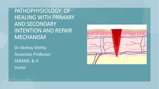
- 1. PATHOPHYSIOLOGY OF HEALING WITH PRIMARY AND SECONDARY INTENTION AND REPAIR MECHANISM Dr Akshay Shetty Associate Professor SSRAMC & H Inchal
- 2. Contents • Objectives • Definition of Healing • Pathophysiology of healing • Primary intention • Secondary intention • Summary • References 25/01/2024 PATHOPHYSIOLOGY OF HEALING WITH PRIMARY AND SECONDARY INTENTION AND REPAIR MECHANISM(Dr Akshay Shetty) 2
- 3. Objectives By the end of the presentation the students must be able to – 1. Define Healing 2. Illustrate Pathophysiology of healing & Repair Mechanism 3. Describe Primary intention 4. Explain Secondary intention 25/01/2024 PATHOPHYSIOLOGY OF HEALING WITH PRIMARY AND SECONDARY INTENTION AND REPAIR MECHANISM(Dr Akshay Shetty) 3
- 4. HEALING Injury to tissue may result in cell death and tissue destruction. Healing on the other hand is the body response to injury in an attempt to restore normal structure and function. 25/01/2024 PATHOPHYSIOLOGY OF HEALING WITH PRIMARY AND SECONDARY INTENTION AND REPAIR MECHANISM(Dr Akshay Shetty) 4
- 5. The process of healing involves two distinct processes: • Regeneration when healing takes place by proliferation of parenchymal cells and usually results in complete restoration of the original tissues. • Repair when the healing takes place by proliferation of connective tissue elements resulting in fibrosis and scarring. At times, both the processes take place simultaneously 25/01/2024 PATHOPHYSIOLOGY OF HEALING WITH PRIMARY AND SECONDARY INTENTION AND REPAIR MECHANISM(Dr Akshay Shetty) 5
- 6. Regeneration & Repair Some parenchymal cells are short lived while others have a longer lifespan. In order to maintain proper structure of tissues, these cells are under the constant regulatory control of their cell cycle. These include growth factors such as: • Epidermal growth factor, • Fibroblast growth factor, • Platelet -derived growth factor, • Endothelial growth factor, • Transforming growth factor-B. 25/01/2024 PATHOPHYSIOLOGY OF HEALING WITH PRIMARY AND SECONDARY INTENTION AND REPAIR MECHANISM(Dr Akshay Shetty) 6
- 7. Cell cycle is defined as the period between two successive cell divisions and is divided into 4 unequal chases: M (mitosis) phase: phase of mitosis. G1, (gap i) phase: The daughter cell enters G1, phase after mitosis. S (synthesis) phase: During this phase, the synthesis of nuclear DNA takes place. G2, (gap 2) phase: After completion of nuclear DNA duplication, the cell enters G, phase. G0, (gap 0) phase: This is the quiescent or resting phase of the cell after an M phase. 25/01/2024 PATHOPHYSIOLOGY OF HEALING WITH PRIMARY AND SECONDARY INTENTION AND REPAIR MECHANISM(Dr Akshay Shetty) 7
- 8. Not all cells of the body divide at the same pace. Some mature cells do not divide at all while others complete a cell cycle every 16-24 hours. The main difference between slowly- dividing and rapidly-dividing cells is the duration of G, phase. Depending upon their capacity to divide, the cells of the body can be divided into 3 groups: Labile cells Stable cells Permanent cells. 25/01/2024 PATHOPHYSIOLOGY OF HEALING WITH PRIMARY AND SECONDARY INTENTION AND REPAIR MECHANISM(Dr Akshay Shetty) 8
- 9. Labile cells. These cells continue to multiply throughout life under normal physiologic conditions. These include: • Surface epithelial cells of epidermis, • Alimentary tract, • Respiratory tract, • Urinary tract, • Vagina , cervix, uterine endometrium, • Haematopoietic cells of bone marrow and cells of lymph nodes and spleen. 25/01/2024 PATHOPHYSIOLOGY OF HEALING WITH PRIMARY AND SECONDARY INTENTION AND REPAIR MECHANISM(Dr Akshay Shetty) 9
- 10. Stable cells. These cells decrease or lose their ability to proliferate after adolescence but retain the capacity to multiply in response to stimuli throughout adult life. These include: • Parenchymal cells of organs like liver, pancreas, kidneys, adrenal and thyroid; • Mesenchymal cells like smooth muscle cells, fibroblasts, vascular endo-thelium, bone and cartilage cells. 25/01/2024 PATHOPHYSIOLOGY OF HEALING WITH PRIMARY AND SECONDARY INTENTION AND REPAIR MECHANISM(Dr Akshay Shetty) 10
- 11. Permanent cells. These cells lose their ability to proliferate around the time of birth. These include: • Neurons of nervous system • Skeletal muscle • Cardiac muscle cells 25/01/2024 PATHOPHYSIOLOGY OF HEALING WITH PRIMARY AND SECONDARY INTENTION AND REPAIR MECHANISM(Dr Akshay Shetty) 11
- 12. RELATIONSHIP OF PARENCHYMAL CELLS WITH CELL CYCLE If the three types of parenchymal cells described above are correlated with the phase of cell cycle , the following inferences can be derived: • Labile cells which are continuously dividing cells remain in the cell cycle from one mitosis to the next. • Stable cells are in the resting phase (G) but can be stimulated to enter the cell cycle. • Permanent cells are non-dividing cells which have left the cell cycle and die after injury. 25/01/2024 PATHOPHYSIOLOGY OF HEALING WITH PRIMARY AND SECONDARY INTENTION AND REPAIR MECHANISM(Dr Akshay Shetty) 12
- 13. 25/01/2024 PATHOPHYSIOLOGY OF HEALING WITH PRIMARY AND SECONDARY INTENTION AND REPAIR MECHANISM(Dr Akshay Shetty) 13
- 14. Regeneration of any type of parenchymal cells involves the following two processes: Proliferation of original cells from the margin of injury with migration so as to cover the gap. Proliferation of migrated cells with subsequent differentiation and maturation so as to reconstitute the original tissue. 25/01/2024 PATHOPHYSIOLOGY OF HEALING WITH PRIMARY AND SECONDARY INTENTION AND REPAIR MECHANISM(Dr Akshay Shetty) 14
- 15. REPAIR Repair is the replacement of injured tissue by fibrous tissue. Two processes are involved in repair: 1. Granulation tissue formation, and 2. Contraction of wounds. Repair response takes place by participation of mesenchymal cells (consisting of connective tissue stem cells, fibrocytes and histiocytes), endothelial cells, macrophages, platelets, and the parenchymal cells of the injured organ. 25/01/2024 PATHOPHYSIOLOGY OF HEALING WITH PRIMARY AND SECONDARY INTENTION AND REPAIR MECHANISM(Dr Akshay Shetty) 15
- 16. WOUND HEALING Healing of skin wounds provides a classical example of combination of regeneration and repair described above. This can be accomplished in one of the following two ways: • Healing by first intention (primary union) • Healing by second intention (secondary union). 25/01/2024 PATHOPHYSIOLOGY OF HEALING WITH PRIMARY AND SECONDARY INTENTION AND REPAIR MECHANISM(Dr Akshay Shetty) 16
- 17. 25/01/2024 PATHOPHYSIOLOGY OF HEALING WITH PRIMARY AND SECONDARY INTENTION AND REPAIR MECHANISM(Dr Akshay Shetty) 17
- 18. Healing by First Intention (Primary Union) • Clean and uninfected • Surgically incised • If without much loss of cells and tissue • Edges of wound are approximated by surgical sutures. This is defined as healing of a wound which has the following characteristics: 25/01/2024 PATHOPHYSIOLOGY OF HEALING WITH PRIMARY AND SECONDARY INTENTION AND REPAIR MECHANISM(Dr Akshay Shetty) 18
- 19. The sequence of events in primary unions is illustrated in and described below: • INITIAL HAEMORRHAGE. Immediately after injury, the space between the approximated surfaces of incised wound is filled with blood which them clots and seals the wound against dehydration and infection. • ACUTE INFLAMMATORY RESPONSE. This occurs within 24 hours with appearance of polymorph is from the margins of incision. By 3rd day, polymorphs are replaced by macrophages. 25/01/2024 PATHOPHYSIOLOGY OF HEALING WITH PRIMARY AND SECONDARY INTENTION AND REPAIR MECHANISM(Dr Akshay Shetty) 19
- 20. III. EPITHELIAL CHANGES. The basal cells of epidermis from both the cut margins start proliferating and migrating towards incisional space in the form of epithelial spurs. A well-approximated wound is covered by a layer of epithelium in 48 hours. The migrated epidermal cells separate the underlying viable dermis from the overlying necrotic material and clot, forming scab which is cast off. The basal cells from the margins continue to divide. By 5th day, a multilayered new epidermis is formed which is differentiated into superficial and deeper layers. 25/01/2024 PATHOPHYSIOLOGY OF HEALING WITH PRIMARY AND SECONDARY INTENTION AND REPAIR MECHANISM(Dr Akshay Shetty) 20
- 21. IV. ORGANISATION. By 3rd day, fibroblasts also invade the wound area. By 5th day, new collagen fibrils start forming which dominate till healing is completed. In 4 weeks, the scar tissue with scanty cellular and vascular elements, a few inflammatory cells aid epithelialized surface is formed. 25/01/2024 PATHOPHYSIOLOGY OF HEALING WITH PRIMARY AND SECONDARY INTENTION AND REPAIR MECHANISM(Dr Akshay Shetty) 21
- 22. V. SUTURE TRACKS. Each suture track is a separate wound and incites the same phenomena as in healing of the primary wound i.e. filling the space with haemorrhage, some inflammatory cell reaction, epithelial cell proliferation along the suture track from both margins, fibro-blastic proliferation and formation of young collagen. . 25/01/2024 PATHOPHYSIOLOGY OF HEALING WITH PRIMARY AND SECONDARY INTENTION AND REPAIR MECHANISM(Dr Akshay Shetty) 22
- 23. When sutures are removed around 7th day, much of epithelialized suture track is avulsed and the remaining epithelial tissue in the track is absorbed. However, sometimes the suture track gets infected (stitch abscess), or the epithelial cells may persist in the track (implantation or epidermal cysts). Thus, the scar formed in a sutured wound is neat due to close apposition of the margins of wound; the use of adhesive tapes avoids removal of stitches and its complications 25/01/2024 PATHOPHYSIOLOGY OF HEALING WITH PRIMARY AND SECONDARY INTENTION AND REPAIR MECHANISM(Dr Akshay Shetty) 23
- 24. Healing by Second Intention (Secondary Union) This is defined as healing of a wound having the following characteristics: • Open with a large tissue defect, at times infected • Having extensive loss of cells and tissues; and • The wound is not approximated by surgical sutures but is left open. 25/01/2024 PATHOPHYSIOLOGY OF HEALING WITH PRIMARY AND SECONDARY INTENTION AND REPAIR MECHANISM(Dr Akshay Shetty) 24
- 25. The basic events in secondary union are similar to primary union but differ in having a larger tissue defect which has to be bridged. Hence healing takes place from the base upwards as well as from the margins inwards. The healing by second intention is slow and results in a large, at times ugly, scar as compared to rapid healing and neat scar of primary union. 25/01/2024 PATHOPHYSIOLOGY OF HEALING WITH PRIMARY AND SECONDARY INTENTION AND REPAIR MECHANISM(Dr Akshay Shetty) 25
- 26. The sequence of events in secondary union is described below: • Initial haemorrhage. As a result of injury, the wound space is filled with blood and fibrin clot which dries • Inflammatory phase. There is an initial acute inflammatory response followed by appearance of macrophages which clear off the debris as in primary union. 25/01/2024 PATHOPHYSIOLOGY OF HEALING WITH PRIMARY AND SECONDARY INTENTION AND REPAIR MECHANISM(Dr Akshay Shetty) 26
- 27. III. Epithelial changes. As in primary healing, the epidermal cells from both the margins of wound proliferate and migrate into the wound in the form of epithelial spurs till they meet in the middle and re-epithelialise the gap completely. However, the proliferating epithelial cells do not cover the surface fully until granulation tissue from base has started filling the wound space. In this way, pre-existing viable connective tissue is separated from necrotic material and clot on the surface, forming scab which is cast off. In time, the regenerated epidermis becomes stratified and keratinised. 25/01/2024 PATHOPHYSIOLOGY OF HEALING WITH PRIMARY AND SECONDARY INTENTION AND REPAIR MECHANISM(Dr Akshay Shetty) 27
- 28. IV. Granulation tissue. The main bulk of secondary healing is by granulations. Granulation tissue is formed by proliferation of fibroblasts and neovascularisation from the adjoining viable elements. The newly- formed granulation tissue is deep red, granular and very fragile. With time, the scar on maturation becomes pale and white due to increase in collagen and decrease in vascularity. The specialised structures of skin like hair follicles and sweat glands are not replaced unless their viable residues remain which may regenerate. 25/01/2024 PATHOPHYSIOLOGY OF HEALING WITH PRIMARY AND SECONDARY INTENTION AND REPAIR MECHANISM(Dr Akshay Shetty) 28
- 29. V. Wound contraction. Contraction of wound is an important feature of secondary healing, not seen in primary healing. Due to the action of myofibroblasts present in granulation tissue, the wound contracts to one- third to one-fourth of its original size. Wound contraction occurs at a time when active granulation tissue is being formed. VI. Presence of infection. Bacterial contamination of an open wound delays the process of healing due to release of bacterial toxins that provoke necrosis, suppuration and thrombosis. Surgical removal of dead and necrosed tissue, debridement, helps in preventing the bacterial infection of open wounds 25/01/2024 PATHOPHYSIOLOGY OF HEALING WITH PRIMARY AND SECONDARY INTENTION AND REPAIR MECHANISM(Dr Akshay Shetty) 29
- 30. 25/01/2024 PATHOPHYSIOLOGY OF HEALING WITH PRIMARY AND SECONDARY INTENTION AND REPAIR MECHANISM(Dr Akshay Shetty) 30
- 31. 25/01/2024 PATHOPHYSIOLOGY OF HEALING WITH PRIMARY AND SECONDARY INTENTION AND REPAIR MECHANISM(Dr Akshay Shetty) 31
- 32. Summary 1.Define Healing Illustrate Pathophysiology of healing & Repair Mechanism Describe Primary intention Explain Secondary intention 25/01/2024 PATHOPHYSIOLOGY OF HEALING WITH PRIMARY AND SECONDARY INTENTION AND REPAIR MECHANISM(Dr Akshay Shetty) 32
- 33. References Harsh Mohan Text Book of Pathology https://youtu.be/G3B0ApUsYag ?feature=shared 25/01/2024 PATHOPHYSIOLOGY OF HEALING WITH PRIMARY AND SECONDARY INTENTION AND REPAIR MECHANISM(Dr Akshay Shetty) 33
- 34. 25/01/2024 PATHOPHYSIOLOGY OF HEALING WITH PRIMARY AND SECONDARY INTENTION AND REPAIR MECHANISM(Dr Akshay Shetty) 34