SlideShare a Scribd company logo
1 of 77
Download to read offline
1 Biological Classification
All right copy reserved. No part of the material can be produced without prior permission
2 Biological Classification
Biological Classification
Biological classification is the scientific procedure to classify the organisms into different groups on
the basis of their similarities and dissimilarities and placing the groups in a hierarchy of categories.
KINGDOM SYSTEMS OF CLASSIFICATION
The earlier systems of classification of organisms were simple and based on one or two characters.
First scientific attempt for classification was performed by Aristotle in following manner:
(1) Two Kingdom Classification: It was given by Linnaeus.
Traditionally all the organisms of the world were divided into two
kingdoms -the animal kingdom (Animalia) and the plant kingdom
(Plantae). The major criterion of classification was the presence or
absence of cell wall. Other criterias were locomotion, mode of
nutrition, response to external stimuli etc.
All right copy reserved. No part of the material can be produced without prior permission
3 Biological Classification
Shortcomings of two-kingdom system of classification:
 This system did not distinguish between the eukaryotes and prokaryotes, unicelled and
multicelled organisms, photosynthetic and non-photosynthetic organisms.
 There are few organisms like Chlamydomonas, Euglena and the slime moulds which have
been claimed by both zoologists and botanists (organisms which share characteristics of both
animals and plants).
 Since there are certain organisms that do not fall naturally into either plant or animal
kingdom, it was proposed that a new kingdom is to be established to accommodate such
organisms.
(2) Three Kingdom Classification:
 Haeckel, a German zoologist (1866), suggested that a third kingdom Protista, be created to
include all unicellular microorganisms.
 This includes a wide variety of unicellular, mostly aquatic eukaryotes like -Fungi, Protozoa,
Algae, Bacteria and Slime moulds.
 Thus, he proposed three kingdoms, namely -Plantae, Protista and Animalia.
(3) Four Kingdom Classification:
 Copeland (1956) gave four kingdom of
classification and included Monera as fourth
kingdom.
 Copeland originally called it as kingdom
'Mychota'.
 It was called 'Monera' by Daugherty and Allen.
 Kingdom Monera includes all the prokaryotic
organisms i.e., eubacteria (including
cyanobacteria, formerly known as blue-green
algae) and archaebacteria.
 The actinomycetes (filamentous bacteria) are also included in this kingdom.
All right copy reserved. No part of the material can be produced without prior permission
4 Biological Classification
(4) Five Kingdom Classification:
 According to five-kingdom concept proposed by R.H. Whittaker.
 Whittaker (1969), the organisms are divided into five kingdoms
namely Monera, Protista, Fungi, Plantae, Animalia, on the basis of the
following criteria:
(a) Complexity of cell structure: prokaryotic vs eukaryotic organisation of
cells.
(b) Complexity of body organisation: unicellularity vs multicellularity;
simple multicellular forms to complex multicellular forms.
(c) Mode of nutrition: Autotrophic vs heterotrophic (parasitic or saprobic or ingestive
organisms). It was the major criteria of this classification system.
(d) Reproduction.
(e) Phylogenetic or evolutionary interrelations
All right copy reserved. No part of the material can be produced without prior permission
5 Biological Classification
Table 1: Comparative account of different characteristics of the Five Kingdoms
(5) Six Kingdom Classification:
 Carl Woese proposed six kingdom classification.
 These six kingdoms are Archaebacteria, Eubacteria, Protista, Fungi, Plantae and Animalia.
 He separated the archaebacteria from eubacteria on the basis of some major differences such
as the absence of peptidoglycan in the cell walls of the former and the occurrence of
branched chain lipids (a monolayer instead of a phospholipid bilayer) in the membrane.
 Based on the sequence of 16S ribosomal RNA genes, Woese found that the six kingdoms
naturally cluster into three main categories.
 He called these categories as domains of life.
 These domains are Bacteria, Archae and Eukarya and are believed to have originated from
common ancestor called progenote.
Three Domains of life
 Based on the sequence of 16 S ribosomal RNA genes, woese found that the six kingdoms
naturally cluster into three domains.
 These domains are Archae, Bacteria and Eukarya, and are believed to be originated from
All right copy reserved. No part of the material can be produced without prior permission
6 Biological Classification
common ancestor called Progenate. Domain is a category higher than kingdom.
KINGDOM: MONERA
 The Kingdom Monera includes all prokaryotes.
 Monerans are the most primitive forms of life, originating from more ancient living stock
termed progenote.
 The kingdom Monera includes eubacteria and archaebacteria.
 Eubacteria includes Cyanobacteria, Actinomycetes, Mycoplasma, Rickettsiae, Chlamydiae and
Spirochaetes etc.
Classification of Monera
(1) In 4 kingdom system, a new kingdom was created to accommodate all prokaryotic organisms
i.e, eubacteria and archaebacteria. Copeland called it kingdom Mychota. It was called
'Monera' by Daugherty and Allen.
All right copy reserved. No part of the material can be produced without prior permission
7 Biological Classification
(2) Actually, archaebacteria differ from eubacteria in many respects and resemble eukaryotes in
some ways.
(3) (i) Carl Woese separated the archaebacteria from eubacteria on the basis of some major
differences such as the absence of peptidoglycan in the cell walls of the former and the
occurrence of branched chain lipids (a monolayer instead of a phospholipid bilayer) in the
membrane.
(ii) Therefore, 6 kingdoms given by Carl Woese are
Kingdom-1 -Archaebacteria Kingdom-2 -Eubacteria
Kingdom-3 -Protista Kingdom-4 -Fungi
Kingdom-5 -Plantae Kingdom-6 -Animalia
Salient Features of Monera
1. These are unicellular, colonial, multicellular prokaryotic organisms without nuclear
membrane, nucleolus, chromatin and histone proteins.
2. Cell wall is made up of peptidoglycan (exceptions are Archaebacteria and Mycoplasma).
3. Membrane bound organelles are absent.
4. Cyclosis is absent and ribosomes are of 70 S type .
5. Respiratory enzymes are found associated with plasma membrane.
6. Nucleoid or genophore or incipient nucleus or prochromosome is composed of naked DNA,
RNA and nonhistone proteins.
7. Reproduction by asexual method.
8. Cell division is amitotic type and lacks spindle formation.
Let us discuss various monerans in detail:
1. EUBACTERIA
All right copy reserved. No part of the material can be produced without prior permission
8 Biological Classification
 Bacteria are cosmopolitan and occur in every habitat wherever living or dead organic matter
is present.
 Anton Von Leeuwenhoek discovered bacteria in rain water which had been allowed to stand
for many days and tartar scrapped from teeth.
 In 1695, he published his work "The Secrets of Nature".
 A.V. Leeuwenhoek termed these microorganisms as dierkens which was later translated as
animalcules by the Royal society.
 Term microbe for animalcules was coined by Se' dillot, but the term microorganism was
proposed by Pasteur.
 Ehrenberg first of all coined the word 'bacteria'
 Louis Pasteur is considered father of modern microbiology. He introduced the term aerobic
and anaerobic for the life in the presence or absence of oxygen respectively.
 Robert Koch, a German doctor, demonstrated that the anthrax disease of sheep was caused
by bacteria. Koch had followed four experimental steps (Koch's postulates) which help to
establish a relationship between a microorganism and a disease.
 Smallest bacterium : Dialister pneumosintes
 Largest filamentous bacterium : Beggiatoa mirabilis
Shapes of Bacteria
 Bacteria occur in four basic forms or shapes.
 These are spherical (Cocci), rod shaped (Bacilli), Vibrio and Spiral.
 Though most bacterial species have cells that are of a fairly constant and characteristic shape,
some species are pleomorphic (i.e., these can exhibit a variety of shapes), e.g., Rhizobium
leguminosarum.
(a) Coccus: Spherical or nearly spherical, aflagellate, sub-divided into six groups on the basis of
cell arrangement:
 Monococcus -Only single cell represents the bacterium, e.g., Micrococcus luteus, M. roseus.
 Diplococcus -Cocci divide in one plane and remain attached in pairs, e.g., Meningococcus,
Gonococcus, Diplococcus pneumoniae.
 Streptococcus -Cocci remain attached to form chains of different lengths, e.g., Streptococcus
Concept Builder
All right copy reserved. No part of the material can be produced without prior permission
9 Biological Classification
lactis.
 Tetracoccus -Cocci divide in two planes at right angles to one another and form groups of
four, e.g., Tetracoccus, Neisseria.
 Staphylococcus -Cocci divide in several planes resulting in formation of irregular bunches of
cells, sometimes resembling a cluster of grapes, e.g., Staphylococcus aureus.
 Sarcinae -Cocci divide in 3 planes at right angles to one another and resemble cubical packets
of 8 or more cells forming three dimensional geometrical figures, e.g., Sarcina lutea.
(b) Bacillus: Rod-like forms, either singly or may be arranged differently. They are generally
flagellate. It is the most common of all the shapes. They are of following types:
(i) Monobacillus -The bacteria occur singly, e.g., Bacillus anthracis, Lactobacillus.
(ii) Diplobacillus -Bacteria are arranged in pairs. e.g., Bacillus subtilis
(iii) Streptobacillus -Bacteria form a chain of rods, e.g., Streptobacillus.
(iv) Palisade-like -If the cells are lined side by side like match sticks and at angles to one another.
e.g., Corynebacterium diphtheriae.
(c) Spiral bacteria: Coiled forms of bacteria exhibiting twists with one or more turns are called
spirilla, e.g., Spirillum volutans.
(d) Vibrio: Bacteria with less than one complete twist or turn are called vibrio. These resemble a
comma (,) in appearance, e.g., Vibrio cholerae.
(e) Stalked bacteria: The body of bacterium possesses a stalk, e.g., Caulobacter.
(f) Budding bacteria: The body is swollen at places, e.g., Rhodomicrobium.
All right copy reserved. No part of the material can be produced without prior permission
10 Biological Classification
Bacterial Cell Structure
Bacterial cell structure is very simple although they are very complex in behaviour. They show the
most extensive metabolic diversity. Electron microscope can only reveal the detailed structure of
bacterial cell. It consists of following structures:
1. Glycocalyx:
(i) It is outermost part of cell envelope (Glycocalyx, cell wall, plasma membrane)
(ii) Represented by either slime layer or capsule
(a) Slime layer is composed of dextran, dextrin and lavan sugars and protect the cell against
desiccation and loss of nutrients.
(b) Capsule is made up of polysaccharides and D-glutamic acid. It provides gummy or sticky
character and virulent property to the cell.
2. Cell wall:
 It is present outside the cell membrane and is a rigid structure.
 Due to its rigidity, it protects the internal structures of the cell and provides shape to the cell.
 However, its main function is to prevent the cell from expanding and bursting because most
bacteria live in hypotonic environments, and are likely to take in much water and eventually
burst.
 The cell walls of almost all the eubacteria (true bacteria) are made up of peptidoglycan, also
called murein or mucopeptide.
 It is found only in prokaryotes.
 As the name suggests, the peptidoglycan consists of two components-a peptide portion
which is composed of amino acids connected by peptide linkages, and a glycan or sugar
portion.
 The glycan portion, which forms the backbone of peptidoglycan, is composed of alternating
units of amino sugars N-acetyl-glucosamine (NAG) and N-acetylmuramic acid (NAM) joined
together by -1, 4 linkages.
 The peptidoglycan chains are laterally linked by short chains of four amino acids which are
attached to N-acetylmuramic acid residues.
 The four amino acids of this tetrapeptide are D-alanine, L-alanine, D-glutamic acid and L-
All right copy reserved. No part of the material can be produced without prior permission
11 Biological Classification
Iysine (in Gram +ve bacteria) or diaminopimelic acid (in Gram -ve bacteria).
 The tetrapeptide chains are also interlinked by a peptide bridge between the carboxyl group
of an amino acid in one tetrapeptide chain and amino group of an amino acid in another
tetrapeptide chain.
 The cross linkages can occur between tetrapeptides in different chains, as well as between
adjacent tetrapeptide chains. As a result, peptidoglycan forms a rigid, multilayered sheet.
 Another component, teichoic acid, an acidic polymer consisting of a carbohydrate (e.g.,
glucose), phosphate and an alcohol is found in cell walls of Gram +ve bacteria.
 Teichoic acid has several functions such as binding metals, acting as receptor sites for some
viruses and maintaining cells at low pH to prevent degradation of cell walls by self-produced
enzymes.
 The walls of Gram-positive bacteria contain very little amount of lipids.
 The cell walls of Gram-negative bacteria are much more complex.
 The peptidoglycan layer is very thin making up only 10% or less of the cell wall.
 However, the most interesting feature is the presence of an outer membrane that covers a
thin underlying layer of peptidoglycan.
 The outer membrane is a bilayered structure consisting chiefly of phospholipids, proteins and
lipopolysaccharides (LPS).
 The outer membrane serves as a barrier to prevent the escape of important enzymes from
the space (periplasmic space) between the cytoplasmic membrane and the outer membrane.
 It also prevents the entry of various chemicals that could damage the cell.
 It acts as main surface antigen in cell wall.
 However, permeability of outer membrane to nutrients is provided by proteins called porins
which form channels in the membrane through which substances of hydrophilic nature and
low molecular weight can diffuse.
 Christian Gram (1884) developed a staining method for bacteria, using Gram stain (crystal
violet).
 On the basis of stainability with Gram Stain, bacteria are classified into two groups; Gram
positive and Gram negative.
3. Surface Appendages: These include flagella and fimbriae (or pili).
(a) Flagella are long, fine, wavy, filamentous appendages that protrude through the cell wall,
responsible for the motility of bacteria. These are much thinner than the flagella or cilia of
eukaryotes.
 Structure of Flagella: The entire flagellar apparatus is made up of three distinct regions: basal
body, hook and filament.
 Basal body: It is most complex portion of flagellum and has four rings (L, P, S and M), only two
rings S and M are present in gram +ve bacteria. L and P rings in cell wall constitute distal set,
while S and M rings are present in plasma membrane, forming proximal set.
 Hook: Made up of different protein units.
 Filament: Bacterial flagella are made up of identical spherical subunits of a protein called
flagellin. Longitudinal chains of flagellin molecules run longitudinally around each other to
form a wavy helical or rope-like structure. Therefore, a cross section of the flagellum reveals a
number of flagellin molecules around a central space.
Depending upon the presence or absence, number and position, following types of flagellar arrangements are observed
among bacteria :
 Atrichous: Flagella absent, e.g., Pasteurella, Lactobacillus.
 Monotrichous: Only one flagellum attached at one pole of the organism, e.g.,Thiobacillus, Vibrio.
 Amphitrichous: One flagellum at both ends, e.g., Nitrosomonas.
 Cephalotrichous: Two or more flagella attached at one end, e.g., Pseudomonas fluorescence.
Concept Builder
All right copy reserved. No part of the material can be produced without prior permission
12 Biological Classification
 Lophotrichous: Two or more flagella attached at both ends, e.g., Spirillum volutans.
 Peritrichous: Flagella distributed all over the surface of the cell, e.g., Escherichia coli, Clostridium tetani.
(b) Pili and fimbriae are hollow, non helical, filamentous appendages projecting from the walls of
Gram-negative bacteria. These are thinner and shorter and more in number than the flagella.
These are made up of specific proteins called pilin.
There are different types of pili which serve different functions. One type, known as type I pili,
(somatic pili) play a major role in infection by facilitating the attachment of bacterial cell to
the host cell. Another type, termed sex pili, serve as portals of genetic material from donor to
recipient cell during conjugation.
Differences between Gram positive and Gram negative bacteria
Gram Positive Bacteria Gram Negative Bacteria
1. They retain the blue colour of Gram stain even after
washing with alcohol
2. The cell wall is 150-200 Å thick. The cell wall is not
covered by lipopolysaccharide layer, i.e., it is a single
layer.
3. Cell wall is more rigid due to high percentage (80%) of
peptidoglycan
4. Muramic acid content 70-95%
5. Lipid content is low, i.e. ,2-4%
6. Phospholipid absent in cell wall
7. Teichoic acid present
8. Fewer amino acids in cell wall
9. Diaminopimelic acid (DAPA) is absent in the cell wall,
instead L-Lysine is present
10. Wall is more sensitive to antibiotics such as penicillin
11. Wall is resistant to alkalies and insoluble in 1% KOH
solution
12. Mostly noncapsulated
13. Protoplast is produced by the reaction with lysozyme
Other Structures
14. Mesosomes are very common
15. Pili usually absent
16. Flagellationless common
17. Basal body of flagellum has 2 rings (S, M) only
18. Only few forms are pathogenic and may produce
exotoxins
e.g. Bacillus, Clostridium, Lactobacillus, Streptococcus,
Leuconostoc, Staphylococcus, Corynebacterium
1. They get stained blue with Gram stain initially but lose
it after washing with alcohol
2. The cell is 75-120 Å thick. Cell wall is covered by
lipopolysaccharide layer, i.e. , it is a double layer.
3. Cell wall is less rigid due to low percentage (3-12%) of
peptidoglycan
4. Muramic acid content 5-20%
5. Lipid content is high, i.e. , 20-30%
6. Present
7. Absent
8. Several types of amino acids in cell wall
9. DAPA present in cell wall in place of L-Lysine
10. Wall is-not sensitive to penicillin
11. Wall is sensitive to alkalies and soulble in 1% KOH
solution
12. Mostly capsulated
13. Sphaeroplast is formed by the reaction with lysozyme
(LPS remains unaffected)
14. Mesosomes are rare
15. Pili very common
16. Flagellation very common
17. It has four rings (L, P, S and M)
18. More forms are pathogenic and may produce
endotoxins
e.g. E. coli, Salmonella, Acetobacter, Azotobacter,
Vibrio, Agrobacterium, Shigella, Xanthomonas
4. Protoplast: Cell wall encloses the protoplast, the living matter. It includes (i) Cell membrane
(ii) Cytoplasm, (iii) Nucleoid and may have plasmid and episome.
(i) Cell membrane:
 It lies inner to the cell wall, actually representing the outermost layer of the protoplast.
 It is living and semipermeable, controlling the movements of various dissolved substances in
and out of the cells.
 Functionally, the cell membrane of bacteria resembles mitochondria of eukaryotic cells as
respiratory ETS enzymes and succinate dehydrogenase (Kreb's Cycle) are associated with the
membrane.
All right copy reserved. No part of the material can be produced without prior permission
13 Biological Classification
 The cell membrane gets invaginated and folded to form a structure called mesosome
(chondroid) in some bacteria, particularly the Gram positive bacteria.
 These may be central or peripheral in position and they are supposed to play a role in
replication of DNA during cell division, as these are often attached to the nuclear body.
 Besides, these increase the surface area of absorption and help in septa formation during
binary fission.
(ii) Cytoplasm:
 It is homogenous colloidal mass of carbohydrates, fats, proteins, lipids, nucleic acids, minerals
and water.
 It does not show streaming movements.
 It lacks sap vacuoles and gas vacuoles (may be present in some bacteria which live in aquatic
condition).
 Typical membrane bound organelles of eukaryotic cells like endoplasmic reticulum,
mitochondria, golgi complex and plastids are absent.
 The cytoplasm appears granular due to the presence of ribosomes.
 However, these are 70S type in bacteria as compared to 80S type in eukaryotes.
 Ribosomes lie scattered freely in the cytoplasm, but sometimes may form a small chain of 4-6
ribosomes attached to mRNA constituting polyribosome or polysome.
 Various non-living inclusions like glycogen particles, fat bodies, volutin granules
(polymetaphosphate -source of energy) and lipid molecules acting as food reserve lie
dispersed in the cytoplasm.
 The cytoplasm is usually colourless, lacking pigments.
 However, in photosynthetic bacteria, the cytoplasm contains pigments like
bacteriochlorophyll and bacterioviridin.
 The pigments either lie dispersed in the cytoplasm or present in membrane bound spherical
vesicles called chromatophores.
 These pigments are capable of entrapping solar energy for photosynthesis.
(iii) Nucleoid (Prochromosome, Genophore, Incipient nucleus) –
 Bacterial cell lacks a well-organized nucleus.
 It consists of a long double stranded DNA molecule repeatedly folded with the help of RNA to
form a circular ring.
 DNA has no free ends and not associated with histone proteins (polyamines present). Circular
DNA ring, without histones is often termed bacterial chromosome.
Plasmid (Minichromosome) :
 Term plasmid was given by Lederberg and Hays.
 These are small, extrachromosomal, non-essential, circular, double stranded, free naked DNA
molecules.
 The genes present on them have no vital role in survival and growth of bacteria.
 These perform autonomous replication.
 If plasmids temporarily integrate with bacterial chromosome, then they are called episomes.
Types of plasmids :
(a) F-Plasmid: It forms sex pilus and is responsible for process of conjugation or fertility factor transfer.
(b) R-Plasmid: These plasmids have resistance gene (Resistance Transfer Factor, RTF) for antibiotics like penicillin,
tetracycline.
(c) Col-Plasmid: Genes of this plasmid are responsible for production of colicins (bacteriocin) for killing other
bacteria.
(d) Ti Plasmid: From Agrobacterium tumefaciens, used in genetic engineering
(e) Degradative plasmid of Pseudomonas putida (superbug) helps to decompose hydrocarbons of petroleum in oil
spills.
Concept Builder
All right copy reserved. No part of the material can be produced without prior permission
14 Biological Classification
5. Bacterial Life Processes
Discussion of bacterial life processes revolves around the study of the prominent metabolic
activities like respiration and nutrition.
(A) Respiration: On the basis of mode of respiration, the bacteria are divided into two main
groups: i.e., aerobes and anaerobes. Each group is further of two types i.e. strict or obligate
and facultative.
(a) Obligate or strict aerobes: These bacteria can live only in presence of oxygen as they possess
the enzyme system for aerobic respiration only. In the absence of oxygen, they cannot respire
and thus, die, e.g., Bacillus subtilis.
(b) Facultative anaerobes: They normally respire aerobically. However, they are capable of
switching over to anaerobic mode to get energy for their survival, if sufficient oxygen to
sustain aerobic respiration is not available in the environment, e.g., Pseudomonas.
(c) Obligate or strict anaerobes: These bacteria respire anaerobically only. The growth of such
bacteria will certainly be slower as anaerobic respiration liberates much less amount of
energy as compared to aerobic respiration. They lack enzymes necessary for carrying out
aerobic respiration e.g., Clostridium botulinum.
(d) Facultative aerobes: They normally respire anaerobically, but are capable of respiring
aerobically as well, if oxygen is available. Most of the photosynthetic bacteria are facultative
aerobes e.g., photosynthetic bacteria Chlorobium.
(i) Aerotolerant anaerobes: Bacteria that continue to perform anaerobic respiration even in the presence of
oxygen, e.g., Lactic acid bacteria.
(ii) Anaerotolerant aerobes: Aerobic bacteria continue to perform aerobic respiration even in absence of free
oxygen by using oxygen of oxidised salts, e.g., Denitrifying bacteria.
(B) Nutrition:
 Nutrition in bacteria is of two types i.e. autotrophic and heterotrophic.
 Bacteria having autotrophic mode of nutrition may be photoautotrophs and
chemoautotrophs, carrying out photosynthesis and chemosynthesis, respectively.
Concept Builder
All right copy reserved. No part of the material can be produced without prior permission
15 Biological Classification
(I) Autotrophic Nutrition
(i) Photoautotrophic bacteria:
 These bacteria are capable of entrapping solar energy and utilizing it for the synthesis of
complex food materials due to the presence of pigments like bacteriochlorophyll
(bacteriopurpurin) and bacterioviridin.
 Purple sulpher bacteria (e.g., Thiospirillum) and green sulpher bacteria (Chlorobium limicola)
are the most familiar examples containing pigment bacteriochlorophyll, bacteriopurpurin and
bacterioviridin respectively.
 Bacterial photosynthesis, however, differs from photosynthesis of higher plants in not
liberating oxygen.
 This type of photosynthesis, characteristic of bacteria, is termed as anoxygenic.
 Normal photosynthesis, occurring in higher plants, is termed as oxygenic.
 In bacterial photosynthesis water is not the source of electron that acts as reducing power to
convert CO2 into glucose.
 The bacteria obtain reducing power from various compounds such as hydrogen sulphide,
thiosulphate or even some organic compounds.
 No oxygen is evolved as it does not involve splitting of water.
 Hydrogen released by various compounds mentioned above is picked up by NAD+ which gets
reduced to NADH2 acting as reducing power.
 NADH2 alongwith ATP, produced generally by entrapping solar energy are used to reduce CO2
to glucose.
All right copy reserved. No part of the material can be produced without prior permission
16 Biological Classification
 Simple equation for anoxygenic photosynthesis may be written as follows :
CO2 +
–
2
(or any other
compound acting
as e donor)
H S
Solar energy
enzymes
⎯⎯⎯⎯⎯
→ Sugar + (Sulphur or other oxidised compound) + H2O + energy
 Most of the photosynthetic bacteria are anaerobes (facultative aerobes).
(ii) Chemoautotrophic Bacteria:
 Bacteria belonging to this category obtain energy for the synthesis of food by oxidising certain
inorganic substances like ammonia, nitrates, nitrites, ferrous ions etc.
 Thus, they do not utilise light as energy source.
 The chemical energy thus obtained, is trapped in ATP molecules.
 This energy is then used in carbon assimilation with the help of hydrogen from some source other
than water, e.g., hydrogen bacteria, nitrifying bacteria, sulphur bacteria, etc.
 They play a great role in recycling nutrients like nitrogen, phosphorous, iron, sulphur.
(a) Hydrogen bacteria. These bacteria oxidise hydrogen in the presence of oxygen, e.g.,
Hydrogenomonas.
(b) Nitrifying bacteria obtain energy by oxidizing ammonia into nitrate, oxidation of ammonia occurs in 2
steps. Each step is carried out by a specialised group of bacteria.
• In the first step, ammonia is oxidised into nitrites by the species of the genus Nitrosomonas and
Nitrococcus.
• In the second step, the nitrite is converted into nitrate. This is brought about by species of the genus
Nitrobacter and Nitrocystis which use this energy for chemosynthesis.
(c) Sulpher bacteria. These bacteria obtain energy either by the oxidation of elemental sulphur or
oxidation of H2S.
• Oxidation of elemental sulpher. Sulpher bacteria (Thiobacillus thioxidans) oxidise elemental sulpher
to sulphuric acid and utilise energy produced in this process. These bacteria can survive even in
extreme acidic environment.
• Oxidation of H2S to S. Some bacteria like Beggiatoa use the energy from oxidation of H2S and store
the sulphur so produced in the form of granules.
(d) Iron bacteria. These bacteria (e.g., Ferrobacillus, Leptothrix) inhabit water which contain iron
compounds. These bacteria convert ferrous ions to ferric form. The ferric ion is deposited as insoluble
ferric hydroxide. The energy so released, is utilised in the assimilation of CO2.
(e) Methane bacteria. Methanomonas, is one example which oxidises methane to carbon dioxide.
(II) Heterotrophic bacteria:
 These bacteria are most abundant in nature and are incapable of synthesizing their own food from
simple raw materials.
 They obtain nourishment either from dead and decaying organic matter or directly from a living host.
 All heterotrophic bacteria are segregated into three main categories, i.e., saprophytic, symbiotic and
parasitic forms.
(i)Saprophytic bacteria:
 They are free living bacteria, obtaining nourishment from organic remains such as dead
animals, animal excreta, fallen leaves, decaying vegetables, fruits, bread and other products
of animal and plant origin.
 These bacteria secrete digestive enzymes into the substrate and the complex insoluble
Substances are converted into simple soluble compounds like water, hydrogen sulphide,
ammonia, CO2 etc.
S
Sa
al
lm
mo
on
ne
el
ll
la
a t
ty
yp
ph
hi
i
V
Vi
ib
br
ri
io
o c
ch
ho
ol
le
er
ra
ae
e
R
Rh
hi
iz
zo
ob
bi
iu
um
m
All right copy reserved. No part of the material can be produced without prior permission
17 Biological Classification
 Some of the simpler substances are absorbed and assimilated by the bacteria, whereas the
others are added to the soil and atmosphere to complete the nature's material cycle.
 Anaerobic breakdown of carbohydrates and proteins is termed fermentation and putrefaction
respectively.
 Aerobic breakdown of organic compounds is called decay.
(ii) Symbiotic bacteria:
 They are mainly Gram-negative type.
 A familiar example of symbiotic bacteria is Rhizobium leguminosarum, associated with roots
of leguminous plants.
 They are capable of fixing atmospheric nitrogen as ammonia, inside the nodule only and not
in free state.
 However, some bacteria like Azotobacter, Beijerinckia, Klebsiella are free living, aerobic and
capable of nitrogen fixation in free state, enriching the soil.
 Clostridium pasteurianum is anaerobic N2 fixing bacteria.
(iii) Parasitic bacteria:
 These bacteria draw nourishment and obtain special organic compounds required for growth
from living organisms, either plants or animals, called hosts.
 The disease-causing bacteria are termed pathogenic and the ones not causing any disease are
termed as non-pathogenic.
6. Reproduction
Bacteria reproduce mainly by asexual method and also show sexual recombination (True
sexual reproduction is absent).
A. Asexual Reproduction:
 Bacteria produce several types of asexual spores like, sporangiospores, oidia, conidia and
endospores. However, the most common mode of asexual reproduction is binary fission.
 Under favourable conditions of nutrient availability, moisture and temperature, daughter
cells may repeat binary fission many times and may forms a large population.
 Fortunately, such a rapid rate is seldom achieved.
 The process gradually slows down and ultimately stops because of:
(i) Shortage of space.
(ii) Lack of nutrient availability.
(iii) Accumulation of waste products (making environmental conditions unfavourable for growth).
(iv) Development of bacteriophages, destroying bacteria.
(a) Binary Fission:
 It is the most common method under favourable conditions
of temperature, moisture and availability of nutrients.
 Mature bacterial cell divides into two daughter cells.
 In this process the cell division is amitotic type i.e., not
involving the spindle formation.
(b) Endospores:
 Cells of certain bacteria, e.g., Bacillus, Clostridium etc. form thick-walled, highly resistant
bodies within the cell, called endospores.
All right copy reserved. No part of the material can be produced without prior permission
18 Biological Classification
 One bacterial cell normally produces only a single endospore.
 The endospores may be spherical or oval in shape and are terminal or central in position.
 Anticoagulant nature of endospore is due to the presence of Ca-dipicolinic acid in cortex layer
of wall.
B. Sexual Recombination (Genetic Recombination) :
 The bacteria exhibit a primitive form of sexual reproduction which differs from eukaryotic
sexual reproduction because there is no gamete formation and fusion.
 However, the essential feature of sexual reproduction, i.e., exchange of genetic material does
take place and is called genetic recombination.
 Three methods are known by which genetic recombination is achieved by bacteria.
 In the order of their discovery, these are transformation, conjugation and transduction.
(a) Transformation:
 Griffith (1928) worked on the effect of Diplococcus or Streptococcus pneumoniae bacteria on
mice and discovered the process of transformation.
 In transformation, the donor and recipient do not come in contact.
 The donor cell releases a piece of DNA which is actively taken up by the recipient cell from the
solution.
 This ability to pick up DNA from the solution is called competence.
 Two strains of D. pneumoniae are :
Capsulated or S-III (Virulent strain) and Non-capsulated or R-II (non-virulent strain).
 Four steps were performed in experiment:
(i) S-III bacteria Injectedinto
⎯⎯⎯⎯⎯
→ Healthy mice ⎯⎯
→ Mice died.
(ii) R-II bacteria Injectedinto
⎯⎯⎯⎯⎯
→ Healthy mice ⎯⎯
→ Mice survived.
(iii) S-III bacteria Injectedinto
⎯⎯⎯⎯⎯
→ Healthy mice ⎯⎯
→ Mice survived.
(Heat killed)
(iv) R-II (living) + S-III (heat killed) bacteria Injectedinto
⎯⎯⎯⎯⎯
→ Healthy mice ⎯⎯
→ Mice died.
(But R-II is not virulent and S-III lost its virulent capacity upon heating).
 Griffith concluded that 'something' passed from heat killed S-III to R-II bacteria, so that non
virulent strain changed or transformed into virulent bacterial strain.
 Avery, Macleod and McCarty (1948) repeated this experiment using various enzymes and
proved that the transformation principle is DNA of heat killed S-III strain.
 They proved that DNA is a genetic material.
(b) Conjugation:
 Lederberg and Tatum (1946) demonstrated in E. coli that during conjugation, one cell
containing F-plasmid acts as donor (F+ or male) cell and the other lacking F-plasmid as
recipient (F– or female) cell.
 The plasmid contains fertility factor or F gene which produces protrusions termed sex pili.
All right copy reserved. No part of the material can be produced without prior permission
19 Biological Classification
 These help the donor F+ cell in attaching to the recipient cell.
 The plasmid replicates and a replica is transferred to recipient cell, changing it into F+.
 Often the plasmid integrates with bacterial chromosome, converting it into Hfr (High
frequency of recombination) cell or super male and a part or whole of bacterial chromosome
is transferred to recipient cell through-conjugation tube.
 Such association of episome with the endogenote increases the efficiency of genetic transfer.
 The number of genes transferred depends upon the time for which the two cells remain
joined together.
 When F-conjugates with super male, the frequency of recombination increases by 1000 times,
that is why it is called as Hfr (super male).
(c) Transduction:
 During transduction, a small double stranded piece of DNA is transferred from donor to
recipient by a bacteriophage.
 This mode of genetic recombination in bacteria was first demonstrated by Zinder and
Lederberg (1952) while working with Salmonella typhimurlum.
 Some viruses have the ability to integrate their DNA with bacterial DNA, which is replicated at
the same time as the host DNA and is passed from one bacterial generation to the next.
 Such bacteria carrying phage (viral) DNA with their own DNA are called lysogenic bacteria.
 Occasionally, the phage DNA becomes active and codes for the production of new virus
particles.
 A number of phage particles are synthesised followed by the destruction of the host cell and
release of phage particles.
 Upon release, the phage particles attack sensitive bacterial cells, multiply and release more
phage particles.
 However, sometimes faulty deatchment of phage DNA from bacterial DNA results in the
incorporation of a small amount of bacterial DNA into the phage DNA.
 Subsequent infection of another bacterium with this aberrant phage called transducing
phage, introduces the piece of foreign bacterial DNA into the recipient's chromosomes,
producing a genetic change.
Types of transductions:
 The ability of the bacteriophage to carry the genetic material from any region of bacterial
DNA is called generalised transduction, e.g., T4-phage.
All right copy reserved. No part of the material can be produced without prior permission
20 Biological Classification
 On the other hand, there are bacteriophages such as lambda phage () of E coli which can
carry only a specific region of the bacterial DNA to a recipient.
 This is called specialised transduction (or restricted transduction).
 Sometimes, the DNA brought by the phage does not integrate with the genome of the
recipient bacterium and is lost after one or two generations.
 Such a transduction is called abortive transduction.
7.Economic Importance of Bacteria
Bacteria play significant role in day-to-day activities of human beings.
A. Beneficial activities
(a) Role in agriculture
(i) Decay and decomposition of organic matter :
 They bring about decay and decomposition of dead remains of plants and animals.
 These are the most important for mineral cycling to occur.
(ii) Sewage disposal:
 The bacteria decompose the organic matter present in the sewage, converting into simpler
inorganic substances.
 The inorganic substances thus formed, being soluble, pass out through filter alongwith water
which is highly useful for irrigation purposes, e.g., Clostridium, E. coli.
(iii) Nitrogen cycle:
 The proteins present in dead remains of living organisms are converted into amino acids by
different types of saprophytic bacteria.
 Amino acids are converted into ammonia by ammonifying bacteria (Bacillus vulgaris, B.
ramosus).
 Nitrifying bacteria convert ammonium salts first into nitrites (Nitrosomonas, Nitrococcus)
which are subsequently converted into nitrates (Nitrobacter).
 In presence of denitrifying bacteria (Pseudomonas denitrificans), nitrates and nitrites of soil
are converted to gaseous nitrogen.
(iv) Nitrogen fixation:
 It is the biological process in which atmospheric nitrogen is converted into nitrogenous
compounds by nitrogen fixing bacteria.
 These bacteria are of 2 types :
(a) Free living
(b) Symbiotic
(a) FreelivingbacteriaareAzotobacterandBeijerinckia(aerobic)&Clostridium (anaerobic).
(b) Common symbiotic bacteria are Rhizobium leguminosarum and Xanthomonas. These
convert nitrogen into ammonia which is directly converted into amino acids by plants.
(v) Manure preparation:
 Saprotrophic bacteria help in preparation of farmyard manure by converting farm refuse,
dung and other organic wastes into humus.
(b) Role in Industry:
 Man has utilized the metabolic activities of bacteria in preparation of a number of industrial
products as listed below :
(i) Butter milk and sour cream
(ii) Yoghurt
(iii) Cheese
(iv) Vinegar
(v) Retting of fibres : Retting is a controlled microbial decomposition for separation of fibres.
All right copy reserved. No part of the material can be produced without prior permission
21 Biological Classification
The tissues are immersed in water tanks where anaerobic butyric acid bacteria dissolve the
pectin of middle lamella of cells, thus, separating the fibres. Clostridium perfringens and
Pseudomonas fluorescence are useful in this process.
(vi) Curing of leaves: To improve the flavour and taste in tea, using Micrococcus candidans and in
tobacco leaves by Bacillus megatherium.
(vii) Single cell proteins (SCP): Like -Methylophilus methylotropus and Rhodopseudomonas
capsulata.
(c) Role in Medicine:
 Bacteria have been used extensively in preparation of antibiotics, vaccines, serums and
vitamins.
(i) Antibiotics :
 The term antibiotic was given by Waksman, who discovered streptomycin.
 These are the organic substances produced by microorganisms which inhibit the growth of
other organisms (mostly pathogens) but do not affect the growth of organisms secreting
these.
 The first commercial antibiotic penicillin was discovered by Flemming (1959) from a fungus
called Penicillium.
Some antibiotics of eubacterial origin:
(a) Bacitracin ........................... Bacillus licheniformis
(b) Polymixin ........................... Bacillus polymyxa
(c) Gramicidin ........................... B. brevis
(d) Subtilin ........................... B. subtilis
(ii) Vaccine production
 Vaccines and serums against typhoid, cholera, TB, pertussis, tetanus and diphtheria are made
with the help of bacteria or their toxins e.g., DPT (against diphtheria, pertussis and tetanus),
TI(against tetanus), BCG (Bacille of Calmette-Guerin against TB), DT (against diphtheria and
tetanus).
(iii) Vitamins :
 Escherichia coli present in human intestine produces large quantities of B-complex vitamins
and vitamin K. Bacteria are utilized in industrial production of a number of vitamins like
riboflavin from Clostridium butylicum, Cobalamine (B12) from Bacillus megatherium and
Pseudomonas denitrificans.
(iv) Pollution control :
 Pseudomonas putida degrades petroleum wastes.
 Flavobacterium can decompose 2, 4-D. DDT can be decomposed by Acetobacter aerogens.
 Ganga's water contains Bdellovibrio bacteriovorus that maintains purity of its water.
(v) Poly--hydroxybutyrate is used to produce biodegradable plastic.
B.Harmful activities
(i) Spoilage of food: Saprophytic bacteria cause decay of vegetables, fruit, meat, bread and
other foods, making these unfit for human consumption. Some bacteria even produce strong
toxins in the infected food stuffs which cause food poisoning when consumed.
(ii) Deterioration of Domestic Articles: Some saprophytic bacteria like Cellulomonas, Spirochaete
cytophaga cause deterioration of domestic articles of daily use such as leather, woolen,
canvas articles etc.
(iii) Denitrification of Soils: Denitrifying bacteria like Thiobacillus denitrificans, Micrococcus
denitrificans and Pseudomonas convert nitrates and nitrites present in the soil into gaseous
nitrogen, thus depleting the soil nitrogen, thereby decreasing soil fertility.
All right copy reserved. No part of the material can be produced without prior permission
22 Biological Classification
(iv) Desulphurification: Desulphovibrio desulphuricans.
(v) Diseases: Common disease of humans, animals and plants are listed below respectively –
S.No. Name of causal organism Name of the human diseases
1. Clostridium botulinum Botulism
2. Vibrio cholerae Cholera
3. Corynebacterium diphtheriae Diphtheria
4. Salmonella typhimurium Staphylococcus aureus Food poisoning
5. Clostridium perfringens Gangrene
6. Neisseria gonorrhoeae or Salmonella schottmulleri Gonorrhoea
7. Mycobacterium leprae (Hensen 's bacillus) Leprosy
8. Neisseria meningitidis Meningitis
9. Yersinia pestis Plague
10. Diplococcus pneumoniae Pneumonia
11. Treponema pallidum Syphilis
12. Clostridium tetani Tetanus
13. Streptococcus pyrogenes Tonsilitis
14. Mycobacterium tuberculosis Tuberculosis
15. Salmonella typhosa / typhi Typhoid
16. Bordetella pertussis / Hemophilous pertussis Whooping cough
17. Salmonella typhimurium Enteric fever (paratyphoid)
S.No. Name of causal organism Name of the plant diseases
1. Pseudomonas solanacearum Potato wilt
2. Xanthomonas citri Citrus canker
3. Agrobacterium tumefaciens Crown gall
4. Erwinia amylovora Fire blight of apple
5. Xanthomonas oryzae Bacterial blight of rice
6. Xanthomonas malvacearum Angular leaf spot of Gossypium
7. Pseudomonas rubrilineans Red stripe of sugarcane
8. Erwinia cartovora Soft rot of carrot
9. Corynebacterium tritici Tundu(bacterial rot) of wheat
10. Xanthomonas campestris Black rot of cabbage
11. Streptomyces scabies Potato scab
II. CYANOBACTERIA
 Cyanobacteria are Gram negative photosynthetic prokaryotes, being the most primitive organisms to
have oxygenic photosynthesis.
 They added oxygen to the atmosphere, which is indispensible for the existence of aerobic forms of
living organisms.
 They are also known as BGA (Blue green algae) and are classified variously under cyanophyceae or
myxophyceae.
Occurrence
 They are mainly fresh water forms, though few are marine.
 Red sea is named so because of abundant occurrence of a cyanobacterium Trichodesmium
erythraeum, which imparts red colouration to water.
 They occur in symbiotic association with almost every group of eukaryotes i.e. green algae, fungi,
bryophytes like mosses and Anthoceros, ferns, gymnosperms, angiosperms, sponge, shrimps,
mammals etc.
 Anabaena azollae is associated with Azolla, an aquatic fern.
 Anabaena cycadeae is associated with coralloid roots of Cycas.
 In many lichens (symbiotic association of algae and fungi), the algal partner may be a cyanobacterium.
All right copy reserved. No part of the material can be produced without prior permission
23 Biological Classification
 When they live endozoically in protozoans they are called cyanelle.
Structural Organization
 These may be unicelled or multicelled. The latter
may be filamentous or colonial.
 Filamentous form consists of one or more cellular
strands, called trichomes, surrounded by
mucilagenous sheath.
 Cyanobacteria are characterised by the absence of
flagellum throughout life cycle.
Cell Structure
 The cell structure in cyanobacteria is typically prokaryotic.
 The cell lacks a well-defined nucleus and the chromatin material is centrally located,
resembling the bacterial chromosome.
 The cell wall is 4 layered and is invariably covered by mucilagenous sheath, composed largely
of mucopeptides.
 Protoplasm in cyanobacterial cell can be distinctly divided into two parts the centroplasm and
chromoplasm.
 The central colourless centroplasm contains the chromatin material.
 The peripheral protoplasm is coloured or pigmented because of the presence of thylakoids,
called as chromoplasm.
 The protoplast lacks membrane-bound organelles like endoplasmic reticulum, golgi bodies,
mitochondria, lysosomes, plastids and contains 70S ribosomes.
 Similar to the mesosome of bacteria, a group of coiled membrane called lamellasome is
found which connects nucleoid to the cell membrane.
 It helps in respiration and replication of DNA.
 The cell membrane lack sterols. The sap vacuoles are absent.
 Instead, the cell may contain gas filled vacuoles which help to regulate the buoyancy of the
organism in water.
 The characteristic feature of cyanobacterium cell is the presence of a system of
photosynthetic lamellae called thylakoids.
 The characteristic photosynthetic pigments present in the thylakoids are chlorophyll a and
phycobilins i.e., phycocyanin (blue coloured), phycoerythrin (red coloured) and
allophycocyanin (light blue coloured).
The cyanobacterial cell contains reserve food material in the following forms
Nostoc
All right copy reserved. No part of the material can be produced without prior permission
24 Biological Classification
(i) Cyanophycean granules (Protein)
(ii) -granule (Fat droprets)
(iii) Cyano-or myxophycean starch or -granule (Similar to glycogen but negative to iodine
test)
(iv) Volutin body (Reserve phosphate)
(v) Polyhedral body (Rubisco rich)
Metabolism
 They are the most self-dependent organisms, because most of these are capable of
converting atmospheric nitrogen into ammonium compounds besides utilizing atmospheric
CO2 for synthesis of organic food during photosynthesis.
 Biological nitrogen fixation is an anaerobic process as nitrogenase enzyme required for the
process acts efficiently in the absence of oxygen.
 Nitrogen fixation under anaerobic conditions occurs mainly in large, specialized cells called
heterocysts as in Nostoc.
 Heterocyst has terminal pores which at maturity develops a polar granule.
 Thickened cell wall of these cells is impermeable to oxygen so this creates anaerobic
environment in the cell even under aerobic conditions.
 Besides this, heterocysts also lack PS II activities and CO2 fixation is done only by vegetative
cells.
 Oxygen is not evolved due to absence of PSII.
 However, PSI remains active in heterocyst which generates ATP required to fix nitrogen.
 Besides N2 fixation heterocyst promotes fragmentation.
 This property of nitrogen fixation, most of the BGA enrich the soil by releasing nitrogenous
compounds in the surroundings.
Reproduction
 Cyanobacteria reproduce asexually. Typical sexual reproduction is absent.
 Asexual reproduction occurs by following methods :
(i) Binary fission: It occurs in unicellular forms. The daughter cells formed by amitotic division
separate immediately after the division.
(ii) Fragmentation: It occurs in filamentous forms. The filament breaks up into short pieces or
fragments which grow to form new filaments.
(iii) Heterocysts : Under special conditions, the heterocysts germinate to form new filaments.
(iv) Hormogonia: Due to the formation of biconcave, mucilage filled dead cells called necridia, in
between living cells of trichome, the filament breaks into hormogonia.
(v) Akinetes: Vegetative cells are transformed into thick walled akinetes due to the deposition of
All right copy reserved. No part of the material can be produced without prior permission
25 Biological Classification
food material followed by the thickening of wall. On the arrival of favourable conditions, they
germinate to form new filaments.
Importance of Cyanobacteria
(i) They are the most ancient organisms having oxygenic photosynthesis and thus, played a
significant role in the evolution of aerobic forms of life.
(ii) They convert atmospheric nitrogen into ammonium compounds and excess of these
compounds is excreted out, enriching the soil. The death and decay of these also increase the
soil fertility, particularly the nitrogen content of the soil. Tolypothrix and Aulosira fix N2 non-
symbiotically in rice fields .
Cyanobacteria like Nostoc and Anabaena have been used for reclaiming usar soils. As they can
live in damp or aquatic habitat, they enrich the root environment in any wetland condition as
in rice fields.
(iii) Cyanobacteria are associated in symbiotic relationship with almost every group of plants.
They benefit the partner by providing nitrogenous compounds because of their capability of
nitrogen fixation.
(iv) Some cyanobacteria serve as food to several aquatic animals. Spirulina is edible, non-toxic,
fast growing cyanobacterium. It is cultivated in tanks as source of protein rich animal food
(SCP).
(v) Extract of Lyngbya is used for the manufacture of antibiotic.
(vi) Some cyanobacteria like Microcystis aeruginosa, Anabaena flos-aquae, Aphanizomenon flos-
aquae are known to cause algal blooms in water bodies. These also secrete toxins into the
surroundings, which are harmful to aquatic animals and even to human beings. Water from
such sources is harmful and may even prove fatal for organisms drinking it. They also deplete
the oxygen from the water reservoir and thereby, cause large scale death of the fishes and
other aquatic animals.
III. MYCOPLASMA
 E. Nocard and E.R. Roux (1898)-two French Scientists, discovered these organisms from
pleural fluid of cattles suffering from pleuropneumonia.
 These are pleomorphic and were called PPLO (Pleuropneumonia Like Organisms) or Jokers of
plant kingdom.
 This organism was later on given the name Asterococcus mycoides by Borrel et al. (1910).
 Nowak (1929) placed Asterococcus mycoides under the genus Mycoplasma.
 All such organisms are now called Mycoplasma, or MLO's (Mollicutes like organisms).
 These are sometimes placed in a separate class called Mollicuta.
 Mycoplasma infects animals (e.g., dog, sheep, mice and man) and plants (e.g., potato, corn,
brinjal etc.).
 They are generally found in soil, sewage water, plants and animals.
Structure:
 These are unicellular, simplest free-living prokaryotes.
 They do not have cell wall so they are highly pleomorphic and can assume various shapes like
spherical, granular, filamentous, coccoid etc.
 Cell membrane is the outermost limiting layer.
 It is trilamellar unit membrane structure.
 In culture, colonies of mycoplasma show a characteristic fried egg appearance with an opaque central
area and translucent peripheral zone.
 The cells are generally non motile, but a few are gliding type.
All right copy reserved. No part of the material can be produced without prior permission
26 Biological Classification
 The protoplasmic matrix contains ribosomes (70S type), fatty acid substances and proteins.
 Organized nucleus, endoplasmic reticulum, plastids, mitochondria, golgi bodies, Iysosomes, centrioles,
flagella, etc. are absent.
 Mycoplasma has both RNA and DNA.
 RNA is single stranded, present in both ribosomes and cytoplasm and DNA is double stranded, long
coiled thread extending almost throughout the cell.
 Enzymes are present freely in the cytoplasm as well as associated with the plasma membrane.
 Replicating disc assist in replication and separation of the genetic material.
Nature of Mycoplasma:
 Mycoplasma can pass through bacteriological filters and lack cell wall.
 This shows that they are not bacteria.
 Since they can multiply in abiotic medium having sterols, so they are not considered as virus.
 Due to similarities with bacteria, they are said to be "Bacteria with their coats off".
 Mode of nutrition is heterotrophic.
 Some are saprophytic, but mostly they are parasitic.
 They are parasitic, because they are unable to synthesize required growth factors, e.g., M.
gallisepticum (0.3 to 0.5 µm, smallest prokaryote).
 They can survive without oxygen.
Sensitivity to Antibiotics:
 Mycoplasma are Gram negative, insensitive to penicillin but
sensitive to streptomycin, erythromycin, chloramphenicol
(metabolic inhibitors) etc.,
 They are insensitive to penicillin because they are wall less
and penicillin interferes in the synthesis of peptidoglycan, a
component of cell wall of bacteria.
 Reproduction - Much is not known about reproduction of
Mycoplasma but they mainly reproduce by means of
elementary bodies.
Mycoplasma
IV. ARCHAEBACTERIA
 They are believed to have evolved immediately after the origin of life on earth, as even now these are
living under extremely adverse conditions such as extreme salty areas (halophiles), hot springs
(thermoacidophiles) and marshy areas (methanogens).
All right copy reserved. No part of the material can be produced without prior permission
27 Biological Classification
 Very few other organisms can survive under such environmental conditions. So these are
termed as "living fossils".
 These possess introns in DNA, their ribosomal proteins are highly acidic, these prokaryotes
possess histone proteins different from that of eukaryotes.
 These being the most primitive and ancient most bacteria.
 Archaebacteria differ from other bacteria in having a different cell wall structure and this
feature is responsible for their survival in extreme conditions.
 The cell wall in archaebacteria contains proteins and non-cellulosic polysaccharides.
 It lacks peptidoglycan, the characteristic cell wall material in bacteria and cyanobacteria.
 It consists of glycoprotein, pseudomurein and non cellulosic polysaccharide.
 Pseudomurein is like bacterial peptidoglycan, but contain N-acetyltalosaminuronic acid
instead of NAM and lacks D-Amino acid.
 The cell membrane contains branched chain lipids (phytanyl side chains) which decreases
membrane fluidity.
 This chemical composition of the cell membrane enables these organisms to withstand
extremes of temperature and pH.
 Archaebacteria are divided into three groups· methanogens, halophiles and
thermoacidophiles.
(a) Methanogens:
 They are obligate anaerobes occurring in marshy habitats.
 They are capable of converting CO2, methanol and formic acid (HCOOH) into methane and
hence the name methanogens.
 This property is exploited commercially in the production of fuel gas and methane in gobar
gas plants (biogas fermenters).
 Some of the methanogens live in rumen of herbivorous animals like buffalo, cow etc.
(ruminants).
 These microorganisms assist in fermentation of cellulose in such animals, e.g.,
Methanococcus, Methanobacterium, Methanosarcina, Methanospirillum.
(b) Halophiles:
 They are aerobic chemoheterotrophic coccoid forms and are Gram negative. They occur in
high salt concentration medium like sea, salt lake, brines, marshes, salted field etc.
 In high light intensity a reddish pigment bacteriorhodopsin develops in their membrane to
trap sun light to produce ATP, but they cannot use this ATP in food synthesis.
 Sap vacuoles are absent in halophiles, hence, they cannot get plasmolysed in high salt
concentration.
 They maintain a high osmotic concentration of KCl in their cells.
 These bacteria get lysed if NaCl level falls below 10%, e.g., Halococcus, Halobacterium.
 These can grow well in a medium containing 25-30% of NaCl.
(c) Thermoacidophiles:
 They are capable of tolerating high temperature as well as high acidity and hence, the name
thermoacidophiles.
 They often live in hot-water springs where the temperature is as high as 80°C and the pH as
low as 2.
 They oxidise sulphur to sulphuric acid under aerobic conditions and the energy obtained in
this reaction is utilized for the synthesis of organic food.
 The medium becomes highly acidic due to the production of sulphuric acid. Under anaerobic
conditions sulphur is reduced to H2S, e.g., Thermoplasma, Thermoproteus, Thermococcus.
Hence, these are chemosynthetic in nature.
All right copy reserved. No part of the material can be produced without prior permission
28 Biological Classification
V. ACTINOMYCETES (Ray Fungi)
(1) These are branched, filamentous bacteria and
are considered as intermediate form between
bacteria and fungi, e.g., Nocardia,
Mycobacterium, Corynebacterium, Frankia,
Streptomyces.
(2) These filaments form radiating colonies in
cultures so are also called mycelial bacteria.
(3) Cell wall contains peptidoglycan and is Gram
positive in nature. It contains mycolic acid.
(4) They are facultative anaerobic, saprophytic,
non-motile and are important decomposers of
dead organic matter.
(5) They reproduce commonly by conidia,
sporangiospore, oidia and fragmentation.
(6) They are acid fast in nature.
KINGDOM: PROTISTA
All unicellular eukaryotes, irrespective of their mode of nutrition, are included in the kingdom
Protista in Whittaker's system. The term protista was coined by Ernst Haeckel. This kingdom forms a
All right copy reserved. No part of the material can be produced without prior permission
29 Biological Classification
link between kingdom Monera on one hand and other three kingdoms i.e. , Plantae, Fungi and
Animalia on the other hand. Protistans are ancestors of all multicellular eukaryotes (plants, fungi and
animals).
Kingdom Protista includes
General Characteristics of Protista
1. Unicellular, eukaryotic organisms. Some are colonial without much cellular differentiation.
Organisation at tissue level is absent.
2. Mostly aquatic organisms.
3. Cell structure is eukaryotic type having all kinds of membrane bound organelles and 80 S
cytoplasmic ribosomes and cells may possess cellulosic cell wall.
4. Flagella and cilia have (9+2) pattern of microtubule organization consisting of tubulin protein.
5. Movement by pseudopodia, flagella or cilia where ciliary mode is fastest.
6. Mode of nutrition may be photosynthetic (holophytic), holozoic (ingestive), saprobic or
parasitic (absorptive). Some have mixotrophic nutrition (photosynthetic and saprobic) as in
Euglena.
7. Reproduction occurs by asexual and sexual means.
8. Life cycle is of 2 types-(i) Showing zygotic meiosis (ii) Showing gametic meiosis.
9. These are decomposers, photosynthetic or parasites. Parasitic protists may cause diseases
like dysentery, malaria, sleeping sickness etc.
Photosynthetic protists and Slime moulds are described below:
(1) Photosynthetic Protists
These are popularly called protistan algae. Protistan algae constitute the major portion of the
phytoplanktons.
A. Diatoms
Diatoms are golden brown photosynthetic protists and are called Chrysophytes (including
both diatoms and desmids). They are both aquatic and terrestrial. Some are marine. They
support much of marine life. Their important characters are
 These are microscopic organisms possessing varying colours.
 They are basically unicellular, but may form pseudofilament and colonies, lacking flagella
except in the reproductive stage. They may be free floating (phytoplanktonic) due to
presence of light weight lipids.
 The cellulosic cell wall is impregnated with silica to form transparent siliceous shell, known as
All right copy reserved. No part of the material can be produced without prior permission
30 Biological Classification
frustule. Depending upon the symmetry, diatoms may be pennate type, having bilateral
symmetry (e.g., Navicula) and centric type, having radial symmetry (e.g., Melosira).
 The cell wall is characteristic, made up of two halves; one half covering the other (epitheca
over hypotheca) resembling a soap box.
 The cell wall encloses the peripheral layer of cytoplasm (primordial utricle) surrounding a large
central vacuole.
 Nucleus lies in the central vacuole, suspended with the help of cytoplasmic strands.
 Mode of nutrition is holophytic (photoautotrophic), photosynthetic pigments are chlorophyll a,
chlorophyll c, -carotene and special carotenoids containing fucoxanthin; xanthophylls like
diatoxanthin, diadinoxanthin.
 The reserve food is oil and a polysaccharide called leucosin (chrysolaminarin), volutin granules are also
present.
 They are responsible for almost 50% of the total organic matter synthesized in the biosphere.
 Movement occurs by mucilage propulsion.
 They mainly undergo asexual reproduction. The common mode of asexual reproduction is binary
fission.
 During binary fission, one half of the cell wall is retained by each of the daughter cells formed. The
other half of the cell wall is secreted afresh.
 Resting spores are called statospores (centric diatoms).
 They reproduce sexually as well. Sexual reproduction varies from isogamy to oogamy. It involves
gametic meiosis as diatoms are generally diploid (diplontic life cycle).
B. Dinoflagellates
 Dinoflagellates are golden brown photosynthetic protists, belonging to class Dinophyceae
(Pyrrophyta). They are mainly marine, though few are fresh water forms.
 They may appear red, yellow, green, brown or blue depending upon the main pigment present in cell.
General characters:
 Unicellular, motile, biflagellate, golden brown photosynthetic protists (some are non-motile,
amoeboid, palmelloid or filamentous).
 They are mostly marine, some are found in fresh water.
 The body is enclosed by a rigid coat called theca or lorica consisting of 2 to many articulated or
sculptured plates of cellulose and pectin , hence are also called armoured dinoflagellates.
 Theca has generally two grooves i.e., longitudinal called sulcus and transverse called cingulum or
annulus or girdle.
 Flagella are heterokont (different). One is longitudinal and other is transverse. The flagella pass out
through the pores in the lorica and lie in the grooves. The transverse flagellum lies in the circular
groove and the longitudinal flagellum in the longitudinal groove. The longitudinal flagellum is narrow,
smooth directed posteriorly and the transverse flagellum is ribbon like.
Both are oriented at right angle to each other producing spinning movements. Therefore, these
protists are also called 'whirling whips'.
 Most of the species have brown, green or yellow chromatophores with chlorophyll a, c, -carotene,
All right copy reserved. No part of the material can be produced without prior permission
31 Biological Classification
xanthophyll (e.g., Peridinin). Plastids are generally surrounded by 3-membrane envelope and contain
3-thylakoid lamellae. They are autotrophic or photosynthetic (Ceratium), a few are saprobic or
parasitic.
 Reserve food is carbohydrate and oils.
 Nucleus is relatively larger in size, has condensed chromosomes even in interphase, chromosomes do
not have histone. Nuclear envelope and nucleolus remain present even during cell division. This
organisation is called Mesokaryon (Dodge, 1966).
 A non-contractile vacuole called pusule is present near the flagellar base. It may have one or more
vesicle and takes part in floatation and osmoregulation.
 Some dinoflagellates possess trichocysts and cnidoblasts like those of coelentrates.
 Reproduction is commonly asexual and occurs through cell division.
 Isogamous and anisogamous sexual reproduction is reported from some dinoflagellates
e.g., Ceratium.
 Life cycle involves zygotic meiosis (Ceratium, Gymnodinium). Gametic meiosis occurs in Noctiluca.
C. Euglenoid (Euglena-like)
It is a group of chlorophyllous and non chlorophyllous flagellate protists. Largest genera being Euglena
amongst them.
 Euglenoids are unicellular, flagellate protists found in water or damp soil. Majority of them are fresh
water organisms found in stagnant water.
 Body is spindle shaped with blunt anterior end and pointed posterior end.
 Cell wall is absent but a covering periplast or pellicle is present which is proteinaceous (elastic) in
structure.
 Locomotory organs are flagella.
 The cell bears a single long tinsel type flagellum (stichonematic) arising at the anterior end. Actually,
there are two flagella but one of these is reduced. The longer flagellum has two branches at the base
each having its own basal granule. In the area of union of two flagella is present a photosensitive
paraflagellar body.
 Myonemes are oblique but parallely arranged strips in pellicle. Euglenoids perform creeping
movement of contraction and expansion with the help of myonemes which is called metaboly or
euglenoid movement.
 The apical end of the cell bears an invagination with three distinct parts, i.e., mouth (cytostome), canal
(gullet or cytopharynx) and reservoir. It helps in the ingestion of solid food particles.
 Stigma or an eye spot is attached to the membrane of the reservoir at the level of paraflagellar body
and along with it seems to be involved in perception of light stimulus. It contains photosensitive red-
orange pigment called astaxanthin.
All right copy reserved. No part of the material can be produced without prior permission
32 Biological Classification
 A contractile vacuole occurs in the anterior end of the cell just below the reservoir, meant for
osmoregulation and excretion.
 Single large nucleus lies near the centre of the protoplast.
 Nutrition in Euglena viridis is photoautotrophic. However, it is capable of getting nourishment
from dead and decaying organic matter in the substrate by secreting digestive enzymes
(saprophytic nutrition) in the absence of light. This dual mode of nutrition is termed as
mixotrophic. Holozoic nutrition is absent in Euglena. Some forms are holozoic (Paranema) or
saprobic (Rhabdomonas).
 Photosynthetic pigments are chlorophyll a, chlorophyll b, xanthophyll and -carotene.
 Reserve food material is paramylon, stored in cytoplasm in the form of paramylum granules.
They are chemically  1,3-glucans.
 Under favourable conditions, they mainly reproduce by longitudinal binary fission. During
unfavourable conditions, palmella stage and cysts are formed for perennation. Sexual
reproduction is not known to occur in euglenoids, e.g., Euglena and Paranema.
(2) Slime moulds or consumer-decomposer protists
They were included in class myxomycetes of fungi in two-kingdom classification. They were
called mycetozoa by DeBary as they are closely related to animals. Mycologists include them
in gymnomycota. Because of their nature they are called protistan fungi.
Slime Mould Sporangia in Slime Moulds
General characteristics of the slime moulds are :
All right copy reserved. No part of the material can be produced without prior permission
33 Biological Classification
 They are usually free-living, creeping over debris like fallen leaves and rotting logs of wood.
 They have naked protoplast, not covered by any cell wall in vegetative stage.
 They lack chlorophyll and have saprobic or phagotrophic mode of nutrition.
 During life cycle they are amoeboid and non-cellulosic, but spores have cellulosic wall so that
their vegetative phase resembles with animals while reproductive phase resembles with
plants.
 Amoeboid plasmodial stage resembles protozoa and spore forming nature is like fungi.
 Spores are extremely resistant and survive for many years, even under adverse conditions.
The spores are dispersed by air currents.
 Reproduction is both asexual and sexual. This group is represented by two separate types of
organisms i.e. acellular and cellular.
A. Acellular or Plasmodial slime moulds
General characters:
 Slimy masses found on decaying leaves and lumber.
 Somatic body is free living, multinucleate, naked, diploid mass called Plasmodium. Movement
occurs by means of pseudopodia.
 During unfavourable conditions, entire plasmodium forms many fructifications/fruting bodies
(polycentric). The fruiting body is called sporocarp which contains a stalk having a
sporangium at its tip. The wall of sporangium is called peridium.
 Sporangium has an intricate network of cytoplasmic threads called capillitium.
 DiploId protoplast forms haploid spores by meiosis.
 Spore wall is double, outer wall is spiny and sculptured.
 On germination, spores produce biflagellate swarm cells or non-motile myxamoebae which
act as gametes.
 Sexual reproduction is isogamous.
 Diploid zygote directly forms the plasmodium which becomes multinucleate by repeated
mitotic divisions of the diploid nucleus.
 Chief mode of nutrition is saprotrophic.
 Vegetative reproduction is by fission .
e.g., Physarum, Physarella, Fuligo, Dictydium, Lycogala.
B. Cellular slime moulds or communal slime moulds
General characters:
 Wall less, uninucleate myxamoebae present. Complete absence of flagellated cells during life
All right copy reserved. No part of the material can be produced without prior permission
34 Biological Classification
cycle.
 Formation of pseudoplasmodium stage as a result of chemotactic movement of myxamoebae
due to release of cAMP and acrasin.
 Sporangia are naked.
 Spores have cellulosic wall.
 Sexual reproduction is anisogamous.
 Common cellular slime mould, Dictyostelium, is a colonial form in which hundreds of
uninucleate, haploid amoeboid cells are aggregated without any fusion to form a colony.
 The colony gives the appearance of single multinucleate mass of protoplasm and thus, called
pseudoplasmodium.
 Under exhausted food supply and stimulation by cAMP and chemical acrasin, many cells
come close together by chemotactic movement during the formation of pseudoplasmodium.
 Pseudoplasmodium exhibits primitive form of multicellularity and division of labour.
 So these are also called as communal slime moulds.
 On these basis cellular slime moulds are regarded as advanced protists and primitive fungi.
 During unfavourable conditions, the myxamoebae may form a cyst called microcyst for
perennation and dispersal.
 Under dry conditions, the pseudoplasmodium produces stalked sporocarp, which may be
branched or unbranched, each branch bearing single sporangium terminally (monocentric).
 Sporangium is wall less. Within the sporangium, amoeboid cells become rounded to secrete a
spore wall around.
 On the approach of favourable conditions, spores are liberated.
 Each spore germinates by rupturing cellulosic wall to form myxamoeba and the myxamoebae
may live independently, multiply by repeated mitotic divisions or get aggregated to form
pseudoplasmodium.
 Sexual reproduction is anisogamous type. During sexual reproduction, a number of
myxamoebae form a clump.
 One of the myxamoeba becomes larger and engulfs the surrounding smaller myxamoebae.
 The plasmogamy occurs and the fused protoplast secretes a thick wall around to form
macrocyst.
 In the macrocyst, karyogamy occurs and it thus, becomes zygote.
 It is followed by meiosis and several mitotic divisions to form a large number of haploid
myxamoebae, which are released by rupture of macrocyst wall.
e.g., Dictyostelium, Polysphondylium.
All right copy reserved. No part of the material can be produced without prior permission
35 Biological Classification
KINGDOM: FUNGI
 This kingdom contains achlorophyllous, eukaryotic, heterotrophic, spore producing, thalloid
organisms. The study of fungi is called mycology.
 Pier Antonio Micheli is considered as father or founder of mycology.
 Mycologist H.A. de Bary is the father of modern mycology.
 The father of Indian mycology is E.J. Butler.
General Characters
 They are cosmopolitan and occur in air, water, soil and on animals and plants. They are
mostly terrestrial. They prefer to grow in warm and humid places.
 They may grow on tree bark, dung, wood, burnt wood and keratinous material (e.g., hair,
horns) and are called corticolous (bark), coprophilous
(cow dung), epixylic (wood), xylophilous (burnt wood)
and keratinophilous (keratin) respectively.
 The body is haploid (n) and thalloid, i.e., not
differentiated into root, stem and leaves. They are
multicellular (except Yeast and Synchytrium).
 The fungal body is made up of thread like elongated
tubular structures, called hyphae. These cris-cross with one
another to form a network known as mycelium.
 The hyphae may be aseptate and multinucleate. Such a hypha is termed
coenocytic. In most of the fungi, the mycelium is septate.
Penicillium
Y
Ye
ea
as
st
t P
Pu
uc
cc
ci
in
ni
ia
a
All right copy reserved. No part of the material can be produced without prior permission
36 Biological Classification
 The septum, however, is not complete, but has a pore through which continuity of the cytoplasm of
the adjoining cells is maintained.
 The septum may have simple central pore as in ascomycetes, but in higher fungi (class
basidiomycetes), the septum is dolipore septum, in which central pore possesses a barrel shaped
inflation.
 In septate mycelium, individual cell may contain single nucleus (monokaryotic -feature of primary
mycelium) or an intermediate phase of two nuclei (dikaryotic -feature of secondary mycelium).
 The cell wall of the hyphae is made up of chitin or fungal cellulose, which is a polysaccharide
containing nitrogenous compound and it is basically made up of acetylglucosamine.
 In some fungi, the cell wall is made up of cellulose (e.g., Phytophthora, Pythium and other
oomycetes). Reserve food material is stored in the form of oil and glycogen.
 Cells have unicisternal golgi bodies.
 Mitosis in somatic cells is Karyochorisis type (mitosis with intranuclear spindle formation).
 Nutrition is heterotrophic which includes saprophytes, parasites and symbionts.
 In most of the fungi, there are two distinct phases in the life cycle, the vegetative or assimilative
phase and the reproductive phase.
 In vegetative phase, fungus is microscopic hidden in the substratum and is hardly visible to the naked
eyes.
 The fungus enters into reproductive phase after attaining maturity in the vegetative phase.
 In unicellular yeasts, the same cell performs both assimilative and reproductive functions.
 Such type of fungal bodies in which entire cell gets transformed into reproductive structures are
known as holocarpic.
 Fungal body is termed eucarpic in which a part of mycelium is used up in the development of
reproductive structures.
Reproduction in Fungi
 Fungi reproduce by all the three modes, i.e., vegetative, asexual and sexual.
All right copy reserved. No part of the material can be produced without prior permission
37 Biological Classification
1. Vegetative reproduction: It occurs by the following methods:
(a) Fragmentation: The mycelium breaks up into two or more fragments due to mechanical
injury, decay or some other reasons. Each fragment grows into independent mycelium.
(b) Fission: Here, simple splitting of vegetative cells into two daughter cells takes place by simple
constriction.
(c) Budding: Some fungi like yeast produce small outgrowths, i.e., buds from their vegetative
body. Eventually, the buds are cut off from parent cell and mature to form new individuals.
2. Asexual reproduction:
 It occurs through spores.
 These are single celled specialized structures which separate from the organism, get
dispersed and germinate to produce new mycelium after falling on suitable substrate.
 The spores produced during asexual reproduction in fungi are formed by mitotic division and
are thus termed, mitospores.
 The various means of asexual reproduction are as follows :
(a) Zoospore:
 Many fungi, especially aquatic members produce these types of spores.
 Zoospore may be uniflagellate, e.g., Synchytrium or biflagellate, e.g., Saprolegnia, Pythium
and are naked uninucleate structures formed in zoosporangia.
 They germinate to give rise to new mycelium.
 Biflagellate zoospores are of two kinds (e.g., Saprolegnia) pear shaped or pyriform with 2
All right copy reserved. No part of the material can be produced without prior permission
38 Biological Classification
flagella placed at anterior end (primary zoospore) and kidney shaped or bean shaped,
bearing two laterally inserted flagella (secondary zoospore).
 This phenomenon of having two types of zoospores is called diplanetism.
(b) Sporangiospore:
 Sporangiospores are thin-walled non-motile
spores produced endogenously in a
sporangium during favourable conditions,
which after liberation give rise to new
mycelium, e.g., Rhizopus, Mucor.
(c) Conidia:
 Conidia are non motile, thin walled exogenous
spores produced at the tips of erect hyphae
called conidiophore.
 They are arranged in chains upon the
conidiophore, e.g., Aspergillus and Penicillium.
(d) Chlamydospore:
 In some fungi the hyphae under unfavourable
conditions, forms thick-walled resting resistant
spores which later get separated from each
other.
 They may be terminal or intercalary.
 They may remain viable for several years.
 On return to favourable conditions, they
germinate to give rise to new individuals.
 Thus, chlamydospores are structures for perennation also, e.g., Rhizopus.
(e) Oidia:
 Non-motile thin walled spores developing under sugar rich conditions in medium.
 Their budding condition is called torula stage.
3. Sexual reproduction:
 Sexual reproduction is reduced in fungi and takes place by two fusing gametes.
It includes 3 stages :
(a) Plasmogamy:
 There is union between two haploid protoplasts which results in bringing the fusing nuclei of
different parents close together.
 In some fungi, plasmogamy is immediately followed by karyogamy.
 However, in Ascomycetes and Basidiomycetes, an intermediate dikaryotic (n + n) condition
occurs. This phase is called dikaryophase.
(b) Karyogamy: The two haploid nuclei which come together in plasmogamy fuse and thus, a
diploid zygote is produced.
All right copy reserved. No part of the material can be produced without prior permission
39 Biological Classification
(c) Meiosis: Reduction division takes place in the zygote thus, reducing the number of
chromosomes to half.
All right copy reserved. No part of the material can be produced without prior permission
40 Biological Classification
Plasmogamy occurs by the following methods:
(a) Planogametic copulation / Gametic fusion:
 This is the simplest form of sexual reproduction.
 In this process, fusion of two gametes of opposite sex or strains takes place.
 One or both of the fusing gametes are motile.
 It results in the formation of a diploid zygote, e.g., Allomyces.
 This process is usually of three types: Isogamy, Anisogamy, Oogamy.
(b) Gametangial contact:
 In this process two gametangia come in contact with one another.
 A fertilization tube is developed to facilitate the migration of entire contents of male
gametangium into the female gametangium.
 Both the gametangia never fuse together losing their identity, e.g., Pythium, Albugo
All right copy reserved. No part of the material can be produced without prior permission
41 Biological Classification
(Oomycetes).
(b) Gametangial contact, (c) Gametangial copulation
Different modes of plasmogamy in fungi
(c) Gametangial copulation:
 In this process, direct fusion of entire contents of two gametangia is accomplished by
dissolution of their common walls resulting in the formation of a single cell, in which
protoplasts of two gametangia fuse, e.g., Mucor, Rhizopus (Zygomycetes).
(d) Spermatization:
 Some fungi produce many minute, spore like, single-celled structures called spermatia (non
motile male gametes) on spermatiophores (hyphae).
 These structures are transferred through agencies like water, wind and insects to special
female receptive, hyphae (Basidiomycetes).
 The contents migrate into receptive structure.
 Thus, dikaryotic condition is established, e.g., Puccinia.
(e) Somatogamy :
 This takes place in most of the higher true fungi, where formation of gametes is absent.
 In such fungi, direct fusion of somatic hyphal cells occur to establish dikaryophase, e.g.,
Agaricus.
Classification of Fungi
 A number of criteria are used for classifying fungi.
 Morphology of mycelium, mode of spore formation and fruiting bodies form the basis for the
classification of this kingdom.
 A common systematic presentation is given below :
I. Oomycetes : The algal fungi
 Hyphal wall contains cellulose and other glucans in many members.
 The mycelium is coenocytic (multinucleate and aseptate).
 Asexual reproduction involves the formation of spore containing sacs or sporangia. In aquatic
forms, the sporangia produce zoospores.
All right copy reserved. No part of the material can be produced without prior permission
42 Biological Classification
 Zoospores generally have two laterally inserted flagella with heterokont condition, in which
one flagellum is smooth (whiplash) while the other is of tinsel type (having fine surface
outgrowths called mastigonemes).
 Sexual reproduction is by planogametic fusion or gametangial contact.
 The product of sexual reproduction and site of meiosis is oospore.
II. Zygomycetes: The conjugation fungi
 It is class of terrestrial fungi which are mostly saprotrophic and rarely parasitic.
 Hyphal wall contains chitin or fungal cellulose.
 The mycelium is coenocytic (multinucleate, aseptate) like the one found in Oomycetes.
 Motile cells (zoospores or planogametes) are absent.
 Mitospores are non motile. They are called sporangiospores as the spores are formed inside
sporangia that are borne at the tips of special hyphae called sporangiophores.
 Sexual reproduction occurs through gametangial copulation or conjugation. Because of it,
zygomycetes are also called conjugation fungi.
 The gametes are commonly multinucleate and are called coenogametes.
 Sexual reproduction produces a resting diploid spore called zygospore. Because of the
presence of zygospore, the group of fungi is called zygomycetes. Zygospore differs from
oospore in that, for its formation a distinct food laden, non motile, large female gamete is not
produced.
 Zygospore is the site of meiosis and does not give rise to new mycelium directly. Instead it
produces a new sporangium called germ sporangium (previously called zygosporangium).
Germ sporangium forms meiospores called germ spores.
 Sometimes, gametangia fail to fuse. Gametangia become surrounded by a thick wall resulting
in formation of azygospore (parthenogenetically produced zygospore).
Life Cycle of Rhizopus
 Rhizopus is a saprophytic fungus, commonly found on dead organic matter rich in
carbohydrates.
 Mycelium is made up of white narrow thread like hyphae growing on the surface of
substratum.
 Two types of vegetative hypha (Rhizoidal and Stoloniferous) arise from definite points called
hold fast / apparent nodes.
 Third hypha is asexual, called Sporangiophore and fourth type is sexual hypha called
zygophore.
 The absorptive hyphae are called rhizoidal hyphae which penetrate into the substratum.
 The hyphae appeared over the surface of substratum are in the form of stolons.
 From the nodes of stolons arise branched rhizoidal hyphae on the under surface and a group
of aerial structures called sporangiophore (asexual hypha) from the upper surface of
'apparent nodes'.
 The apical portion of each sporangiophore ends into a swollen structure called sporangium
which is filled with spores.
 The hyphae are coenocytic, aseptate and branched. It has many nuclei, oil drops, glycogen
bodies and vacuoles in the cytoplasm.
 The reproduction in Rhizopus is vegetative, asexual and sexual.
1. Vegetative reproduction: This takes place by fragmentation of hyphae.
2. Asexual reproduction :
All right copy reserved. No part of the material can be produced without prior permission
43 Biological Classification
 Asexual reproduction takes place by the formation of non-motile spores inside a sporangium.
 The tip of aerial hypha swells in which cytoplasm migrates with nuclei.
 The tip swells considerably and the nuclei divide repeatedly.
 The contents of the young swollen tip differentiate into a central zone called columellaplasm
mainly filled with vacuolated cytoplasm surrounded by a peripheral zone called
sporangioplasm containing dense cytoplasm and many nuclei.
 Vacuoles ultimately form a continuous vacuolated layer by fusing laterally one after the other
and ultimately develop into a dome shaped septum known as columella (sterile part).
 In the meantime, cleavage of sporangioplasm takes place resulting into innumerable, small 2-
10 nucleate portions which round up, become invested with spore membranes, and develop
into nonflagellate spores, the sporangiospores.
 These are formed under most favourable conditions. Thus, the sporangium is large, globose
and contains many spores.
 Spores are dispersed by bursting of the thin wall of the sporangium due to pressure that is set
up in the columella.
 Other two asexual spores are oidia and chlamydospores (formed under unfavourable
conditions).
 The spores on germination produce a germ tube giving rise to new mycelium.
3. Sexual Reproduction :
 Sexual reproduction in Rhizopus takes place by the formation of two multinucleate
gametangia.
 Both the gametangia are similar externally but are different physiologically i.e. they are of (+)
and (-) type.
 It is called heterothallism (This was discovered by Blakeslee in Rhizopus stolonifei ).
 Another species called Rhizopus sexualis is a homothallic species.
 When two mycelia of opposite strains, (+) behaving as male and (-) behaving as female
respectively, come near one another, under influence of a chemical called trisporic acid, this
stimulates the formation of special sub-aerial hypha called zygophores.
 This hypha produces small outgrowths, called progametangia.
 Their apical ends are swollen and filled with multinucleate protoplasm.
 This apical portion of these progametangia, comes in contact with one another.
 A septum is laid down, separating the terminal portion which is now termed gametangium.
The remaining part of progametangia is called suspensor.
 The multinucleate undifferentiated protoplast of each gametangium is termed as
coenogamete.
 As the gametangia mature, the separating wall dissolves from the middle to outward and
intermingling of the contents of two gametangia takes place (Gametangial copulation).
 Nuclear pairing and fusion of one (+) and another (-) nuclei gives rise to a large number of
diploid nuclei.
 The young zygospore lying within the parent gametangial wall enlarges considerably and
secretes several layers of thick wall (5 layered, 2 layered exosporium and 3 layered
endosporium) around it.
 The zygospore matures it breaks up the original gametangial wall into small pieces that fall
apart exposing the outer thick, spiny and black exosporium.
All right copy reserved. No part of the material can be produced without prior permission
44 Biological Classification
 Meiosis occurs at the time of germination of zygospore.
 The zygote germinates after rest period.
 On germination, the exosporium cracks and endosporium produces a germsporangiophore
(promycelium) that terminally develops a germ sporangium (zygosporangium) which bears
large number of spores.
 The meiosis produces 4 haploid nuclei where only one remain functional.
 This divides repeatedly to produce coenocytic mycelium with many haploid nuclei.
 Occasionally, failure of gametangial copulation results in parthenogenetic development of
zygospore, which are called azygospores (parthenospores).
III. Ascomycetes: The sac fungi
 The mycelium consists of septate hyphae. (Yeasts are an exception in that they are basically
unicellular).
 They are saprophytic, decomposers, parasitic or coprophilous (growing on dung).
 The septa possess central pores called septal pores. The pores allow communication and
transport between adjacent cens.
 Cell wall contains chitin.
 Motile structures do not occur in the life cycle.
 In majority of ascomycetes, the common mode of asexual reproduction is through the
formation of conidia . Conidia are borne on branched or unbranched hyphae called
conidiophores , e.g., Penicillium, Aspergillus.
 Female sex organ is called ascogonium.
 Plasmogamy occurs by means of -
(i) Gametangial contact (e.g., pyronema)
(ii) Conjugation (e.g., Yeast)
(iii) Spermatization (e.g., Ascobolus)
(iv) Somatogamy (e.g., Peziza)
(v) Autogamy (e.g., Morchella).
 Karyogamy is delayed after plasmogamy. A new transitional phase appears in the life cycle. It
is called dikaryophase. The cells of dikaryophase are called dikaryotic cells. Each such cell
possesses two different nuclei (Dikaryon). This forms a shorter phase of life cycle.
 Once a cell becomes dikaryotic, it transfers the nucleus to other cells by the crozier method
(method of dikaryotization) to make them dikaryotic.
 Some dikaryotic cells function as ascus mother cells. This converts the cells into asci (singular
ascus). Ascus is a sporangial sac peculiar to Ascomycetes. Ascus is the site of karyogamy and
meiosis. 4 to 8 haploid meiospores named ascospores are produced endogenously in each
All right copy reserved. No part of the material can be produced without prior permission
45 Biological Classification
ascus. In most of the cases, half the number of ascospores belong to one mating type(+) while
the other half belong to the second mating type (-).
 Ascospores may be arranged linearly (Neurospora) or unorderly (yeast).
 The asci may occur freely or get aggregated into specific fructifications called ascocarps.
Ascocarps are of many types : cup-like (apothecium, e.g., Peziza), flask-shaped (perithecium,
e.g., Neurospora, Claviceps) , elongated with a slit (hysterothecium), closed (cleistothecium ,
e.g., Penicillium) cushion like, chambered (Ascostroma, e.g., Pleospora). The fructifications of
some ascomycetes are edible, e.g., morels, truffles.
Life cycle events of (A) Yeast and (B) Penicillium are described below:
(A) Yeast
 Yeasts are a group of non mycelial or pseudomycelial ascomycetes which multiply asexually
by budding or fission and where asci are not organised into ascocarps.
 These are facultative aerobes.
 Depending upon the mode of asexual reproduction, yeasts are of three types -budding yeast
(e.g., Saccharomyces), fission yeast (e.g., Schizosaccharomyces) and helobial yeast (both
budding and fission, e.g., Saccharomycoides).
 Yeasts in which ascus formation is known are named as true yeasts.
 Related forms which resemble yeasts in main characterstics, but where ascus formation is not
reported, are called false yeasts, e.g., Candida, Mycoderma, Geotrichum, Cryptococcus (false
yeasts belong to deuteromycetes).
Life cycle of Yeast
 It is a saprophytic fungus found on substratum which is rich in sugars, e.g., sugarcane juice,
fruits (banana, plums, grapes), milk etc.
Structure:
 This is a circular or elliptical, unicellular, colourless fungi not having a typical mycelium.
 Their cell wall contains mannans, glucans, lipids, proteins and chitin.
 Protoplasm of yeast can be differentiated into two regions.
 Outer region called ectoplasm in the form a thin layer and endoplasm which is granular.
 The protoplasm has stored foods in the form of glycogen bodies and volutin granules and fats.
 Mitochondria and ribosomes are found in the cytoplasm.
All right copy reserved. No part of the material can be produced without prior permission
46 Biological Classification
Structure of a yeast cell
1. Vegetative Reproduction : Yeast reproduces vegetatively either by Transverse wall fission or
by budding. Depending on this character, they are grouped as fission yeast
(Schizosaccharomyces) and budding yeast (Saccharomyces).
(a) By fission:
 During reproduction by fission the parent cell elongates, the nucleus divides into two
daughter nuclei and gradually a transverse partition wall is laid down somewhat near the
middle, starting from periphery to the centre dividing the mother cell into daughter cells.
 The two daughter cells so formed, may remain together for sometime and begin to divide
again or they may separate soon and then divide.
(b) By budding:
 Budding yeasts are rather common than the fission yeast. At the commencement of budding
a small portion of the cell wall, usually near the end, softens.
 The protoplast of the mother cell covered by a thin membrane bulges out in the form of a
bud which ultimately develops into a daughter cell.
 Meanwhile, the nucleus of the mother cell divides mitotically, (according to some, the
division is amitotic).
 One of the two daughter nuclei migrates into the enlarging bud.
 The bud grows until it attains the size of the mother cell.
 The daughter cell then becomes separated from the mother cell and the process may be
repeated indefinitely.
Budding in yeast
 Under conditions of rapid growth, the daughter
cell also starts producing buds before being
detached from the mother cell and the process
may be repeated several times, giving rise to
All right copy reserved. No part of the material can be produced without prior permission
47 Biological Classification
chains or groups of yeast cells.
 This results in the formation of branched or unbranched pseudomycelium. The cells in chains
of pseudomycelium are loosely joined together.
 Sooner or 'A later, however, the chains break into their constituent cells.
2. Sexual Reproduction:
 It takes place by the union of two cells (more often similar in size).
 The copulating pair of cells may be vegetative cells or ascospores.
 Yeasts may be homothallic or heterothallic and stages of fusion are extremely variable.
 Three life cycle patterns are distinguishable among yeasts. These are :
(a) Haplontic life cycle: This is exhibited by Schizosaccharomyces octosporus (fission yeast).
(b) Diplontic life cycle: This is exhibited by Saccharomycoides ludwigii (Helobial yeast).
(c) Diplohaplontic life cycle: This is exhibited by Saccharomyces cerevisiae (Budding yeast).
(B) Penicillium
Important characters
 Facultative parasite and saprophytic fungi.
 Mycelium is branched septate, with simple septal pore and each cell is uni or multinucleate
depending upon the species.
 Asexual reproduction occurs by conidia.
 Conidiophores are often branched.
 The first branch level is called rami and second or ultimate branches are called metulae
having bottle shaped sterigmata.
 Each sterigmata produces a chain of conidia.
 The conidia in chain are arranged in basipetal order.
 Each conidium is uninucleate, non motile, two layered, dispersed by air and germinates to
form new mycelium.
 Sexual reproduction: It exhibits dikaryophase and produces ascocarp.
 The ascocarp is cleistothecium type.
 Each ascus has 8 ascospores.
 Ascospore germinates to form new mycelium.
IV. Basidiomycetes: The Club Fungi
 They are the most advanced and most commonly seen fungi. Their fructifications are often
large and conspicuous, e.g., mushrooms, toadstools, puff balls, bracket fungi etc.
 Basidiomycetes are among the best decomposers of wood. They are able to decompose both
cellulose and lignin. Lignin is not metabolised by most other fungi and even bacteria.
Ganoderma species causes decay of wood even on standing trees.
 Motile structures or cells are absent.
 Mycelia are of two types, primary and secondary. Primary mycelium contains monokaryotic
cells and is short lived.
 Monokaryotic phase or primary mycelium may multiply by oidia, conidia-like spores and
pycniospores.
 Secondary mycelium is long lived and dominant phase of life cycle. It is represented as
dikaryophase. It consists of profusely branched septate hyphae.
 Septa possess dolipores or central pores with barrel-shaped outgrowths (except rusts and
All right copy reserved. No part of the material can be produced without prior permission
48 Biological Classification
smuts).
 Handle like outgrowths are found on the sides of septa. They are called clamp connections.
Clamp connections are meant for proper distribution of dikaryons at the time of cell division.
 Secondary mycelium can perennate in the soil or wood by means of sclerotia or rhizomorphs.
 Dikaryophase or secondary myceium may multiply by different types of spores chlamydo-
spores, aecidiospores, uredospores, teleutospores etc.
 There is often differentiation of two mating types (+) and (-) in thallus.
 Sexual reproduction does not involve sex organs. Instead fusion occurs between
basidiospores and other monokaryotic spores, between a spore or spermatium and a hypha
or between two hyphal cells of primary mycelia.
 Karyogamy and meiosis occur in club-shaped structures known as basidia (singular -
basidium). The name of the class is based after them.
 A basidium commonly produces four meiospores or basidiospores exogenously at the tips of
fine outgrowths called sterigmata or directly on the basidium.
 The fungi may or may not produce fructifications called basidiocarps. The basidiocarps vary
from microscopic forms to large macroscopic structures. Some puff balls and brackets can be
over 50 cm in diameter.
Life History of a Mushroom
 Agaricus campestris is the common field mushroom which has edible basidiocarp. The fungus
is saprotrophic.
 The vegetative or assimilative part of mycelium is subterranean. It is found in moist humus
All right copy reserved. No part of the material can be produced without prior permission
NEET Biology-Class 11 Biological Classification Study Material pdf download

More Related Content

What's hot

Prokaryotic and eukaryotic cells
Prokaryotic and eukaryotic cellsProkaryotic and eukaryotic cells
Prokaryotic and eukaryotic cellskhushbushastri
 
Kingdom monera characteristics
Kingdom monera characteristicsKingdom monera characteristics
Kingdom monera characteristicsJessi Dildy
 
Chapter 2 : Cell Structure and Functions
Chapter 2 : Cell Structure and FunctionsChapter 2 : Cell Structure and Functions
Chapter 2 : Cell Structure and FunctionsSimple ABbieC
 
Cell structure and function for class 8
Cell structure and function for class 8Cell structure and function for class 8
Cell structure and function for class 8Aashuutoshh
 
19 Bacteria And Viruses
19  Bacteria And Viruses19  Bacteria And Viruses
19 Bacteria And VirusesZBTHS
 
Kingdom Eubacteria
Kingdom EubacteriaKingdom Eubacteria
Kingdom EubacteriaDee Barreto
 
Diversity in living organisms ppt
Diversity in living organisms pptDiversity in living organisms ppt
Diversity in living organisms pptaafiya1994
 
Introduccion a la biologia
Introduccion a la biologiaIntroduccion a la biologia
Introduccion a la biologiaIsmael Corales
 
GENERAL BIOLOGY Unit 1 lesson 1 ppt
GENERAL BIOLOGY Unit 1 lesson 1 pptGENERAL BIOLOGY Unit 1 lesson 1 ppt
GENERAL BIOLOGY Unit 1 lesson 1 pptSherry (Cross) Smith
 
Los dominios biológicos
Los dominios biológicosLos dominios biológicos
Los dominios biológicosGiuliana Tinoco
 
Classification and Keys
Classification and KeysClassification and Keys
Classification and KeysMrs Parker
 

What's hot (20)

Prokaryotic and eukaryotic cells
Prokaryotic and eukaryotic cellsProkaryotic and eukaryotic cells
Prokaryotic and eukaryotic cells
 
Kingdom monera characteristics
Kingdom monera characteristicsKingdom monera characteristics
Kingdom monera characteristics
 
Chapter 2 : Cell Structure and Functions
Chapter 2 : Cell Structure and FunctionsChapter 2 : Cell Structure and Functions
Chapter 2 : Cell Structure and Functions
 
Prokaryotes Vs Eukaryotes
Prokaryotes Vs EukaryotesProkaryotes Vs Eukaryotes
Prokaryotes Vs Eukaryotes
 
Cell structure and function for class 8
Cell structure and function for class 8Cell structure and function for class 8
Cell structure and function for class 8
 
Kingdoms and classification
Kingdoms and classificationKingdoms and classification
Kingdoms and classification
 
Introduction to biology
Introduction to biologyIntroduction to biology
Introduction to biology
 
19 Bacteria And Viruses
19  Bacteria And Viruses19  Bacteria And Viruses
19 Bacteria And Viruses
 
Kingdom protista
Kingdom protistaKingdom protista
Kingdom protista
 
Cell theory
Cell theoryCell theory
Cell theory
 
8. Cell: The Unit of Life
8. Cell: The Unit of Life8. Cell: The Unit of Life
8. Cell: The Unit of Life
 
cell structure biology.pdf
cell structure biology.pdfcell structure biology.pdf
cell structure biology.pdf
 
Kingdom Eubacteria
Kingdom EubacteriaKingdom Eubacteria
Kingdom Eubacteria
 
Diversity in living organisms ppt
Diversity in living organisms pptDiversity in living organisms ppt
Diversity in living organisms ppt
 
Introduccion a la biologia
Introduccion a la biologiaIntroduccion a la biologia
Introduccion a la biologia
 
Kingdom monera
Kingdom moneraKingdom monera
Kingdom monera
 
GENERAL BIOLOGY Unit 1 lesson 1 ppt
GENERAL BIOLOGY Unit 1 lesson 1 pptGENERAL BIOLOGY Unit 1 lesson 1 ppt
GENERAL BIOLOGY Unit 1 lesson 1 ppt
 
Los dominios biológicos
Los dominios biológicosLos dominios biológicos
Los dominios biológicos
 
Classification and Keys
Classification and KeysClassification and Keys
Classification and Keys
 
History of Cell biology.pptx
History of Cell biology.pptxHistory of Cell biology.pptx
History of Cell biology.pptx
 

Similar to NEET Biology-Class 11 Biological Classification Study Material pdf download

Biological classification chapter 2
Biological classification   chapter 2Biological classification   chapter 2
Biological classification chapter 2GAMPA kumar
 
Microbiology chapter 1 lect(2)
Microbiology chapter 1 lect(2)Microbiology chapter 1 lect(2)
Microbiology chapter 1 lect(2)Star Reddy
 
Classification of microorganism & their taxonomy
Classification of microorganism & their taxonomyClassification of microorganism & their taxonomy
Classification of microorganism & their taxonomyRavikumar Patil
 
Classification of microorganism & their taxonomy
Classification of microorganism & their taxonomyClassification of microorganism & their taxonomy
Classification of microorganism & their taxonomyRavikumar Patil
 
Chapter 2 biological classification
Chapter 2 biological classificationChapter 2 biological classification
Chapter 2 biological classificationSP EduMo
 
Chapter 18.3
Chapter 18.3Chapter 18.3
Chapter 18.3fj560
 
Introduction of microbiology
Introduction of microbiologyIntroduction of microbiology
Introduction of microbiologymanisha murgude
 
Comparing The 18Th Century Physician And Botanist Linnaeus...
Comparing The 18Th Century Physician And Botanist Linnaeus...Comparing The 18Th Century Physician And Botanist Linnaeus...
Comparing The 18Th Century Physician And Botanist Linnaeus...Natasha Barnett
 
1.1 The Beginnings of Microbiology1. • The discovery of mic.docx
 1.1 The Beginnings of Microbiology1. • The discovery of mic.docx 1.1 The Beginnings of Microbiology1. • The discovery of mic.docx
1.1 The Beginnings of Microbiology1. • The discovery of mic.docxjoyjonna282
 
classsifcation of living beings calss 9.ppt
classsifcation of living beings calss 9.pptclasssifcation of living beings calss 9.ppt
classsifcation of living beings calss 9.pptgovindasunar3
 
Chapter 2 - 7th Grade.pptx
Chapter 2 - 7th Grade.pptxChapter 2 - 7th Grade.pptx
Chapter 2 - 7th Grade.pptxtutiulamrina
 
Biology notes for summative assessment
Biology notes for summative assessmentBiology notes for summative assessment
Biology notes for summative assessmentAPEX INSTITUTE
 
Classification of organisms
Classification of organismsClassification of organisms
Classification of organismssaraswatimedidi
 
Classification of micro organisms
Classification of micro organismsClassification of micro organisms
Classification of micro organismsDarshanPatil116
 

Similar to NEET Biology-Class 11 Biological Classification Study Material pdf download (20)

Biological classification chapter 2
Biological classification   chapter 2Biological classification   chapter 2
Biological classification chapter 2
 
Microbiology chapter 1 lect(2)
Microbiology chapter 1 lect(2)Microbiology chapter 1 lect(2)
Microbiology chapter 1 lect(2)
 
Classification of microorganism & their taxonomy
Classification of microorganism & their taxonomyClassification of microorganism & their taxonomy
Classification of microorganism & their taxonomy
 
Classification of microorganism & their taxonomy
Classification of microorganism & their taxonomyClassification of microorganism & their taxonomy
Classification of microorganism & their taxonomy
 
Chapter 2 biological classification
Chapter 2 biological classificationChapter 2 biological classification
Chapter 2 biological classification
 
B10vrv5183
B10vrv5183B10vrv5183
B10vrv5183
 
Chapter 18.3
Chapter 18.3Chapter 18.3
Chapter 18.3
 
Introduction of microbiology
Introduction of microbiologyIntroduction of microbiology
Introduction of microbiology
 
Comparing The 18Th Century Physician And Botanist Linnaeus...
Comparing The 18Th Century Physician And Botanist Linnaeus...Comparing The 18Th Century Physician And Botanist Linnaeus...
Comparing The 18Th Century Physician And Botanist Linnaeus...
 
1.1 The Beginnings of Microbiology1. • The discovery of mic.docx
 1.1 The Beginnings of Microbiology1. • The discovery of mic.docx 1.1 The Beginnings of Microbiology1. • The discovery of mic.docx
1.1 The Beginnings of Microbiology1. • The discovery of mic.docx
 
Introduction to Bacteriology
Introduction to Bacteriology Introduction to Bacteriology
Introduction to Bacteriology
 
Biology.pdf
Biology.pdfBiology.pdf
Biology.pdf
 
classsifcation of living beings calss 9.ppt
classsifcation of living beings calss 9.pptclasssifcation of living beings calss 9.ppt
classsifcation of living beings calss 9.ppt
 
Chapter 2 - 7th Grade.pptx
Chapter 2 - 7th Grade.pptxChapter 2 - 7th Grade.pptx
Chapter 2 - 7th Grade.pptx
 
Biology notes for summative assessment
Biology notes for summative assessmentBiology notes for summative assessment
Biology notes for summative assessment
 
Classification of organisms
Classification of organismsClassification of organisms
Classification of organisms
 
Level of classification
Level of classificationLevel of classification
Level of classification
 
Level of classification
Level of classificationLevel of classification
Level of classification
 
Classification of micro organisms
Classification of micro organismsClassification of micro organisms
Classification of micro organisms
 
Microbiology-1
Microbiology-1Microbiology-1
Microbiology-1
 

More from Vivekanand Anglo Vedic Academy

Plant Growth and Development Class 11 Biology Free Study Material
Plant Growth and Development Class 11 Biology Free Study MaterialPlant Growth and Development Class 11 Biology Free Study Material
Plant Growth and Development Class 11 Biology Free Study MaterialVivekanand Anglo Vedic Academy
 
Neural Control and Coordination Class 11 Biology Free Study Material PDF
Neural Control and Coordination Class 11 Biology Free Study Material PDFNeural Control and Coordination Class 11 Biology Free Study Material PDF
Neural Control and Coordination Class 11 Biology Free Study Material PDFVivekanand Anglo Vedic Academy
 
Locomotion and Movement Class 11 Biology Free Study Material in PDF
Locomotion and Movement Class 11 Biology Free Study Material in PDFLocomotion and Movement Class 11 Biology Free Study Material in PDF
Locomotion and Movement Class 11 Biology Free Study Material in PDFVivekanand Anglo Vedic Academy
 
Excretory products and their elimination class 11 Biology Free Study material...
Excretory products and their elimination class 11 Biology Free Study material...Excretory products and their elimination class 11 Biology Free Study material...
Excretory products and their elimination class 11 Biology Free Study material...Vivekanand Anglo Vedic Academy
 
Body Fluids and Circulation Class 11 NCERT Solutions Study Material Free PDF
Body Fluids and Circulation Class 11 NCERT Solutions Study Material Free PDFBody Fluids and Circulation Class 11 NCERT Solutions Study Material Free PDF
Body Fluids and Circulation Class 11 NCERT Solutions Study Material Free PDFVivekanand Anglo Vedic Academy
 
Breathing and exchange of gases class 11 ncert study material pdf
Breathing and exchange of gases class 11 ncert study material pdfBreathing and exchange of gases class 11 ncert study material pdf
Breathing and exchange of gases class 11 ncert study material pdfVivekanand Anglo Vedic Academy
 
Respiration in Plants Class 11 Free Study Material pdf download
Respiration in Plants Class 11 Free Study Material pdf downloadRespiration in Plants Class 11 Free Study Material pdf download
Respiration in Plants Class 11 Free Study Material pdf downloadVivekanand Anglo Vedic Academy
 
Photosynthesis in Higher Plants Class 11 Free Study Material PDF
Photosynthesis in Higher Plants Class 11 Free Study Material PDFPhotosynthesis in Higher Plants Class 11 Free Study Material PDF
Photosynthesis in Higher Plants Class 11 Free Study Material PDFVivekanand Anglo Vedic Academy
 
Cell Cycle and Cell Division Class 11 Important Notes for NEET Biology pdf
Cell Cycle and Cell Division Class 11 Important Notes for NEET Biology pdfCell Cycle and Cell Division Class 11 Important Notes for NEET Biology pdf
Cell Cycle and Cell Division Class 11 Important Notes for NEET Biology pdfVivekanand Anglo Vedic Academy
 
Chemical Coordination and Integration in Class 11 Biology Free Study Material...
Chemical Coordination and Integration in Class 11 Biology Free Study Material...Chemical Coordination and Integration in Class 11 Biology Free Study Material...
Chemical Coordination and Integration in Class 11 Biology Free Study Material...Vivekanand Anglo Vedic Academy
 
Mechanical Properties Of Fluids Class 11 free pdf download
Mechanical Properties Of Fluids Class 11 free pdf downloadMechanical Properties Of Fluids Class 11 free pdf download
Mechanical Properties Of Fluids Class 11 free pdf downloadVivekanand Anglo Vedic Academy
 
electromagnetic waves class 12 physics free study material
electromagnetic waves class 12 physics free study materialelectromagnetic waves class 12 physics free study material
electromagnetic waves class 12 physics free study materialVivekanand Anglo Vedic Academy
 
magnetism and matter class 12 ncert pdf study material
magnetism and matter class 12 ncert pdf study materialmagnetism and matter class 12 ncert pdf study material
magnetism and matter class 12 ncert pdf study materialVivekanand Anglo Vedic Academy
 
Class 12 Physics Study Material on Alternating Current
Class 12 Physics Study Material on Alternating CurrentClass 12 Physics Study Material on Alternating Current
Class 12 Physics Study Material on Alternating CurrentVivekanand Anglo Vedic Academy
 
Physics Class 12 Current Electricity Study material pdf download
Physics Class 12 Current Electricity Study material pdf downloadPhysics Class 12 Current Electricity Study material pdf download
Physics Class 12 Current Electricity Study material pdf downloadVivekanand Anglo Vedic Academy
 
ELECTROSTAT ELECTROSTATIC POTENTIAL AND CAPACITANCE Class 12 Study material i...
ELECTROSTAT ELECTROSTATIC POTENTIAL AND CAPACITANCE Class 12 Study material i...ELECTROSTAT ELECTROSTATIC POTENTIAL AND CAPACITANCE Class 12 Study material i...
ELECTROSTAT ELECTROSTATIC POTENTIAL AND CAPACITANCE Class 12 Study material i...Vivekanand Anglo Vedic Academy
 
electric charges and fields class 12 study material pdf download
electric charges and fields class 12 study material pdf downloadelectric charges and fields class 12 study material pdf download
electric charges and fields class 12 study material pdf downloadVivekanand Anglo Vedic Academy
 
MECHANICAL PROPERTIES OF BULK MATTER STUDY MATERIAL PDF DOWNLOAD
MECHANICAL PROPERTIES OF BULK MATTER STUDY MATERIAL PDF DOWNLOADMECHANICAL PROPERTIES OF BULK MATTER STUDY MATERIAL PDF DOWNLOAD
MECHANICAL PROPERTIES OF BULK MATTER STUDY MATERIAL PDF DOWNLOADVivekanand Anglo Vedic Academy
 

More from Vivekanand Anglo Vedic Academy (20)

Plant Growth and Development Class 11 Biology Free Study Material
Plant Growth and Development Class 11 Biology Free Study MaterialPlant Growth and Development Class 11 Biology Free Study Material
Plant Growth and Development Class 11 Biology Free Study Material
 
Neural Control and Coordination Class 11 Biology Free Study Material PDF
Neural Control and Coordination Class 11 Biology Free Study Material PDFNeural Control and Coordination Class 11 Biology Free Study Material PDF
Neural Control and Coordination Class 11 Biology Free Study Material PDF
 
Locomotion and Movement Class 11 Biology Free Study Material in PDF
Locomotion and Movement Class 11 Biology Free Study Material in PDFLocomotion and Movement Class 11 Biology Free Study Material in PDF
Locomotion and Movement Class 11 Biology Free Study Material in PDF
 
Excretory products and their elimination class 11 Biology Free Study material...
Excretory products and their elimination class 11 Biology Free Study material...Excretory products and their elimination class 11 Biology Free Study material...
Excretory products and their elimination class 11 Biology Free Study material...
 
Body Fluids and Circulation Class 11 NCERT Solutions Study Material Free PDF
Body Fluids and Circulation Class 11 NCERT Solutions Study Material Free PDFBody Fluids and Circulation Class 11 NCERT Solutions Study Material Free PDF
Body Fluids and Circulation Class 11 NCERT Solutions Study Material Free PDF
 
Breathing and exchange of gases class 11 ncert study material pdf
Breathing and exchange of gases class 11 ncert study material pdfBreathing and exchange of gases class 11 ncert study material pdf
Breathing and exchange of gases class 11 ncert study material pdf
 
Respiration in Plants Class 11 Free Study Material pdf download
Respiration in Plants Class 11 Free Study Material pdf downloadRespiration in Plants Class 11 Free Study Material pdf download
Respiration in Plants Class 11 Free Study Material pdf download
 
Photosynthesis in Higher Plants Class 11 Free Study Material PDF
Photosynthesis in Higher Plants Class 11 Free Study Material PDFPhotosynthesis in Higher Plants Class 11 Free Study Material PDF
Photosynthesis in Higher Plants Class 11 Free Study Material PDF
 
Cell Cycle and Cell Division Class 11 Important Notes for NEET Biology pdf
Cell Cycle and Cell Division Class 11 Important Notes for NEET Biology pdfCell Cycle and Cell Division Class 11 Important Notes for NEET Biology pdf
Cell Cycle and Cell Division Class 11 Important Notes for NEET Biology pdf
 
Biomolecules Class 11 Free Study Material pdf
Biomolecules Class 11  Free Study Material pdfBiomolecules Class 11  Free Study Material pdf
Biomolecules Class 11 Free Study Material pdf
 
Chemical Coordination and Integration in Class 11 Biology Free Study Material...
Chemical Coordination and Integration in Class 11 Biology Free Study Material...Chemical Coordination and Integration in Class 11 Biology Free Study Material...
Chemical Coordination and Integration in Class 11 Biology Free Study Material...
 
Mechanical Properties Of Fluids Class 11 free pdf download
Mechanical Properties Of Fluids Class 11 free pdf downloadMechanical Properties Of Fluids Class 11 free pdf download
Mechanical Properties Of Fluids Class 11 free pdf download
 
electromagnetic waves class 12 physics free study material
electromagnetic waves class 12 physics free study materialelectromagnetic waves class 12 physics free study material
electromagnetic waves class 12 physics free study material
 
magnetism and matter class 12 ncert pdf study material
magnetism and matter class 12 ncert pdf study materialmagnetism and matter class 12 ncert pdf study material
magnetism and matter class 12 ncert pdf study material
 
Class 12 Physics Study Material on Alternating Current
Class 12 Physics Study Material on Alternating CurrentClass 12 Physics Study Material on Alternating Current
Class 12 Physics Study Material on Alternating Current
 
moving charges and magnetism class 12 pdf download
moving charges and magnetism class 12 pdf downloadmoving charges and magnetism class 12 pdf download
moving charges and magnetism class 12 pdf download
 
Physics Class 12 Current Electricity Study material pdf download
Physics Class 12 Current Electricity Study material pdf downloadPhysics Class 12 Current Electricity Study material pdf download
Physics Class 12 Current Electricity Study material pdf download
 
ELECTROSTAT ELECTROSTATIC POTENTIAL AND CAPACITANCE Class 12 Study material i...
ELECTROSTAT ELECTROSTATIC POTENTIAL AND CAPACITANCE Class 12 Study material i...ELECTROSTAT ELECTROSTATIC POTENTIAL AND CAPACITANCE Class 12 Study material i...
ELECTROSTAT ELECTROSTATIC POTENTIAL AND CAPACITANCE Class 12 Study material i...
 
electric charges and fields class 12 study material pdf download
electric charges and fields class 12 study material pdf downloadelectric charges and fields class 12 study material pdf download
electric charges and fields class 12 study material pdf download
 
MECHANICAL PROPERTIES OF BULK MATTER STUDY MATERIAL PDF DOWNLOAD
MECHANICAL PROPERTIES OF BULK MATTER STUDY MATERIAL PDF DOWNLOADMECHANICAL PROPERTIES OF BULK MATTER STUDY MATERIAL PDF DOWNLOAD
MECHANICAL PROPERTIES OF BULK MATTER STUDY MATERIAL PDF DOWNLOAD
 

Recently uploaded

ENGLISH6-Q4-W3.pptxqurter our high choom
ENGLISH6-Q4-W3.pptxqurter our high choomENGLISH6-Q4-W3.pptxqurter our high choom
ENGLISH6-Q4-W3.pptxqurter our high choomnelietumpap1
 
Proudly South Africa powerpoint Thorisha.pptx
Proudly South Africa powerpoint Thorisha.pptxProudly South Africa powerpoint Thorisha.pptx
Proudly South Africa powerpoint Thorisha.pptxthorishapillay1
 
How to do quick user assign in kanban in Odoo 17 ERP
How to do quick user assign in kanban in Odoo 17 ERPHow to do quick user assign in kanban in Odoo 17 ERP
How to do quick user assign in kanban in Odoo 17 ERPCeline George
 
What is Model Inheritance in Odoo 17 ERP
What is Model Inheritance in Odoo 17 ERPWhat is Model Inheritance in Odoo 17 ERP
What is Model Inheritance in Odoo 17 ERPCeline George
 
Types of Journalistic Writing Grade 8.pptx
Types of Journalistic Writing Grade 8.pptxTypes of Journalistic Writing Grade 8.pptx
Types of Journalistic Writing Grade 8.pptxEyham Joco
 
ROOT CAUSE ANALYSIS PowerPoint Presentation
ROOT CAUSE ANALYSIS PowerPoint PresentationROOT CAUSE ANALYSIS PowerPoint Presentation
ROOT CAUSE ANALYSIS PowerPoint PresentationAadityaSharma884161
 
Computed Fields and api Depends in the Odoo 17
Computed Fields and api Depends in the Odoo 17Computed Fields and api Depends in the Odoo 17
Computed Fields and api Depends in the Odoo 17Celine George
 
ECONOMIC CONTEXT - PAPER 1 Q3: NEWSPAPERS.pptx
ECONOMIC CONTEXT - PAPER 1 Q3: NEWSPAPERS.pptxECONOMIC CONTEXT - PAPER 1 Q3: NEWSPAPERS.pptx
ECONOMIC CONTEXT - PAPER 1 Q3: NEWSPAPERS.pptxiammrhaywood
 
ENGLISH 7_Q4_LESSON 2_ Employing a Variety of Strategies for Effective Interp...
ENGLISH 7_Q4_LESSON 2_ Employing a Variety of Strategies for Effective Interp...ENGLISH 7_Q4_LESSON 2_ Employing a Variety of Strategies for Effective Interp...
ENGLISH 7_Q4_LESSON 2_ Employing a Variety of Strategies for Effective Interp...JhezDiaz1
 
Difference Between Search & Browse Methods in Odoo 17
Difference Between Search & Browse Methods in Odoo 17Difference Between Search & Browse Methods in Odoo 17
Difference Between Search & Browse Methods in Odoo 17Celine George
 
Gas measurement O2,Co2,& ph) 04/2024.pptx
Gas measurement O2,Co2,& ph) 04/2024.pptxGas measurement O2,Co2,& ph) 04/2024.pptx
Gas measurement O2,Co2,& ph) 04/2024.pptxDr.Ibrahim Hassaan
 
Employee wellbeing at the workplace.pptx
Employee wellbeing at the workplace.pptxEmployee wellbeing at the workplace.pptx
Employee wellbeing at the workplace.pptxNirmalaLoungPoorunde1
 
Introduction to AI in Higher Education_draft.pptx
Introduction to AI in Higher Education_draft.pptxIntroduction to AI in Higher Education_draft.pptx
Introduction to AI in Higher Education_draft.pptxpboyjonauth
 
Atmosphere science 7 quarter 4 .........
Atmosphere science 7 quarter 4 .........Atmosphere science 7 quarter 4 .........
Atmosphere science 7 quarter 4 .........LeaCamillePacle
 
Roles & Responsibilities in Pharmacovigilance
Roles & Responsibilities in PharmacovigilanceRoles & Responsibilities in Pharmacovigilance
Roles & Responsibilities in PharmacovigilanceSamikshaHamane
 
Procuring digital preservation CAN be quick and painless with our new dynamic...
Procuring digital preservation CAN be quick and painless with our new dynamic...Procuring digital preservation CAN be quick and painless with our new dynamic...
Procuring digital preservation CAN be quick and painless with our new dynamic...Jisc
 
call girls in Kamla Market (DELHI) 🔝 >༒9953330565🔝 genuine Escort Service 🔝✔️✔️
call girls in Kamla Market (DELHI) 🔝 >༒9953330565🔝 genuine Escort Service 🔝✔️✔️call girls in Kamla Market (DELHI) 🔝 >༒9953330565🔝 genuine Escort Service 🔝✔️✔️
call girls in Kamla Market (DELHI) 🔝 >༒9953330565🔝 genuine Escort Service 🔝✔️✔️9953056974 Low Rate Call Girls In Saket, Delhi NCR
 

Recently uploaded (20)

ENGLISH6-Q4-W3.pptxqurter our high choom
ENGLISH6-Q4-W3.pptxqurter our high choomENGLISH6-Q4-W3.pptxqurter our high choom
ENGLISH6-Q4-W3.pptxqurter our high choom
 
Proudly South Africa powerpoint Thorisha.pptx
Proudly South Africa powerpoint Thorisha.pptxProudly South Africa powerpoint Thorisha.pptx
Proudly South Africa powerpoint Thorisha.pptx
 
How to do quick user assign in kanban in Odoo 17 ERP
How to do quick user assign in kanban in Odoo 17 ERPHow to do quick user assign in kanban in Odoo 17 ERP
How to do quick user assign in kanban in Odoo 17 ERP
 
What is Model Inheritance in Odoo 17 ERP
What is Model Inheritance in Odoo 17 ERPWhat is Model Inheritance in Odoo 17 ERP
What is Model Inheritance in Odoo 17 ERP
 
Types of Journalistic Writing Grade 8.pptx
Types of Journalistic Writing Grade 8.pptxTypes of Journalistic Writing Grade 8.pptx
Types of Journalistic Writing Grade 8.pptx
 
ROOT CAUSE ANALYSIS PowerPoint Presentation
ROOT CAUSE ANALYSIS PowerPoint PresentationROOT CAUSE ANALYSIS PowerPoint Presentation
ROOT CAUSE ANALYSIS PowerPoint Presentation
 
Computed Fields and api Depends in the Odoo 17
Computed Fields and api Depends in the Odoo 17Computed Fields and api Depends in the Odoo 17
Computed Fields and api Depends in the Odoo 17
 
ECONOMIC CONTEXT - PAPER 1 Q3: NEWSPAPERS.pptx
ECONOMIC CONTEXT - PAPER 1 Q3: NEWSPAPERS.pptxECONOMIC CONTEXT - PAPER 1 Q3: NEWSPAPERS.pptx
ECONOMIC CONTEXT - PAPER 1 Q3: NEWSPAPERS.pptx
 
ENGLISH 7_Q4_LESSON 2_ Employing a Variety of Strategies for Effective Interp...
ENGLISH 7_Q4_LESSON 2_ Employing a Variety of Strategies for Effective Interp...ENGLISH 7_Q4_LESSON 2_ Employing a Variety of Strategies for Effective Interp...
ENGLISH 7_Q4_LESSON 2_ Employing a Variety of Strategies for Effective Interp...
 
Difference Between Search & Browse Methods in Odoo 17
Difference Between Search & Browse Methods in Odoo 17Difference Between Search & Browse Methods in Odoo 17
Difference Between Search & Browse Methods in Odoo 17
 
Gas measurement O2,Co2,& ph) 04/2024.pptx
Gas measurement O2,Co2,& ph) 04/2024.pptxGas measurement O2,Co2,& ph) 04/2024.pptx
Gas measurement O2,Co2,& ph) 04/2024.pptx
 
Employee wellbeing at the workplace.pptx
Employee wellbeing at the workplace.pptxEmployee wellbeing at the workplace.pptx
Employee wellbeing at the workplace.pptx
 
Introduction to AI in Higher Education_draft.pptx
Introduction to AI in Higher Education_draft.pptxIntroduction to AI in Higher Education_draft.pptx
Introduction to AI in Higher Education_draft.pptx
 
Model Call Girl in Tilak Nagar Delhi reach out to us at 🔝9953056974🔝
Model Call Girl in Tilak Nagar Delhi reach out to us at 🔝9953056974🔝Model Call Girl in Tilak Nagar Delhi reach out to us at 🔝9953056974🔝
Model Call Girl in Tilak Nagar Delhi reach out to us at 🔝9953056974🔝
 
Atmosphere science 7 quarter 4 .........
Atmosphere science 7 quarter 4 .........Atmosphere science 7 quarter 4 .........
Atmosphere science 7 quarter 4 .........
 
TataKelola dan KamSiber Kecerdasan Buatan v022.pdf
TataKelola dan KamSiber Kecerdasan Buatan v022.pdfTataKelola dan KamSiber Kecerdasan Buatan v022.pdf
TataKelola dan KamSiber Kecerdasan Buatan v022.pdf
 
Roles & Responsibilities in Pharmacovigilance
Roles & Responsibilities in PharmacovigilanceRoles & Responsibilities in Pharmacovigilance
Roles & Responsibilities in Pharmacovigilance
 
Procuring digital preservation CAN be quick and painless with our new dynamic...
Procuring digital preservation CAN be quick and painless with our new dynamic...Procuring digital preservation CAN be quick and painless with our new dynamic...
Procuring digital preservation CAN be quick and painless with our new dynamic...
 
call girls in Kamla Market (DELHI) 🔝 >༒9953330565🔝 genuine Escort Service 🔝✔️✔️
call girls in Kamla Market (DELHI) 🔝 >༒9953330565🔝 genuine Escort Service 🔝✔️✔️call girls in Kamla Market (DELHI) 🔝 >༒9953330565🔝 genuine Escort Service 🔝✔️✔️
call girls in Kamla Market (DELHI) 🔝 >༒9953330565🔝 genuine Escort Service 🔝✔️✔️
 
Raw materials used in Herbal Cosmetics.pptx
Raw materials used in Herbal Cosmetics.pptxRaw materials used in Herbal Cosmetics.pptx
Raw materials used in Herbal Cosmetics.pptx
 

NEET Biology-Class 11 Biological Classification Study Material pdf download

  • 1. 1 Biological Classification All right copy reserved. No part of the material can be produced without prior permission
  • 2. 2 Biological Classification Biological Classification Biological classification is the scientific procedure to classify the organisms into different groups on the basis of their similarities and dissimilarities and placing the groups in a hierarchy of categories. KINGDOM SYSTEMS OF CLASSIFICATION The earlier systems of classification of organisms were simple and based on one or two characters. First scientific attempt for classification was performed by Aristotle in following manner: (1) Two Kingdom Classification: It was given by Linnaeus. Traditionally all the organisms of the world were divided into two kingdoms -the animal kingdom (Animalia) and the plant kingdom (Plantae). The major criterion of classification was the presence or absence of cell wall. Other criterias were locomotion, mode of nutrition, response to external stimuli etc. All right copy reserved. No part of the material can be produced without prior permission
  • 3. 3 Biological Classification Shortcomings of two-kingdom system of classification:  This system did not distinguish between the eukaryotes and prokaryotes, unicelled and multicelled organisms, photosynthetic and non-photosynthetic organisms.  There are few organisms like Chlamydomonas, Euglena and the slime moulds which have been claimed by both zoologists and botanists (organisms which share characteristics of both animals and plants).  Since there are certain organisms that do not fall naturally into either plant or animal kingdom, it was proposed that a new kingdom is to be established to accommodate such organisms. (2) Three Kingdom Classification:  Haeckel, a German zoologist (1866), suggested that a third kingdom Protista, be created to include all unicellular microorganisms.  This includes a wide variety of unicellular, mostly aquatic eukaryotes like -Fungi, Protozoa, Algae, Bacteria and Slime moulds.  Thus, he proposed three kingdoms, namely -Plantae, Protista and Animalia. (3) Four Kingdom Classification:  Copeland (1956) gave four kingdom of classification and included Monera as fourth kingdom.  Copeland originally called it as kingdom 'Mychota'.  It was called 'Monera' by Daugherty and Allen.  Kingdom Monera includes all the prokaryotic organisms i.e., eubacteria (including cyanobacteria, formerly known as blue-green algae) and archaebacteria.  The actinomycetes (filamentous bacteria) are also included in this kingdom. All right copy reserved. No part of the material can be produced without prior permission
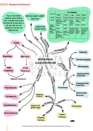
  • 4. 4 Biological Classification (4) Five Kingdom Classification:  According to five-kingdom concept proposed by R.H. Whittaker.  Whittaker (1969), the organisms are divided into five kingdoms namely Monera, Protista, Fungi, Plantae, Animalia, on the basis of the following criteria: (a) Complexity of cell structure: prokaryotic vs eukaryotic organisation of cells. (b) Complexity of body organisation: unicellularity vs multicellularity; simple multicellular forms to complex multicellular forms. (c) Mode of nutrition: Autotrophic vs heterotrophic (parasitic or saprobic or ingestive organisms). It was the major criteria of this classification system. (d) Reproduction. (e) Phylogenetic or evolutionary interrelations All right copy reserved. No part of the material can be produced without prior permission
  • 5. 5 Biological Classification Table 1: Comparative account of different characteristics of the Five Kingdoms (5) Six Kingdom Classification:  Carl Woese proposed six kingdom classification.  These six kingdoms are Archaebacteria, Eubacteria, Protista, Fungi, Plantae and Animalia.  He separated the archaebacteria from eubacteria on the basis of some major differences such as the absence of peptidoglycan in the cell walls of the former and the occurrence of branched chain lipids (a monolayer instead of a phospholipid bilayer) in the membrane.  Based on the sequence of 16S ribosomal RNA genes, Woese found that the six kingdoms naturally cluster into three main categories.  He called these categories as domains of life.  These domains are Bacteria, Archae and Eukarya and are believed to have originated from common ancestor called progenote. Three Domains of life  Based on the sequence of 16 S ribosomal RNA genes, woese found that the six kingdoms naturally cluster into three domains.  These domains are Archae, Bacteria and Eukarya, and are believed to be originated from All right copy reserved. No part of the material can be produced without prior permission
  • 6. 6 Biological Classification common ancestor called Progenate. Domain is a category higher than kingdom. KINGDOM: MONERA  The Kingdom Monera includes all prokaryotes.  Monerans are the most primitive forms of life, originating from more ancient living stock termed progenote.  The kingdom Monera includes eubacteria and archaebacteria.  Eubacteria includes Cyanobacteria, Actinomycetes, Mycoplasma, Rickettsiae, Chlamydiae and Spirochaetes etc. Classification of Monera (1) In 4 kingdom system, a new kingdom was created to accommodate all prokaryotic organisms i.e, eubacteria and archaebacteria. Copeland called it kingdom Mychota. It was called 'Monera' by Daugherty and Allen. All right copy reserved. No part of the material can be produced without prior permission
  • 7. 7 Biological Classification (2) Actually, archaebacteria differ from eubacteria in many respects and resemble eukaryotes in some ways. (3) (i) Carl Woese separated the archaebacteria from eubacteria on the basis of some major differences such as the absence of peptidoglycan in the cell walls of the former and the occurrence of branched chain lipids (a monolayer instead of a phospholipid bilayer) in the membrane. (ii) Therefore, 6 kingdoms given by Carl Woese are Kingdom-1 -Archaebacteria Kingdom-2 -Eubacteria Kingdom-3 -Protista Kingdom-4 -Fungi Kingdom-5 -Plantae Kingdom-6 -Animalia Salient Features of Monera 1. These are unicellular, colonial, multicellular prokaryotic organisms without nuclear membrane, nucleolus, chromatin and histone proteins. 2. Cell wall is made up of peptidoglycan (exceptions are Archaebacteria and Mycoplasma). 3. Membrane bound organelles are absent. 4. Cyclosis is absent and ribosomes are of 70 S type . 5. Respiratory enzymes are found associated with plasma membrane. 6. Nucleoid or genophore or incipient nucleus or prochromosome is composed of naked DNA, RNA and nonhistone proteins. 7. Reproduction by asexual method. 8. Cell division is amitotic type and lacks spindle formation. Let us discuss various monerans in detail: 1. EUBACTERIA All right copy reserved. No part of the material can be produced without prior permission
  • 8. 8 Biological Classification  Bacteria are cosmopolitan and occur in every habitat wherever living or dead organic matter is present.  Anton Von Leeuwenhoek discovered bacteria in rain water which had been allowed to stand for many days and tartar scrapped from teeth.  In 1695, he published his work "The Secrets of Nature".  A.V. Leeuwenhoek termed these microorganisms as dierkens which was later translated as animalcules by the Royal society.  Term microbe for animalcules was coined by Se' dillot, but the term microorganism was proposed by Pasteur.  Ehrenberg first of all coined the word 'bacteria'  Louis Pasteur is considered father of modern microbiology. He introduced the term aerobic and anaerobic for the life in the presence or absence of oxygen respectively.  Robert Koch, a German doctor, demonstrated that the anthrax disease of sheep was caused by bacteria. Koch had followed four experimental steps (Koch's postulates) which help to establish a relationship between a microorganism and a disease.  Smallest bacterium : Dialister pneumosintes  Largest filamentous bacterium : Beggiatoa mirabilis Shapes of Bacteria  Bacteria occur in four basic forms or shapes.  These are spherical (Cocci), rod shaped (Bacilli), Vibrio and Spiral.  Though most bacterial species have cells that are of a fairly constant and characteristic shape, some species are pleomorphic (i.e., these can exhibit a variety of shapes), e.g., Rhizobium leguminosarum. (a) Coccus: Spherical or nearly spherical, aflagellate, sub-divided into six groups on the basis of cell arrangement:  Monococcus -Only single cell represents the bacterium, e.g., Micrococcus luteus, M. roseus.  Diplococcus -Cocci divide in one plane and remain attached in pairs, e.g., Meningococcus, Gonococcus, Diplococcus pneumoniae.  Streptococcus -Cocci remain attached to form chains of different lengths, e.g., Streptococcus Concept Builder All right copy reserved. No part of the material can be produced without prior permission
  • 9. 9 Biological Classification lactis.  Tetracoccus -Cocci divide in two planes at right angles to one another and form groups of four, e.g., Tetracoccus, Neisseria.  Staphylococcus -Cocci divide in several planes resulting in formation of irregular bunches of cells, sometimes resembling a cluster of grapes, e.g., Staphylococcus aureus.  Sarcinae -Cocci divide in 3 planes at right angles to one another and resemble cubical packets of 8 or more cells forming three dimensional geometrical figures, e.g., Sarcina lutea. (b) Bacillus: Rod-like forms, either singly or may be arranged differently. They are generally flagellate. It is the most common of all the shapes. They are of following types: (i) Monobacillus -The bacteria occur singly, e.g., Bacillus anthracis, Lactobacillus. (ii) Diplobacillus -Bacteria are arranged in pairs. e.g., Bacillus subtilis (iii) Streptobacillus -Bacteria form a chain of rods, e.g., Streptobacillus. (iv) Palisade-like -If the cells are lined side by side like match sticks and at angles to one another. e.g., Corynebacterium diphtheriae. (c) Spiral bacteria: Coiled forms of bacteria exhibiting twists with one or more turns are called spirilla, e.g., Spirillum volutans. (d) Vibrio: Bacteria with less than one complete twist or turn are called vibrio. These resemble a comma (,) in appearance, e.g., Vibrio cholerae. (e) Stalked bacteria: The body of bacterium possesses a stalk, e.g., Caulobacter. (f) Budding bacteria: The body is swollen at places, e.g., Rhodomicrobium. All right copy reserved. No part of the material can be produced without prior permission
  • 10. 10 Biological Classification Bacterial Cell Structure Bacterial cell structure is very simple although they are very complex in behaviour. They show the most extensive metabolic diversity. Electron microscope can only reveal the detailed structure of bacterial cell. It consists of following structures: 1. Glycocalyx: (i) It is outermost part of cell envelope (Glycocalyx, cell wall, plasma membrane) (ii) Represented by either slime layer or capsule (a) Slime layer is composed of dextran, dextrin and lavan sugars and protect the cell against desiccation and loss of nutrients. (b) Capsule is made up of polysaccharides and D-glutamic acid. It provides gummy or sticky character and virulent property to the cell. 2. Cell wall:  It is present outside the cell membrane and is a rigid structure.  Due to its rigidity, it protects the internal structures of the cell and provides shape to the cell.  However, its main function is to prevent the cell from expanding and bursting because most bacteria live in hypotonic environments, and are likely to take in much water and eventually burst.  The cell walls of almost all the eubacteria (true bacteria) are made up of peptidoglycan, also called murein or mucopeptide.  It is found only in prokaryotes.  As the name suggests, the peptidoglycan consists of two components-a peptide portion which is composed of amino acids connected by peptide linkages, and a glycan or sugar portion.  The glycan portion, which forms the backbone of peptidoglycan, is composed of alternating units of amino sugars N-acetyl-glucosamine (NAG) and N-acetylmuramic acid (NAM) joined together by -1, 4 linkages.  The peptidoglycan chains are laterally linked by short chains of four amino acids which are attached to N-acetylmuramic acid residues.  The four amino acids of this tetrapeptide are D-alanine, L-alanine, D-glutamic acid and L- All right copy reserved. No part of the material can be produced without prior permission
  • 11. 11 Biological Classification Iysine (in Gram +ve bacteria) or diaminopimelic acid (in Gram -ve bacteria).  The tetrapeptide chains are also interlinked by a peptide bridge between the carboxyl group of an amino acid in one tetrapeptide chain and amino group of an amino acid in another tetrapeptide chain.  The cross linkages can occur between tetrapeptides in different chains, as well as between adjacent tetrapeptide chains. As a result, peptidoglycan forms a rigid, multilayered sheet.  Another component, teichoic acid, an acidic polymer consisting of a carbohydrate (e.g., glucose), phosphate and an alcohol is found in cell walls of Gram +ve bacteria.  Teichoic acid has several functions such as binding metals, acting as receptor sites for some viruses and maintaining cells at low pH to prevent degradation of cell walls by self-produced enzymes.  The walls of Gram-positive bacteria contain very little amount of lipids.  The cell walls of Gram-negative bacteria are much more complex.  The peptidoglycan layer is very thin making up only 10% or less of the cell wall.  However, the most interesting feature is the presence of an outer membrane that covers a thin underlying layer of peptidoglycan.  The outer membrane is a bilayered structure consisting chiefly of phospholipids, proteins and lipopolysaccharides (LPS).  The outer membrane serves as a barrier to prevent the escape of important enzymes from the space (periplasmic space) between the cytoplasmic membrane and the outer membrane.  It also prevents the entry of various chemicals that could damage the cell.  It acts as main surface antigen in cell wall.  However, permeability of outer membrane to nutrients is provided by proteins called porins which form channels in the membrane through which substances of hydrophilic nature and low molecular weight can diffuse.  Christian Gram (1884) developed a staining method for bacteria, using Gram stain (crystal violet).  On the basis of stainability with Gram Stain, bacteria are classified into two groups; Gram positive and Gram negative. 3. Surface Appendages: These include flagella and fimbriae (or pili). (a) Flagella are long, fine, wavy, filamentous appendages that protrude through the cell wall, responsible for the motility of bacteria. These are much thinner than the flagella or cilia of eukaryotes.  Structure of Flagella: The entire flagellar apparatus is made up of three distinct regions: basal body, hook and filament.  Basal body: It is most complex portion of flagellum and has four rings (L, P, S and M), only two rings S and M are present in gram +ve bacteria. L and P rings in cell wall constitute distal set, while S and M rings are present in plasma membrane, forming proximal set.  Hook: Made up of different protein units.  Filament: Bacterial flagella are made up of identical spherical subunits of a protein called flagellin. Longitudinal chains of flagellin molecules run longitudinally around each other to form a wavy helical or rope-like structure. Therefore, a cross section of the flagellum reveals a number of flagellin molecules around a central space. Depending upon the presence or absence, number and position, following types of flagellar arrangements are observed among bacteria :  Atrichous: Flagella absent, e.g., Pasteurella, Lactobacillus.  Monotrichous: Only one flagellum attached at one pole of the organism, e.g.,Thiobacillus, Vibrio.  Amphitrichous: One flagellum at both ends, e.g., Nitrosomonas.  Cephalotrichous: Two or more flagella attached at one end, e.g., Pseudomonas fluorescence. Concept Builder All right copy reserved. No part of the material can be produced without prior permission
  • 12. 12 Biological Classification  Lophotrichous: Two or more flagella attached at both ends, e.g., Spirillum volutans.  Peritrichous: Flagella distributed all over the surface of the cell, e.g., Escherichia coli, Clostridium tetani. (b) Pili and fimbriae are hollow, non helical, filamentous appendages projecting from the walls of Gram-negative bacteria. These are thinner and shorter and more in number than the flagella. These are made up of specific proteins called pilin. There are different types of pili which serve different functions. One type, known as type I pili, (somatic pili) play a major role in infection by facilitating the attachment of bacterial cell to the host cell. Another type, termed sex pili, serve as portals of genetic material from donor to recipient cell during conjugation. Differences between Gram positive and Gram negative bacteria Gram Positive Bacteria Gram Negative Bacteria 1. They retain the blue colour of Gram stain even after washing with alcohol 2. The cell wall is 150-200 Å thick. The cell wall is not covered by lipopolysaccharide layer, i.e., it is a single layer. 3. Cell wall is more rigid due to high percentage (80%) of peptidoglycan 4. Muramic acid content 70-95% 5. Lipid content is low, i.e. ,2-4% 6. Phospholipid absent in cell wall 7. Teichoic acid present 8. Fewer amino acids in cell wall 9. Diaminopimelic acid (DAPA) is absent in the cell wall, instead L-Lysine is present 10. Wall is more sensitive to antibiotics such as penicillin 11. Wall is resistant to alkalies and insoluble in 1% KOH solution 12. Mostly noncapsulated 13. Protoplast is produced by the reaction with lysozyme Other Structures 14. Mesosomes are very common 15. Pili usually absent 16. Flagellationless common 17. Basal body of flagellum has 2 rings (S, M) only 18. Only few forms are pathogenic and may produce exotoxins e.g. Bacillus, Clostridium, Lactobacillus, Streptococcus, Leuconostoc, Staphylococcus, Corynebacterium 1. They get stained blue with Gram stain initially but lose it after washing with alcohol 2. The cell is 75-120 Å thick. Cell wall is covered by lipopolysaccharide layer, i.e. , it is a double layer. 3. Cell wall is less rigid due to low percentage (3-12%) of peptidoglycan 4. Muramic acid content 5-20% 5. Lipid content is high, i.e. , 20-30% 6. Present 7. Absent 8. Several types of amino acids in cell wall 9. DAPA present in cell wall in place of L-Lysine 10. Wall is-not sensitive to penicillin 11. Wall is sensitive to alkalies and soulble in 1% KOH solution 12. Mostly capsulated 13. Sphaeroplast is formed by the reaction with lysozyme (LPS remains unaffected) 14. Mesosomes are rare 15. Pili very common 16. Flagellation very common 17. It has four rings (L, P, S and M) 18. More forms are pathogenic and may produce endotoxins e.g. E. coli, Salmonella, Acetobacter, Azotobacter, Vibrio, Agrobacterium, Shigella, Xanthomonas 4. Protoplast: Cell wall encloses the protoplast, the living matter. It includes (i) Cell membrane (ii) Cytoplasm, (iii) Nucleoid and may have plasmid and episome. (i) Cell membrane:  It lies inner to the cell wall, actually representing the outermost layer of the protoplast.  It is living and semipermeable, controlling the movements of various dissolved substances in and out of the cells.  Functionally, the cell membrane of bacteria resembles mitochondria of eukaryotic cells as respiratory ETS enzymes and succinate dehydrogenase (Kreb's Cycle) are associated with the membrane. All right copy reserved. No part of the material can be produced without prior permission
  • 13. 13 Biological Classification  The cell membrane gets invaginated and folded to form a structure called mesosome (chondroid) in some bacteria, particularly the Gram positive bacteria.  These may be central or peripheral in position and they are supposed to play a role in replication of DNA during cell division, as these are often attached to the nuclear body.  Besides, these increase the surface area of absorption and help in septa formation during binary fission. (ii) Cytoplasm:  It is homogenous colloidal mass of carbohydrates, fats, proteins, lipids, nucleic acids, minerals and water.  It does not show streaming movements.  It lacks sap vacuoles and gas vacuoles (may be present in some bacteria which live in aquatic condition).  Typical membrane bound organelles of eukaryotic cells like endoplasmic reticulum, mitochondria, golgi complex and plastids are absent.  The cytoplasm appears granular due to the presence of ribosomes.  However, these are 70S type in bacteria as compared to 80S type in eukaryotes.  Ribosomes lie scattered freely in the cytoplasm, but sometimes may form a small chain of 4-6 ribosomes attached to mRNA constituting polyribosome or polysome.  Various non-living inclusions like glycogen particles, fat bodies, volutin granules (polymetaphosphate -source of energy) and lipid molecules acting as food reserve lie dispersed in the cytoplasm.  The cytoplasm is usually colourless, lacking pigments.  However, in photosynthetic bacteria, the cytoplasm contains pigments like bacteriochlorophyll and bacterioviridin.  The pigments either lie dispersed in the cytoplasm or present in membrane bound spherical vesicles called chromatophores.  These pigments are capable of entrapping solar energy for photosynthesis. (iii) Nucleoid (Prochromosome, Genophore, Incipient nucleus) –  Bacterial cell lacks a well-organized nucleus.  It consists of a long double stranded DNA molecule repeatedly folded with the help of RNA to form a circular ring.  DNA has no free ends and not associated with histone proteins (polyamines present). Circular DNA ring, without histones is often termed bacterial chromosome. Plasmid (Minichromosome) :  Term plasmid was given by Lederberg and Hays.  These are small, extrachromosomal, non-essential, circular, double stranded, free naked DNA molecules.  The genes present on them have no vital role in survival and growth of bacteria.  These perform autonomous replication.  If plasmids temporarily integrate with bacterial chromosome, then they are called episomes. Types of plasmids : (a) F-Plasmid: It forms sex pilus and is responsible for process of conjugation or fertility factor transfer. (b) R-Plasmid: These plasmids have resistance gene (Resistance Transfer Factor, RTF) for antibiotics like penicillin, tetracycline. (c) Col-Plasmid: Genes of this plasmid are responsible for production of colicins (bacteriocin) for killing other bacteria. (d) Ti Plasmid: From Agrobacterium tumefaciens, used in genetic engineering (e) Degradative plasmid of Pseudomonas putida (superbug) helps to decompose hydrocarbons of petroleum in oil spills. Concept Builder All right copy reserved. No part of the material can be produced without prior permission
  • 14. 14 Biological Classification 5. Bacterial Life Processes Discussion of bacterial life processes revolves around the study of the prominent metabolic activities like respiration and nutrition. (A) Respiration: On the basis of mode of respiration, the bacteria are divided into two main groups: i.e., aerobes and anaerobes. Each group is further of two types i.e. strict or obligate and facultative. (a) Obligate or strict aerobes: These bacteria can live only in presence of oxygen as they possess the enzyme system for aerobic respiration only. In the absence of oxygen, they cannot respire and thus, die, e.g., Bacillus subtilis. (b) Facultative anaerobes: They normally respire aerobically. However, they are capable of switching over to anaerobic mode to get energy for their survival, if sufficient oxygen to sustain aerobic respiration is not available in the environment, e.g., Pseudomonas. (c) Obligate or strict anaerobes: These bacteria respire anaerobically only. The growth of such bacteria will certainly be slower as anaerobic respiration liberates much less amount of energy as compared to aerobic respiration. They lack enzymes necessary for carrying out aerobic respiration e.g., Clostridium botulinum. (d) Facultative aerobes: They normally respire anaerobically, but are capable of respiring aerobically as well, if oxygen is available. Most of the photosynthetic bacteria are facultative aerobes e.g., photosynthetic bacteria Chlorobium. (i) Aerotolerant anaerobes: Bacteria that continue to perform anaerobic respiration even in the presence of oxygen, e.g., Lactic acid bacteria. (ii) Anaerotolerant aerobes: Aerobic bacteria continue to perform aerobic respiration even in absence of free oxygen by using oxygen of oxidised salts, e.g., Denitrifying bacteria. (B) Nutrition:  Nutrition in bacteria is of two types i.e. autotrophic and heterotrophic.  Bacteria having autotrophic mode of nutrition may be photoautotrophs and chemoautotrophs, carrying out photosynthesis and chemosynthesis, respectively. Concept Builder All right copy reserved. No part of the material can be produced without prior permission
  • 15. 15 Biological Classification (I) Autotrophic Nutrition (i) Photoautotrophic bacteria:  These bacteria are capable of entrapping solar energy and utilizing it for the synthesis of complex food materials due to the presence of pigments like bacteriochlorophyll (bacteriopurpurin) and bacterioviridin.  Purple sulpher bacteria (e.g., Thiospirillum) and green sulpher bacteria (Chlorobium limicola) are the most familiar examples containing pigment bacteriochlorophyll, bacteriopurpurin and bacterioviridin respectively.  Bacterial photosynthesis, however, differs from photosynthesis of higher plants in not liberating oxygen.  This type of photosynthesis, characteristic of bacteria, is termed as anoxygenic.  Normal photosynthesis, occurring in higher plants, is termed as oxygenic.  In bacterial photosynthesis water is not the source of electron that acts as reducing power to convert CO2 into glucose.  The bacteria obtain reducing power from various compounds such as hydrogen sulphide, thiosulphate or even some organic compounds.  No oxygen is evolved as it does not involve splitting of water.  Hydrogen released by various compounds mentioned above is picked up by NAD+ which gets reduced to NADH2 acting as reducing power.  NADH2 alongwith ATP, produced generally by entrapping solar energy are used to reduce CO2 to glucose. All right copy reserved. No part of the material can be produced without prior permission
  • 16. 16 Biological Classification  Simple equation for anoxygenic photosynthesis may be written as follows : CO2 + – 2 (or any other compound acting as e donor) H S Solar energy enzymes ⎯⎯⎯⎯⎯ → Sugar + (Sulphur or other oxidised compound) + H2O + energy  Most of the photosynthetic bacteria are anaerobes (facultative aerobes). (ii) Chemoautotrophic Bacteria:  Bacteria belonging to this category obtain energy for the synthesis of food by oxidising certain inorganic substances like ammonia, nitrates, nitrites, ferrous ions etc.  Thus, they do not utilise light as energy source.  The chemical energy thus obtained, is trapped in ATP molecules.  This energy is then used in carbon assimilation with the help of hydrogen from some source other than water, e.g., hydrogen bacteria, nitrifying bacteria, sulphur bacteria, etc.  They play a great role in recycling nutrients like nitrogen, phosphorous, iron, sulphur. (a) Hydrogen bacteria. These bacteria oxidise hydrogen in the presence of oxygen, e.g., Hydrogenomonas. (b) Nitrifying bacteria obtain energy by oxidizing ammonia into nitrate, oxidation of ammonia occurs in 2 steps. Each step is carried out by a specialised group of bacteria. • In the first step, ammonia is oxidised into nitrites by the species of the genus Nitrosomonas and Nitrococcus. • In the second step, the nitrite is converted into nitrate. This is brought about by species of the genus Nitrobacter and Nitrocystis which use this energy for chemosynthesis. (c) Sulpher bacteria. These bacteria obtain energy either by the oxidation of elemental sulphur or oxidation of H2S. • Oxidation of elemental sulpher. Sulpher bacteria (Thiobacillus thioxidans) oxidise elemental sulpher to sulphuric acid and utilise energy produced in this process. These bacteria can survive even in extreme acidic environment. • Oxidation of H2S to S. Some bacteria like Beggiatoa use the energy from oxidation of H2S and store the sulphur so produced in the form of granules. (d) Iron bacteria. These bacteria (e.g., Ferrobacillus, Leptothrix) inhabit water which contain iron compounds. These bacteria convert ferrous ions to ferric form. The ferric ion is deposited as insoluble ferric hydroxide. The energy so released, is utilised in the assimilation of CO2. (e) Methane bacteria. Methanomonas, is one example which oxidises methane to carbon dioxide. (II) Heterotrophic bacteria:  These bacteria are most abundant in nature and are incapable of synthesizing their own food from simple raw materials.  They obtain nourishment either from dead and decaying organic matter or directly from a living host.  All heterotrophic bacteria are segregated into three main categories, i.e., saprophytic, symbiotic and parasitic forms. (i)Saprophytic bacteria:  They are free living bacteria, obtaining nourishment from organic remains such as dead animals, animal excreta, fallen leaves, decaying vegetables, fruits, bread and other products of animal and plant origin.  These bacteria secrete digestive enzymes into the substrate and the complex insoluble Substances are converted into simple soluble compounds like water, hydrogen sulphide, ammonia, CO2 etc. S Sa al lm mo on ne el ll la a t ty yp ph hi i V Vi ib br ri io o c ch ho ol le er ra ae e R Rh hi iz zo ob bi iu um m All right copy reserved. No part of the material can be produced without prior permission
  • 17. 17 Biological Classification  Some of the simpler substances are absorbed and assimilated by the bacteria, whereas the others are added to the soil and atmosphere to complete the nature's material cycle.  Anaerobic breakdown of carbohydrates and proteins is termed fermentation and putrefaction respectively.  Aerobic breakdown of organic compounds is called decay. (ii) Symbiotic bacteria:  They are mainly Gram-negative type.  A familiar example of symbiotic bacteria is Rhizobium leguminosarum, associated with roots of leguminous plants.  They are capable of fixing atmospheric nitrogen as ammonia, inside the nodule only and not in free state.  However, some bacteria like Azotobacter, Beijerinckia, Klebsiella are free living, aerobic and capable of nitrogen fixation in free state, enriching the soil.  Clostridium pasteurianum is anaerobic N2 fixing bacteria. (iii) Parasitic bacteria:  These bacteria draw nourishment and obtain special organic compounds required for growth from living organisms, either plants or animals, called hosts.  The disease-causing bacteria are termed pathogenic and the ones not causing any disease are termed as non-pathogenic. 6. Reproduction Bacteria reproduce mainly by asexual method and also show sexual recombination (True sexual reproduction is absent). A. Asexual Reproduction:  Bacteria produce several types of asexual spores like, sporangiospores, oidia, conidia and endospores. However, the most common mode of asexual reproduction is binary fission.  Under favourable conditions of nutrient availability, moisture and temperature, daughter cells may repeat binary fission many times and may forms a large population.  Fortunately, such a rapid rate is seldom achieved.  The process gradually slows down and ultimately stops because of: (i) Shortage of space. (ii) Lack of nutrient availability. (iii) Accumulation of waste products (making environmental conditions unfavourable for growth). (iv) Development of bacteriophages, destroying bacteria. (a) Binary Fission:  It is the most common method under favourable conditions of temperature, moisture and availability of nutrients.  Mature bacterial cell divides into two daughter cells.  In this process the cell division is amitotic type i.e., not involving the spindle formation. (b) Endospores:  Cells of certain bacteria, e.g., Bacillus, Clostridium etc. form thick-walled, highly resistant bodies within the cell, called endospores. All right copy reserved. No part of the material can be produced without prior permission
  • 18. 18 Biological Classification  One bacterial cell normally produces only a single endospore.  The endospores may be spherical or oval in shape and are terminal or central in position.  Anticoagulant nature of endospore is due to the presence of Ca-dipicolinic acid in cortex layer of wall. B. Sexual Recombination (Genetic Recombination) :  The bacteria exhibit a primitive form of sexual reproduction which differs from eukaryotic sexual reproduction because there is no gamete formation and fusion.  However, the essential feature of sexual reproduction, i.e., exchange of genetic material does take place and is called genetic recombination.  Three methods are known by which genetic recombination is achieved by bacteria.  In the order of their discovery, these are transformation, conjugation and transduction. (a) Transformation:  Griffith (1928) worked on the effect of Diplococcus or Streptococcus pneumoniae bacteria on mice and discovered the process of transformation.  In transformation, the donor and recipient do not come in contact.  The donor cell releases a piece of DNA which is actively taken up by the recipient cell from the solution.  This ability to pick up DNA from the solution is called competence.  Two strains of D. pneumoniae are : Capsulated or S-III (Virulent strain) and Non-capsulated or R-II (non-virulent strain).  Four steps were performed in experiment: (i) S-III bacteria Injectedinto ⎯⎯⎯⎯⎯ → Healthy mice ⎯⎯ → Mice died. (ii) R-II bacteria Injectedinto ⎯⎯⎯⎯⎯ → Healthy mice ⎯⎯ → Mice survived. (iii) S-III bacteria Injectedinto ⎯⎯⎯⎯⎯ → Healthy mice ⎯⎯ → Mice survived. (Heat killed) (iv) R-II (living) + S-III (heat killed) bacteria Injectedinto ⎯⎯⎯⎯⎯ → Healthy mice ⎯⎯ → Mice died. (But R-II is not virulent and S-III lost its virulent capacity upon heating).  Griffith concluded that 'something' passed from heat killed S-III to R-II bacteria, so that non virulent strain changed or transformed into virulent bacterial strain.  Avery, Macleod and McCarty (1948) repeated this experiment using various enzymes and proved that the transformation principle is DNA of heat killed S-III strain.  They proved that DNA is a genetic material. (b) Conjugation:  Lederberg and Tatum (1946) demonstrated in E. coli that during conjugation, one cell containing F-plasmid acts as donor (F+ or male) cell and the other lacking F-plasmid as recipient (F– or female) cell.  The plasmid contains fertility factor or F gene which produces protrusions termed sex pili. All right copy reserved. No part of the material can be produced without prior permission
  • 19. 19 Biological Classification  These help the donor F+ cell in attaching to the recipient cell.  The plasmid replicates and a replica is transferred to recipient cell, changing it into F+.  Often the plasmid integrates with bacterial chromosome, converting it into Hfr (High frequency of recombination) cell or super male and a part or whole of bacterial chromosome is transferred to recipient cell through-conjugation tube.  Such association of episome with the endogenote increases the efficiency of genetic transfer.  The number of genes transferred depends upon the time for which the two cells remain joined together.  When F-conjugates with super male, the frequency of recombination increases by 1000 times, that is why it is called as Hfr (super male). (c) Transduction:  During transduction, a small double stranded piece of DNA is transferred from donor to recipient by a bacteriophage.  This mode of genetic recombination in bacteria was first demonstrated by Zinder and Lederberg (1952) while working with Salmonella typhimurlum.  Some viruses have the ability to integrate their DNA with bacterial DNA, which is replicated at the same time as the host DNA and is passed from one bacterial generation to the next.  Such bacteria carrying phage (viral) DNA with their own DNA are called lysogenic bacteria.  Occasionally, the phage DNA becomes active and codes for the production of new virus particles.  A number of phage particles are synthesised followed by the destruction of the host cell and release of phage particles.  Upon release, the phage particles attack sensitive bacterial cells, multiply and release more phage particles.  However, sometimes faulty deatchment of phage DNA from bacterial DNA results in the incorporation of a small amount of bacterial DNA into the phage DNA.  Subsequent infection of another bacterium with this aberrant phage called transducing phage, introduces the piece of foreign bacterial DNA into the recipient's chromosomes, producing a genetic change. Types of transductions:  The ability of the bacteriophage to carry the genetic material from any region of bacterial DNA is called generalised transduction, e.g., T4-phage. All right copy reserved. No part of the material can be produced without prior permission
  • 20. 20 Biological Classification  On the other hand, there are bacteriophages such as lambda phage () of E coli which can carry only a specific region of the bacterial DNA to a recipient.  This is called specialised transduction (or restricted transduction).  Sometimes, the DNA brought by the phage does not integrate with the genome of the recipient bacterium and is lost after one or two generations.  Such a transduction is called abortive transduction. 7.Economic Importance of Bacteria Bacteria play significant role in day-to-day activities of human beings. A. Beneficial activities (a) Role in agriculture (i) Decay and decomposition of organic matter :  They bring about decay and decomposition of dead remains of plants and animals.  These are the most important for mineral cycling to occur. (ii) Sewage disposal:  The bacteria decompose the organic matter present in the sewage, converting into simpler inorganic substances.  The inorganic substances thus formed, being soluble, pass out through filter alongwith water which is highly useful for irrigation purposes, e.g., Clostridium, E. coli. (iii) Nitrogen cycle:  The proteins present in dead remains of living organisms are converted into amino acids by different types of saprophytic bacteria.  Amino acids are converted into ammonia by ammonifying bacteria (Bacillus vulgaris, B. ramosus).  Nitrifying bacteria convert ammonium salts first into nitrites (Nitrosomonas, Nitrococcus) which are subsequently converted into nitrates (Nitrobacter).  In presence of denitrifying bacteria (Pseudomonas denitrificans), nitrates and nitrites of soil are converted to gaseous nitrogen. (iv) Nitrogen fixation:  It is the biological process in which atmospheric nitrogen is converted into nitrogenous compounds by nitrogen fixing bacteria.  These bacteria are of 2 types : (a) Free living (b) Symbiotic (a) FreelivingbacteriaareAzotobacterandBeijerinckia(aerobic)&Clostridium (anaerobic). (b) Common symbiotic bacteria are Rhizobium leguminosarum and Xanthomonas. These convert nitrogen into ammonia which is directly converted into amino acids by plants. (v) Manure preparation:  Saprotrophic bacteria help in preparation of farmyard manure by converting farm refuse, dung and other organic wastes into humus. (b) Role in Industry:  Man has utilized the metabolic activities of bacteria in preparation of a number of industrial products as listed below : (i) Butter milk and sour cream (ii) Yoghurt (iii) Cheese (iv) Vinegar (v) Retting of fibres : Retting is a controlled microbial decomposition for separation of fibres. All right copy reserved. No part of the material can be produced without prior permission
  • 21. 21 Biological Classification The tissues are immersed in water tanks where anaerobic butyric acid bacteria dissolve the pectin of middle lamella of cells, thus, separating the fibres. Clostridium perfringens and Pseudomonas fluorescence are useful in this process. (vi) Curing of leaves: To improve the flavour and taste in tea, using Micrococcus candidans and in tobacco leaves by Bacillus megatherium. (vii) Single cell proteins (SCP): Like -Methylophilus methylotropus and Rhodopseudomonas capsulata. (c) Role in Medicine:  Bacteria have been used extensively in preparation of antibiotics, vaccines, serums and vitamins. (i) Antibiotics :  The term antibiotic was given by Waksman, who discovered streptomycin.  These are the organic substances produced by microorganisms which inhibit the growth of other organisms (mostly pathogens) but do not affect the growth of organisms secreting these.  The first commercial antibiotic penicillin was discovered by Flemming (1959) from a fungus called Penicillium. Some antibiotics of eubacterial origin: (a) Bacitracin ........................... Bacillus licheniformis (b) Polymixin ........................... Bacillus polymyxa (c) Gramicidin ........................... B. brevis (d) Subtilin ........................... B. subtilis (ii) Vaccine production  Vaccines and serums against typhoid, cholera, TB, pertussis, tetanus and diphtheria are made with the help of bacteria or their toxins e.g., DPT (against diphtheria, pertussis and tetanus), TI(against tetanus), BCG (Bacille of Calmette-Guerin against TB), DT (against diphtheria and tetanus). (iii) Vitamins :  Escherichia coli present in human intestine produces large quantities of B-complex vitamins and vitamin K. Bacteria are utilized in industrial production of a number of vitamins like riboflavin from Clostridium butylicum, Cobalamine (B12) from Bacillus megatherium and Pseudomonas denitrificans. (iv) Pollution control :  Pseudomonas putida degrades petroleum wastes.  Flavobacterium can decompose 2, 4-D. DDT can be decomposed by Acetobacter aerogens.  Ganga's water contains Bdellovibrio bacteriovorus that maintains purity of its water. (v) Poly--hydroxybutyrate is used to produce biodegradable plastic. B.Harmful activities (i) Spoilage of food: Saprophytic bacteria cause decay of vegetables, fruit, meat, bread and other foods, making these unfit for human consumption. Some bacteria even produce strong toxins in the infected food stuffs which cause food poisoning when consumed. (ii) Deterioration of Domestic Articles: Some saprophytic bacteria like Cellulomonas, Spirochaete cytophaga cause deterioration of domestic articles of daily use such as leather, woolen, canvas articles etc. (iii) Denitrification of Soils: Denitrifying bacteria like Thiobacillus denitrificans, Micrococcus denitrificans and Pseudomonas convert nitrates and nitrites present in the soil into gaseous nitrogen, thus depleting the soil nitrogen, thereby decreasing soil fertility. All right copy reserved. No part of the material can be produced without prior permission
  • 22. 22 Biological Classification (iv) Desulphurification: Desulphovibrio desulphuricans. (v) Diseases: Common disease of humans, animals and plants are listed below respectively – S.No. Name of causal organism Name of the human diseases 1. Clostridium botulinum Botulism 2. Vibrio cholerae Cholera 3. Corynebacterium diphtheriae Diphtheria 4. Salmonella typhimurium Staphylococcus aureus Food poisoning 5. Clostridium perfringens Gangrene 6. Neisseria gonorrhoeae or Salmonella schottmulleri Gonorrhoea 7. Mycobacterium leprae (Hensen 's bacillus) Leprosy 8. Neisseria meningitidis Meningitis 9. Yersinia pestis Plague 10. Diplococcus pneumoniae Pneumonia 11. Treponema pallidum Syphilis 12. Clostridium tetani Tetanus 13. Streptococcus pyrogenes Tonsilitis 14. Mycobacterium tuberculosis Tuberculosis 15. Salmonella typhosa / typhi Typhoid 16. Bordetella pertussis / Hemophilous pertussis Whooping cough 17. Salmonella typhimurium Enteric fever (paratyphoid) S.No. Name of causal organism Name of the plant diseases 1. Pseudomonas solanacearum Potato wilt 2. Xanthomonas citri Citrus canker 3. Agrobacterium tumefaciens Crown gall 4. Erwinia amylovora Fire blight of apple 5. Xanthomonas oryzae Bacterial blight of rice 6. Xanthomonas malvacearum Angular leaf spot of Gossypium 7. Pseudomonas rubrilineans Red stripe of sugarcane 8. Erwinia cartovora Soft rot of carrot 9. Corynebacterium tritici Tundu(bacterial rot) of wheat 10. Xanthomonas campestris Black rot of cabbage 11. Streptomyces scabies Potato scab II. CYANOBACTERIA  Cyanobacteria are Gram negative photosynthetic prokaryotes, being the most primitive organisms to have oxygenic photosynthesis.  They added oxygen to the atmosphere, which is indispensible for the existence of aerobic forms of living organisms.  They are also known as BGA (Blue green algae) and are classified variously under cyanophyceae or myxophyceae. Occurrence  They are mainly fresh water forms, though few are marine.  Red sea is named so because of abundant occurrence of a cyanobacterium Trichodesmium erythraeum, which imparts red colouration to water.  They occur in symbiotic association with almost every group of eukaryotes i.e. green algae, fungi, bryophytes like mosses and Anthoceros, ferns, gymnosperms, angiosperms, sponge, shrimps, mammals etc.  Anabaena azollae is associated with Azolla, an aquatic fern.  Anabaena cycadeae is associated with coralloid roots of Cycas.  In many lichens (symbiotic association of algae and fungi), the algal partner may be a cyanobacterium. All right copy reserved. No part of the material can be produced without prior permission
  • 23. 23 Biological Classification  When they live endozoically in protozoans they are called cyanelle. Structural Organization  These may be unicelled or multicelled. The latter may be filamentous or colonial.  Filamentous form consists of one or more cellular strands, called trichomes, surrounded by mucilagenous sheath.  Cyanobacteria are characterised by the absence of flagellum throughout life cycle. Cell Structure  The cell structure in cyanobacteria is typically prokaryotic.  The cell lacks a well-defined nucleus and the chromatin material is centrally located, resembling the bacterial chromosome.  The cell wall is 4 layered and is invariably covered by mucilagenous sheath, composed largely of mucopeptides.  Protoplasm in cyanobacterial cell can be distinctly divided into two parts the centroplasm and chromoplasm.  The central colourless centroplasm contains the chromatin material.  The peripheral protoplasm is coloured or pigmented because of the presence of thylakoids, called as chromoplasm.  The protoplast lacks membrane-bound organelles like endoplasmic reticulum, golgi bodies, mitochondria, lysosomes, plastids and contains 70S ribosomes.  Similar to the mesosome of bacteria, a group of coiled membrane called lamellasome is found which connects nucleoid to the cell membrane.  It helps in respiration and replication of DNA.  The cell membrane lack sterols. The sap vacuoles are absent.  Instead, the cell may contain gas filled vacuoles which help to regulate the buoyancy of the organism in water.  The characteristic feature of cyanobacterium cell is the presence of a system of photosynthetic lamellae called thylakoids.  The characteristic photosynthetic pigments present in the thylakoids are chlorophyll a and phycobilins i.e., phycocyanin (blue coloured), phycoerythrin (red coloured) and allophycocyanin (light blue coloured). The cyanobacterial cell contains reserve food material in the following forms Nostoc All right copy reserved. No part of the material can be produced without prior permission
  • 24. 24 Biological Classification (i) Cyanophycean granules (Protein) (ii) -granule (Fat droprets) (iii) Cyano-or myxophycean starch or -granule (Similar to glycogen but negative to iodine test) (iv) Volutin body (Reserve phosphate) (v) Polyhedral body (Rubisco rich) Metabolism  They are the most self-dependent organisms, because most of these are capable of converting atmospheric nitrogen into ammonium compounds besides utilizing atmospheric CO2 for synthesis of organic food during photosynthesis.  Biological nitrogen fixation is an anaerobic process as nitrogenase enzyme required for the process acts efficiently in the absence of oxygen.  Nitrogen fixation under anaerobic conditions occurs mainly in large, specialized cells called heterocysts as in Nostoc.  Heterocyst has terminal pores which at maturity develops a polar granule.  Thickened cell wall of these cells is impermeable to oxygen so this creates anaerobic environment in the cell even under aerobic conditions.  Besides this, heterocysts also lack PS II activities and CO2 fixation is done only by vegetative cells.  Oxygen is not evolved due to absence of PSII.  However, PSI remains active in heterocyst which generates ATP required to fix nitrogen.  Besides N2 fixation heterocyst promotes fragmentation.  This property of nitrogen fixation, most of the BGA enrich the soil by releasing nitrogenous compounds in the surroundings. Reproduction  Cyanobacteria reproduce asexually. Typical sexual reproduction is absent.  Asexual reproduction occurs by following methods : (i) Binary fission: It occurs in unicellular forms. The daughter cells formed by amitotic division separate immediately after the division. (ii) Fragmentation: It occurs in filamentous forms. The filament breaks up into short pieces or fragments which grow to form new filaments. (iii) Heterocysts : Under special conditions, the heterocysts germinate to form new filaments. (iv) Hormogonia: Due to the formation of biconcave, mucilage filled dead cells called necridia, in between living cells of trichome, the filament breaks into hormogonia. (v) Akinetes: Vegetative cells are transformed into thick walled akinetes due to the deposition of All right copy reserved. No part of the material can be produced without prior permission
  • 25. 25 Biological Classification food material followed by the thickening of wall. On the arrival of favourable conditions, they germinate to form new filaments. Importance of Cyanobacteria (i) They are the most ancient organisms having oxygenic photosynthesis and thus, played a significant role in the evolution of aerobic forms of life. (ii) They convert atmospheric nitrogen into ammonium compounds and excess of these compounds is excreted out, enriching the soil. The death and decay of these also increase the soil fertility, particularly the nitrogen content of the soil. Tolypothrix and Aulosira fix N2 non- symbiotically in rice fields . Cyanobacteria like Nostoc and Anabaena have been used for reclaiming usar soils. As they can live in damp or aquatic habitat, they enrich the root environment in any wetland condition as in rice fields. (iii) Cyanobacteria are associated in symbiotic relationship with almost every group of plants. They benefit the partner by providing nitrogenous compounds because of their capability of nitrogen fixation. (iv) Some cyanobacteria serve as food to several aquatic animals. Spirulina is edible, non-toxic, fast growing cyanobacterium. It is cultivated in tanks as source of protein rich animal food (SCP). (v) Extract of Lyngbya is used for the manufacture of antibiotic. (vi) Some cyanobacteria like Microcystis aeruginosa, Anabaena flos-aquae, Aphanizomenon flos- aquae are known to cause algal blooms in water bodies. These also secrete toxins into the surroundings, which are harmful to aquatic animals and even to human beings. Water from such sources is harmful and may even prove fatal for organisms drinking it. They also deplete the oxygen from the water reservoir and thereby, cause large scale death of the fishes and other aquatic animals. III. MYCOPLASMA  E. Nocard and E.R. Roux (1898)-two French Scientists, discovered these organisms from pleural fluid of cattles suffering from pleuropneumonia.  These are pleomorphic and were called PPLO (Pleuropneumonia Like Organisms) or Jokers of plant kingdom.  This organism was later on given the name Asterococcus mycoides by Borrel et al. (1910).  Nowak (1929) placed Asterococcus mycoides under the genus Mycoplasma.  All such organisms are now called Mycoplasma, or MLO's (Mollicutes like organisms).  These are sometimes placed in a separate class called Mollicuta.  Mycoplasma infects animals (e.g., dog, sheep, mice and man) and plants (e.g., potato, corn, brinjal etc.).  They are generally found in soil, sewage water, plants and animals. Structure:  These are unicellular, simplest free-living prokaryotes.  They do not have cell wall so they are highly pleomorphic and can assume various shapes like spherical, granular, filamentous, coccoid etc.  Cell membrane is the outermost limiting layer.  It is trilamellar unit membrane structure.  In culture, colonies of mycoplasma show a characteristic fried egg appearance with an opaque central area and translucent peripheral zone.  The cells are generally non motile, but a few are gliding type. All right copy reserved. No part of the material can be produced without prior permission
  • 26. 26 Biological Classification  The protoplasmic matrix contains ribosomes (70S type), fatty acid substances and proteins.  Organized nucleus, endoplasmic reticulum, plastids, mitochondria, golgi bodies, Iysosomes, centrioles, flagella, etc. are absent.  Mycoplasma has both RNA and DNA.  RNA is single stranded, present in both ribosomes and cytoplasm and DNA is double stranded, long coiled thread extending almost throughout the cell.  Enzymes are present freely in the cytoplasm as well as associated with the plasma membrane.  Replicating disc assist in replication and separation of the genetic material. Nature of Mycoplasma:  Mycoplasma can pass through bacteriological filters and lack cell wall.  This shows that they are not bacteria.  Since they can multiply in abiotic medium having sterols, so they are not considered as virus.  Due to similarities with bacteria, they are said to be "Bacteria with their coats off".  Mode of nutrition is heterotrophic.  Some are saprophytic, but mostly they are parasitic.  They are parasitic, because they are unable to synthesize required growth factors, e.g., M. gallisepticum (0.3 to 0.5 µm, smallest prokaryote).  They can survive without oxygen. Sensitivity to Antibiotics:  Mycoplasma are Gram negative, insensitive to penicillin but sensitive to streptomycin, erythromycin, chloramphenicol (metabolic inhibitors) etc.,  They are insensitive to penicillin because they are wall less and penicillin interferes in the synthesis of peptidoglycan, a component of cell wall of bacteria.  Reproduction - Much is not known about reproduction of Mycoplasma but they mainly reproduce by means of elementary bodies. Mycoplasma IV. ARCHAEBACTERIA  They are believed to have evolved immediately after the origin of life on earth, as even now these are living under extremely adverse conditions such as extreme salty areas (halophiles), hot springs (thermoacidophiles) and marshy areas (methanogens). All right copy reserved. No part of the material can be produced without prior permission
  • 27. 27 Biological Classification  Very few other organisms can survive under such environmental conditions. So these are termed as "living fossils".  These possess introns in DNA, their ribosomal proteins are highly acidic, these prokaryotes possess histone proteins different from that of eukaryotes.  These being the most primitive and ancient most bacteria.  Archaebacteria differ from other bacteria in having a different cell wall structure and this feature is responsible for their survival in extreme conditions.  The cell wall in archaebacteria contains proteins and non-cellulosic polysaccharides.  It lacks peptidoglycan, the characteristic cell wall material in bacteria and cyanobacteria.  It consists of glycoprotein, pseudomurein and non cellulosic polysaccharide.  Pseudomurein is like bacterial peptidoglycan, but contain N-acetyltalosaminuronic acid instead of NAM and lacks D-Amino acid.  The cell membrane contains branched chain lipids (phytanyl side chains) which decreases membrane fluidity.  This chemical composition of the cell membrane enables these organisms to withstand extremes of temperature and pH.  Archaebacteria are divided into three groups· methanogens, halophiles and thermoacidophiles. (a) Methanogens:  They are obligate anaerobes occurring in marshy habitats.  They are capable of converting CO2, methanol and formic acid (HCOOH) into methane and hence the name methanogens.  This property is exploited commercially in the production of fuel gas and methane in gobar gas plants (biogas fermenters).  Some of the methanogens live in rumen of herbivorous animals like buffalo, cow etc. (ruminants).  These microorganisms assist in fermentation of cellulose in such animals, e.g., Methanococcus, Methanobacterium, Methanosarcina, Methanospirillum. (b) Halophiles:  They are aerobic chemoheterotrophic coccoid forms and are Gram negative. They occur in high salt concentration medium like sea, salt lake, brines, marshes, salted field etc.  In high light intensity a reddish pigment bacteriorhodopsin develops in their membrane to trap sun light to produce ATP, but they cannot use this ATP in food synthesis.  Sap vacuoles are absent in halophiles, hence, they cannot get plasmolysed in high salt concentration.  They maintain a high osmotic concentration of KCl in their cells.  These bacteria get lysed if NaCl level falls below 10%, e.g., Halococcus, Halobacterium.  These can grow well in a medium containing 25-30% of NaCl. (c) Thermoacidophiles:  They are capable of tolerating high temperature as well as high acidity and hence, the name thermoacidophiles.  They often live in hot-water springs where the temperature is as high as 80°C and the pH as low as 2.  They oxidise sulphur to sulphuric acid under aerobic conditions and the energy obtained in this reaction is utilized for the synthesis of organic food.  The medium becomes highly acidic due to the production of sulphuric acid. Under anaerobic conditions sulphur is reduced to H2S, e.g., Thermoplasma, Thermoproteus, Thermococcus. Hence, these are chemosynthetic in nature. All right copy reserved. No part of the material can be produced without prior permission
  • 28. 28 Biological Classification V. ACTINOMYCETES (Ray Fungi) (1) These are branched, filamentous bacteria and are considered as intermediate form between bacteria and fungi, e.g., Nocardia, Mycobacterium, Corynebacterium, Frankia, Streptomyces. (2) These filaments form radiating colonies in cultures so are also called mycelial bacteria. (3) Cell wall contains peptidoglycan and is Gram positive in nature. It contains mycolic acid. (4) They are facultative anaerobic, saprophytic, non-motile and are important decomposers of dead organic matter. (5) They reproduce commonly by conidia, sporangiospore, oidia and fragmentation. (6) They are acid fast in nature. KINGDOM: PROTISTA All unicellular eukaryotes, irrespective of their mode of nutrition, are included in the kingdom Protista in Whittaker's system. The term protista was coined by Ernst Haeckel. This kingdom forms a All right copy reserved. No part of the material can be produced without prior permission
  • 29. 29 Biological Classification link between kingdom Monera on one hand and other three kingdoms i.e. , Plantae, Fungi and Animalia on the other hand. Protistans are ancestors of all multicellular eukaryotes (plants, fungi and animals). Kingdom Protista includes General Characteristics of Protista 1. Unicellular, eukaryotic organisms. Some are colonial without much cellular differentiation. Organisation at tissue level is absent. 2. Mostly aquatic organisms. 3. Cell structure is eukaryotic type having all kinds of membrane bound organelles and 80 S cytoplasmic ribosomes and cells may possess cellulosic cell wall. 4. Flagella and cilia have (9+2) pattern of microtubule organization consisting of tubulin protein. 5. Movement by pseudopodia, flagella or cilia where ciliary mode is fastest. 6. Mode of nutrition may be photosynthetic (holophytic), holozoic (ingestive), saprobic or parasitic (absorptive). Some have mixotrophic nutrition (photosynthetic and saprobic) as in Euglena. 7. Reproduction occurs by asexual and sexual means. 8. Life cycle is of 2 types-(i) Showing zygotic meiosis (ii) Showing gametic meiosis. 9. These are decomposers, photosynthetic or parasites. Parasitic protists may cause diseases like dysentery, malaria, sleeping sickness etc. Photosynthetic protists and Slime moulds are described below: (1) Photosynthetic Protists These are popularly called protistan algae. Protistan algae constitute the major portion of the phytoplanktons. A. Diatoms Diatoms are golden brown photosynthetic protists and are called Chrysophytes (including both diatoms and desmids). They are both aquatic and terrestrial. Some are marine. They support much of marine life. Their important characters are  These are microscopic organisms possessing varying colours.  They are basically unicellular, but may form pseudofilament and colonies, lacking flagella except in the reproductive stage. They may be free floating (phytoplanktonic) due to presence of light weight lipids.  The cellulosic cell wall is impregnated with silica to form transparent siliceous shell, known as All right copy reserved. No part of the material can be produced without prior permission
  • 30. 30 Biological Classification frustule. Depending upon the symmetry, diatoms may be pennate type, having bilateral symmetry (e.g., Navicula) and centric type, having radial symmetry (e.g., Melosira).  The cell wall is characteristic, made up of two halves; one half covering the other (epitheca over hypotheca) resembling a soap box.  The cell wall encloses the peripheral layer of cytoplasm (primordial utricle) surrounding a large central vacuole.  Nucleus lies in the central vacuole, suspended with the help of cytoplasmic strands.  Mode of nutrition is holophytic (photoautotrophic), photosynthetic pigments are chlorophyll a, chlorophyll c, -carotene and special carotenoids containing fucoxanthin; xanthophylls like diatoxanthin, diadinoxanthin.  The reserve food is oil and a polysaccharide called leucosin (chrysolaminarin), volutin granules are also present.  They are responsible for almost 50% of the total organic matter synthesized in the biosphere.  Movement occurs by mucilage propulsion.  They mainly undergo asexual reproduction. The common mode of asexual reproduction is binary fission.  During binary fission, one half of the cell wall is retained by each of the daughter cells formed. The other half of the cell wall is secreted afresh.  Resting spores are called statospores (centric diatoms).  They reproduce sexually as well. Sexual reproduction varies from isogamy to oogamy. It involves gametic meiosis as diatoms are generally diploid (diplontic life cycle). B. Dinoflagellates  Dinoflagellates are golden brown photosynthetic protists, belonging to class Dinophyceae (Pyrrophyta). They are mainly marine, though few are fresh water forms.  They may appear red, yellow, green, brown or blue depending upon the main pigment present in cell. General characters:  Unicellular, motile, biflagellate, golden brown photosynthetic protists (some are non-motile, amoeboid, palmelloid or filamentous).  They are mostly marine, some are found in fresh water.  The body is enclosed by a rigid coat called theca or lorica consisting of 2 to many articulated or sculptured plates of cellulose and pectin , hence are also called armoured dinoflagellates.  Theca has generally two grooves i.e., longitudinal called sulcus and transverse called cingulum or annulus or girdle.  Flagella are heterokont (different). One is longitudinal and other is transverse. The flagella pass out through the pores in the lorica and lie in the grooves. The transverse flagellum lies in the circular groove and the longitudinal flagellum in the longitudinal groove. The longitudinal flagellum is narrow, smooth directed posteriorly and the transverse flagellum is ribbon like. Both are oriented at right angle to each other producing spinning movements. Therefore, these protists are also called 'whirling whips'.  Most of the species have brown, green or yellow chromatophores with chlorophyll a, c, -carotene, All right copy reserved. No part of the material can be produced without prior permission
  • 31. 31 Biological Classification xanthophyll (e.g., Peridinin). Plastids are generally surrounded by 3-membrane envelope and contain 3-thylakoid lamellae. They are autotrophic or photosynthetic (Ceratium), a few are saprobic or parasitic.  Reserve food is carbohydrate and oils.  Nucleus is relatively larger in size, has condensed chromosomes even in interphase, chromosomes do not have histone. Nuclear envelope and nucleolus remain present even during cell division. This organisation is called Mesokaryon (Dodge, 1966).  A non-contractile vacuole called pusule is present near the flagellar base. It may have one or more vesicle and takes part in floatation and osmoregulation.  Some dinoflagellates possess trichocysts and cnidoblasts like those of coelentrates.  Reproduction is commonly asexual and occurs through cell division.  Isogamous and anisogamous sexual reproduction is reported from some dinoflagellates e.g., Ceratium.  Life cycle involves zygotic meiosis (Ceratium, Gymnodinium). Gametic meiosis occurs in Noctiluca. C. Euglenoid (Euglena-like) It is a group of chlorophyllous and non chlorophyllous flagellate protists. Largest genera being Euglena amongst them.  Euglenoids are unicellular, flagellate protists found in water or damp soil. Majority of them are fresh water organisms found in stagnant water.  Body is spindle shaped with blunt anterior end and pointed posterior end.  Cell wall is absent but a covering periplast or pellicle is present which is proteinaceous (elastic) in structure.  Locomotory organs are flagella.  The cell bears a single long tinsel type flagellum (stichonematic) arising at the anterior end. Actually, there are two flagella but one of these is reduced. The longer flagellum has two branches at the base each having its own basal granule. In the area of union of two flagella is present a photosensitive paraflagellar body.  Myonemes are oblique but parallely arranged strips in pellicle. Euglenoids perform creeping movement of contraction and expansion with the help of myonemes which is called metaboly or euglenoid movement.  The apical end of the cell bears an invagination with three distinct parts, i.e., mouth (cytostome), canal (gullet or cytopharynx) and reservoir. It helps in the ingestion of solid food particles.  Stigma or an eye spot is attached to the membrane of the reservoir at the level of paraflagellar body and along with it seems to be involved in perception of light stimulus. It contains photosensitive red- orange pigment called astaxanthin. All right copy reserved. No part of the material can be produced without prior permission
  • 32. 32 Biological Classification  A contractile vacuole occurs in the anterior end of the cell just below the reservoir, meant for osmoregulation and excretion.  Single large nucleus lies near the centre of the protoplast.  Nutrition in Euglena viridis is photoautotrophic. However, it is capable of getting nourishment from dead and decaying organic matter in the substrate by secreting digestive enzymes (saprophytic nutrition) in the absence of light. This dual mode of nutrition is termed as mixotrophic. Holozoic nutrition is absent in Euglena. Some forms are holozoic (Paranema) or saprobic (Rhabdomonas).  Photosynthetic pigments are chlorophyll a, chlorophyll b, xanthophyll and -carotene.  Reserve food material is paramylon, stored in cytoplasm in the form of paramylum granules. They are chemically  1,3-glucans.  Under favourable conditions, they mainly reproduce by longitudinal binary fission. During unfavourable conditions, palmella stage and cysts are formed for perennation. Sexual reproduction is not known to occur in euglenoids, e.g., Euglena and Paranema. (2) Slime moulds or consumer-decomposer protists They were included in class myxomycetes of fungi in two-kingdom classification. They were called mycetozoa by DeBary as they are closely related to animals. Mycologists include them in gymnomycota. Because of their nature they are called protistan fungi. Slime Mould Sporangia in Slime Moulds General characteristics of the slime moulds are : All right copy reserved. No part of the material can be produced without prior permission
  • 33. 33 Biological Classification  They are usually free-living, creeping over debris like fallen leaves and rotting logs of wood.  They have naked protoplast, not covered by any cell wall in vegetative stage.  They lack chlorophyll and have saprobic or phagotrophic mode of nutrition.  During life cycle they are amoeboid and non-cellulosic, but spores have cellulosic wall so that their vegetative phase resembles with animals while reproductive phase resembles with plants.  Amoeboid plasmodial stage resembles protozoa and spore forming nature is like fungi.  Spores are extremely resistant and survive for many years, even under adverse conditions. The spores are dispersed by air currents.  Reproduction is both asexual and sexual. This group is represented by two separate types of organisms i.e. acellular and cellular. A. Acellular or Plasmodial slime moulds General characters:  Slimy masses found on decaying leaves and lumber.  Somatic body is free living, multinucleate, naked, diploid mass called Plasmodium. Movement occurs by means of pseudopodia.  During unfavourable conditions, entire plasmodium forms many fructifications/fruting bodies (polycentric). The fruiting body is called sporocarp which contains a stalk having a sporangium at its tip. The wall of sporangium is called peridium.  Sporangium has an intricate network of cytoplasmic threads called capillitium.  DiploId protoplast forms haploid spores by meiosis.  Spore wall is double, outer wall is spiny and sculptured.  On germination, spores produce biflagellate swarm cells or non-motile myxamoebae which act as gametes.  Sexual reproduction is isogamous.  Diploid zygote directly forms the plasmodium which becomes multinucleate by repeated mitotic divisions of the diploid nucleus.  Chief mode of nutrition is saprotrophic.  Vegetative reproduction is by fission . e.g., Physarum, Physarella, Fuligo, Dictydium, Lycogala. B. Cellular slime moulds or communal slime moulds General characters:  Wall less, uninucleate myxamoebae present. Complete absence of flagellated cells during life All right copy reserved. No part of the material can be produced without prior permission
  • 34. 34 Biological Classification cycle.  Formation of pseudoplasmodium stage as a result of chemotactic movement of myxamoebae due to release of cAMP and acrasin.  Sporangia are naked.  Spores have cellulosic wall.  Sexual reproduction is anisogamous.  Common cellular slime mould, Dictyostelium, is a colonial form in which hundreds of uninucleate, haploid amoeboid cells are aggregated without any fusion to form a colony.  The colony gives the appearance of single multinucleate mass of protoplasm and thus, called pseudoplasmodium.  Under exhausted food supply and stimulation by cAMP and chemical acrasin, many cells come close together by chemotactic movement during the formation of pseudoplasmodium.  Pseudoplasmodium exhibits primitive form of multicellularity and division of labour.  So these are also called as communal slime moulds.  On these basis cellular slime moulds are regarded as advanced protists and primitive fungi.  During unfavourable conditions, the myxamoebae may form a cyst called microcyst for perennation and dispersal.  Under dry conditions, the pseudoplasmodium produces stalked sporocarp, which may be branched or unbranched, each branch bearing single sporangium terminally (monocentric).  Sporangium is wall less. Within the sporangium, amoeboid cells become rounded to secrete a spore wall around.  On the approach of favourable conditions, spores are liberated.  Each spore germinates by rupturing cellulosic wall to form myxamoeba and the myxamoebae may live independently, multiply by repeated mitotic divisions or get aggregated to form pseudoplasmodium.  Sexual reproduction is anisogamous type. During sexual reproduction, a number of myxamoebae form a clump.  One of the myxamoeba becomes larger and engulfs the surrounding smaller myxamoebae.  The plasmogamy occurs and the fused protoplast secretes a thick wall around to form macrocyst.  In the macrocyst, karyogamy occurs and it thus, becomes zygote.  It is followed by meiosis and several mitotic divisions to form a large number of haploid myxamoebae, which are released by rupture of macrocyst wall. e.g., Dictyostelium, Polysphondylium. All right copy reserved. No part of the material can be produced without prior permission
  • 35. 35 Biological Classification KINGDOM: FUNGI  This kingdom contains achlorophyllous, eukaryotic, heterotrophic, spore producing, thalloid organisms. The study of fungi is called mycology.  Pier Antonio Micheli is considered as father or founder of mycology.  Mycologist H.A. de Bary is the father of modern mycology.  The father of Indian mycology is E.J. Butler. General Characters  They are cosmopolitan and occur in air, water, soil and on animals and plants. They are mostly terrestrial. They prefer to grow in warm and humid places.  They may grow on tree bark, dung, wood, burnt wood and keratinous material (e.g., hair, horns) and are called corticolous (bark), coprophilous (cow dung), epixylic (wood), xylophilous (burnt wood) and keratinophilous (keratin) respectively.  The body is haploid (n) and thalloid, i.e., not differentiated into root, stem and leaves. They are multicellular (except Yeast and Synchytrium).  The fungal body is made up of thread like elongated tubular structures, called hyphae. These cris-cross with one another to form a network known as mycelium.  The hyphae may be aseptate and multinucleate. Such a hypha is termed coenocytic. In most of the fungi, the mycelium is septate. Penicillium Y Ye ea as st t P Pu uc cc ci in ni ia a All right copy reserved. No part of the material can be produced without prior permission
  • 36. 36 Biological Classification  The septum, however, is not complete, but has a pore through which continuity of the cytoplasm of the adjoining cells is maintained.  The septum may have simple central pore as in ascomycetes, but in higher fungi (class basidiomycetes), the septum is dolipore septum, in which central pore possesses a barrel shaped inflation.  In septate mycelium, individual cell may contain single nucleus (monokaryotic -feature of primary mycelium) or an intermediate phase of two nuclei (dikaryotic -feature of secondary mycelium).  The cell wall of the hyphae is made up of chitin or fungal cellulose, which is a polysaccharide containing nitrogenous compound and it is basically made up of acetylglucosamine.  In some fungi, the cell wall is made up of cellulose (e.g., Phytophthora, Pythium and other oomycetes). Reserve food material is stored in the form of oil and glycogen.  Cells have unicisternal golgi bodies.  Mitosis in somatic cells is Karyochorisis type (mitosis with intranuclear spindle formation).  Nutrition is heterotrophic which includes saprophytes, parasites and symbionts.  In most of the fungi, there are two distinct phases in the life cycle, the vegetative or assimilative phase and the reproductive phase.  In vegetative phase, fungus is microscopic hidden in the substratum and is hardly visible to the naked eyes.  The fungus enters into reproductive phase after attaining maturity in the vegetative phase.  In unicellular yeasts, the same cell performs both assimilative and reproductive functions.  Such type of fungal bodies in which entire cell gets transformed into reproductive structures are known as holocarpic.  Fungal body is termed eucarpic in which a part of mycelium is used up in the development of reproductive structures. Reproduction in Fungi  Fungi reproduce by all the three modes, i.e., vegetative, asexual and sexual. All right copy reserved. No part of the material can be produced without prior permission
  • 37. 37 Biological Classification 1. Vegetative reproduction: It occurs by the following methods: (a) Fragmentation: The mycelium breaks up into two or more fragments due to mechanical injury, decay or some other reasons. Each fragment grows into independent mycelium. (b) Fission: Here, simple splitting of vegetative cells into two daughter cells takes place by simple constriction. (c) Budding: Some fungi like yeast produce small outgrowths, i.e., buds from their vegetative body. Eventually, the buds are cut off from parent cell and mature to form new individuals. 2. Asexual reproduction:  It occurs through spores.  These are single celled specialized structures which separate from the organism, get dispersed and germinate to produce new mycelium after falling on suitable substrate.  The spores produced during asexual reproduction in fungi are formed by mitotic division and are thus termed, mitospores.  The various means of asexual reproduction are as follows : (a) Zoospore:  Many fungi, especially aquatic members produce these types of spores.  Zoospore may be uniflagellate, e.g., Synchytrium or biflagellate, e.g., Saprolegnia, Pythium and are naked uninucleate structures formed in zoosporangia.  They germinate to give rise to new mycelium.  Biflagellate zoospores are of two kinds (e.g., Saprolegnia) pear shaped or pyriform with 2 All right copy reserved. No part of the material can be produced without prior permission
  • 38. 38 Biological Classification flagella placed at anterior end (primary zoospore) and kidney shaped or bean shaped, bearing two laterally inserted flagella (secondary zoospore).  This phenomenon of having two types of zoospores is called diplanetism. (b) Sporangiospore:  Sporangiospores are thin-walled non-motile spores produced endogenously in a sporangium during favourable conditions, which after liberation give rise to new mycelium, e.g., Rhizopus, Mucor. (c) Conidia:  Conidia are non motile, thin walled exogenous spores produced at the tips of erect hyphae called conidiophore.  They are arranged in chains upon the conidiophore, e.g., Aspergillus and Penicillium. (d) Chlamydospore:  In some fungi the hyphae under unfavourable conditions, forms thick-walled resting resistant spores which later get separated from each other.  They may be terminal or intercalary.  They may remain viable for several years.  On return to favourable conditions, they germinate to give rise to new individuals.  Thus, chlamydospores are structures for perennation also, e.g., Rhizopus. (e) Oidia:  Non-motile thin walled spores developing under sugar rich conditions in medium.  Their budding condition is called torula stage. 3. Sexual reproduction:  Sexual reproduction is reduced in fungi and takes place by two fusing gametes. It includes 3 stages : (a) Plasmogamy:  There is union between two haploid protoplasts which results in bringing the fusing nuclei of different parents close together.  In some fungi, plasmogamy is immediately followed by karyogamy.  However, in Ascomycetes and Basidiomycetes, an intermediate dikaryotic (n + n) condition occurs. This phase is called dikaryophase. (b) Karyogamy: The two haploid nuclei which come together in plasmogamy fuse and thus, a diploid zygote is produced. All right copy reserved. No part of the material can be produced without prior permission
  • 39. 39 Biological Classification (c) Meiosis: Reduction division takes place in the zygote thus, reducing the number of chromosomes to half. All right copy reserved. No part of the material can be produced without prior permission
  • 40. 40 Biological Classification Plasmogamy occurs by the following methods: (a) Planogametic copulation / Gametic fusion:  This is the simplest form of sexual reproduction.  In this process, fusion of two gametes of opposite sex or strains takes place.  One or both of the fusing gametes are motile.  It results in the formation of a diploid zygote, e.g., Allomyces.  This process is usually of three types: Isogamy, Anisogamy, Oogamy. (b) Gametangial contact:  In this process two gametangia come in contact with one another.  A fertilization tube is developed to facilitate the migration of entire contents of male gametangium into the female gametangium.  Both the gametangia never fuse together losing their identity, e.g., Pythium, Albugo All right copy reserved. No part of the material can be produced without prior permission
  • 41. 41 Biological Classification (Oomycetes). (b) Gametangial contact, (c) Gametangial copulation Different modes of plasmogamy in fungi (c) Gametangial copulation:  In this process, direct fusion of entire contents of two gametangia is accomplished by dissolution of their common walls resulting in the formation of a single cell, in which protoplasts of two gametangia fuse, e.g., Mucor, Rhizopus (Zygomycetes). (d) Spermatization:  Some fungi produce many minute, spore like, single-celled structures called spermatia (non motile male gametes) on spermatiophores (hyphae).  These structures are transferred through agencies like water, wind and insects to special female receptive, hyphae (Basidiomycetes).  The contents migrate into receptive structure.  Thus, dikaryotic condition is established, e.g., Puccinia. (e) Somatogamy :  This takes place in most of the higher true fungi, where formation of gametes is absent.  In such fungi, direct fusion of somatic hyphal cells occur to establish dikaryophase, e.g., Agaricus. Classification of Fungi  A number of criteria are used for classifying fungi.  Morphology of mycelium, mode of spore formation and fruiting bodies form the basis for the classification of this kingdom.  A common systematic presentation is given below : I. Oomycetes : The algal fungi  Hyphal wall contains cellulose and other glucans in many members.  The mycelium is coenocytic (multinucleate and aseptate).  Asexual reproduction involves the formation of spore containing sacs or sporangia. In aquatic forms, the sporangia produce zoospores. All right copy reserved. No part of the material can be produced without prior permission
  • 42. 42 Biological Classification  Zoospores generally have two laterally inserted flagella with heterokont condition, in which one flagellum is smooth (whiplash) while the other is of tinsel type (having fine surface outgrowths called mastigonemes).  Sexual reproduction is by planogametic fusion or gametangial contact.  The product of sexual reproduction and site of meiosis is oospore. II. Zygomycetes: The conjugation fungi  It is class of terrestrial fungi which are mostly saprotrophic and rarely parasitic.  Hyphal wall contains chitin or fungal cellulose.  The mycelium is coenocytic (multinucleate, aseptate) like the one found in Oomycetes.  Motile cells (zoospores or planogametes) are absent.  Mitospores are non motile. They are called sporangiospores as the spores are formed inside sporangia that are borne at the tips of special hyphae called sporangiophores.  Sexual reproduction occurs through gametangial copulation or conjugation. Because of it, zygomycetes are also called conjugation fungi.  The gametes are commonly multinucleate and are called coenogametes.  Sexual reproduction produces a resting diploid spore called zygospore. Because of the presence of zygospore, the group of fungi is called zygomycetes. Zygospore differs from oospore in that, for its formation a distinct food laden, non motile, large female gamete is not produced.  Zygospore is the site of meiosis and does not give rise to new mycelium directly. Instead it produces a new sporangium called germ sporangium (previously called zygosporangium). Germ sporangium forms meiospores called germ spores.  Sometimes, gametangia fail to fuse. Gametangia become surrounded by a thick wall resulting in formation of azygospore (parthenogenetically produced zygospore). Life Cycle of Rhizopus  Rhizopus is a saprophytic fungus, commonly found on dead organic matter rich in carbohydrates.  Mycelium is made up of white narrow thread like hyphae growing on the surface of substratum.  Two types of vegetative hypha (Rhizoidal and Stoloniferous) arise from definite points called hold fast / apparent nodes.  Third hypha is asexual, called Sporangiophore and fourth type is sexual hypha called zygophore.  The absorptive hyphae are called rhizoidal hyphae which penetrate into the substratum.  The hyphae appeared over the surface of substratum are in the form of stolons.  From the nodes of stolons arise branched rhizoidal hyphae on the under surface and a group of aerial structures called sporangiophore (asexual hypha) from the upper surface of 'apparent nodes'.  The apical portion of each sporangiophore ends into a swollen structure called sporangium which is filled with spores.  The hyphae are coenocytic, aseptate and branched. It has many nuclei, oil drops, glycogen bodies and vacuoles in the cytoplasm.  The reproduction in Rhizopus is vegetative, asexual and sexual. 1. Vegetative reproduction: This takes place by fragmentation of hyphae. 2. Asexual reproduction : All right copy reserved. No part of the material can be produced without prior permission
  • 43. 43 Biological Classification  Asexual reproduction takes place by the formation of non-motile spores inside a sporangium.  The tip of aerial hypha swells in which cytoplasm migrates with nuclei.  The tip swells considerably and the nuclei divide repeatedly.  The contents of the young swollen tip differentiate into a central zone called columellaplasm mainly filled with vacuolated cytoplasm surrounded by a peripheral zone called sporangioplasm containing dense cytoplasm and many nuclei.  Vacuoles ultimately form a continuous vacuolated layer by fusing laterally one after the other and ultimately develop into a dome shaped septum known as columella (sterile part).  In the meantime, cleavage of sporangioplasm takes place resulting into innumerable, small 2- 10 nucleate portions which round up, become invested with spore membranes, and develop into nonflagellate spores, the sporangiospores.  These are formed under most favourable conditions. Thus, the sporangium is large, globose and contains many spores.  Spores are dispersed by bursting of the thin wall of the sporangium due to pressure that is set up in the columella.  Other two asexual spores are oidia and chlamydospores (formed under unfavourable conditions).  The spores on germination produce a germ tube giving rise to new mycelium. 3. Sexual Reproduction :  Sexual reproduction in Rhizopus takes place by the formation of two multinucleate gametangia.  Both the gametangia are similar externally but are different physiologically i.e. they are of (+) and (-) type.  It is called heterothallism (This was discovered by Blakeslee in Rhizopus stolonifei ).  Another species called Rhizopus sexualis is a homothallic species.  When two mycelia of opposite strains, (+) behaving as male and (-) behaving as female respectively, come near one another, under influence of a chemical called trisporic acid, this stimulates the formation of special sub-aerial hypha called zygophores.  This hypha produces small outgrowths, called progametangia.  Their apical ends are swollen and filled with multinucleate protoplasm.  This apical portion of these progametangia, comes in contact with one another.  A septum is laid down, separating the terminal portion which is now termed gametangium. The remaining part of progametangia is called suspensor.  The multinucleate undifferentiated protoplast of each gametangium is termed as coenogamete.  As the gametangia mature, the separating wall dissolves from the middle to outward and intermingling of the contents of two gametangia takes place (Gametangial copulation).  Nuclear pairing and fusion of one (+) and another (-) nuclei gives rise to a large number of diploid nuclei.  The young zygospore lying within the parent gametangial wall enlarges considerably and secretes several layers of thick wall (5 layered, 2 layered exosporium and 3 layered endosporium) around it.  The zygospore matures it breaks up the original gametangial wall into small pieces that fall apart exposing the outer thick, spiny and black exosporium. All right copy reserved. No part of the material can be produced without prior permission
  • 44. 44 Biological Classification  Meiosis occurs at the time of germination of zygospore.  The zygote germinates after rest period.  On germination, the exosporium cracks and endosporium produces a germsporangiophore (promycelium) that terminally develops a germ sporangium (zygosporangium) which bears large number of spores.  The meiosis produces 4 haploid nuclei where only one remain functional.  This divides repeatedly to produce coenocytic mycelium with many haploid nuclei.  Occasionally, failure of gametangial copulation results in parthenogenetic development of zygospore, which are called azygospores (parthenospores). III. Ascomycetes: The sac fungi  The mycelium consists of septate hyphae. (Yeasts are an exception in that they are basically unicellular).  They are saprophytic, decomposers, parasitic or coprophilous (growing on dung).  The septa possess central pores called septal pores. The pores allow communication and transport between adjacent cens.  Cell wall contains chitin.  Motile structures do not occur in the life cycle.  In majority of ascomycetes, the common mode of asexual reproduction is through the formation of conidia . Conidia are borne on branched or unbranched hyphae called conidiophores , e.g., Penicillium, Aspergillus.  Female sex organ is called ascogonium.  Plasmogamy occurs by means of - (i) Gametangial contact (e.g., pyronema) (ii) Conjugation (e.g., Yeast) (iii) Spermatization (e.g., Ascobolus) (iv) Somatogamy (e.g., Peziza) (v) Autogamy (e.g., Morchella).  Karyogamy is delayed after plasmogamy. A new transitional phase appears in the life cycle. It is called dikaryophase. The cells of dikaryophase are called dikaryotic cells. Each such cell possesses two different nuclei (Dikaryon). This forms a shorter phase of life cycle.  Once a cell becomes dikaryotic, it transfers the nucleus to other cells by the crozier method (method of dikaryotization) to make them dikaryotic.  Some dikaryotic cells function as ascus mother cells. This converts the cells into asci (singular ascus). Ascus is a sporangial sac peculiar to Ascomycetes. Ascus is the site of karyogamy and meiosis. 4 to 8 haploid meiospores named ascospores are produced endogenously in each All right copy reserved. No part of the material can be produced without prior permission
  • 45. 45 Biological Classification ascus. In most of the cases, half the number of ascospores belong to one mating type(+) while the other half belong to the second mating type (-).  Ascospores may be arranged linearly (Neurospora) or unorderly (yeast).  The asci may occur freely or get aggregated into specific fructifications called ascocarps. Ascocarps are of many types : cup-like (apothecium, e.g., Peziza), flask-shaped (perithecium, e.g., Neurospora, Claviceps) , elongated with a slit (hysterothecium), closed (cleistothecium , e.g., Penicillium) cushion like, chambered (Ascostroma, e.g., Pleospora). The fructifications of some ascomycetes are edible, e.g., morels, truffles. Life cycle events of (A) Yeast and (B) Penicillium are described below: (A) Yeast  Yeasts are a group of non mycelial or pseudomycelial ascomycetes which multiply asexually by budding or fission and where asci are not organised into ascocarps.  These are facultative aerobes.  Depending upon the mode of asexual reproduction, yeasts are of three types -budding yeast (e.g., Saccharomyces), fission yeast (e.g., Schizosaccharomyces) and helobial yeast (both budding and fission, e.g., Saccharomycoides).  Yeasts in which ascus formation is known are named as true yeasts.  Related forms which resemble yeasts in main characterstics, but where ascus formation is not reported, are called false yeasts, e.g., Candida, Mycoderma, Geotrichum, Cryptococcus (false yeasts belong to deuteromycetes). Life cycle of Yeast  It is a saprophytic fungus found on substratum which is rich in sugars, e.g., sugarcane juice, fruits (banana, plums, grapes), milk etc. Structure:  This is a circular or elliptical, unicellular, colourless fungi not having a typical mycelium.  Their cell wall contains mannans, glucans, lipids, proteins and chitin.  Protoplasm of yeast can be differentiated into two regions.  Outer region called ectoplasm in the form a thin layer and endoplasm which is granular.  The protoplasm has stored foods in the form of glycogen bodies and volutin granules and fats.  Mitochondria and ribosomes are found in the cytoplasm. All right copy reserved. No part of the material can be produced without prior permission
  • 46. 46 Biological Classification Structure of a yeast cell 1. Vegetative Reproduction : Yeast reproduces vegetatively either by Transverse wall fission or by budding. Depending on this character, they are grouped as fission yeast (Schizosaccharomyces) and budding yeast (Saccharomyces). (a) By fission:  During reproduction by fission the parent cell elongates, the nucleus divides into two daughter nuclei and gradually a transverse partition wall is laid down somewhat near the middle, starting from periphery to the centre dividing the mother cell into daughter cells.  The two daughter cells so formed, may remain together for sometime and begin to divide again or they may separate soon and then divide. (b) By budding:  Budding yeasts are rather common than the fission yeast. At the commencement of budding a small portion of the cell wall, usually near the end, softens.  The protoplast of the mother cell covered by a thin membrane bulges out in the form of a bud which ultimately develops into a daughter cell.  Meanwhile, the nucleus of the mother cell divides mitotically, (according to some, the division is amitotic).  One of the two daughter nuclei migrates into the enlarging bud.  The bud grows until it attains the size of the mother cell.  The daughter cell then becomes separated from the mother cell and the process may be repeated indefinitely. Budding in yeast  Under conditions of rapid growth, the daughter cell also starts producing buds before being detached from the mother cell and the process may be repeated several times, giving rise to All right copy reserved. No part of the material can be produced without prior permission
  • 47. 47 Biological Classification chains or groups of yeast cells.  This results in the formation of branched or unbranched pseudomycelium. The cells in chains of pseudomycelium are loosely joined together.  Sooner or 'A later, however, the chains break into their constituent cells. 2. Sexual Reproduction:  It takes place by the union of two cells (more often similar in size).  The copulating pair of cells may be vegetative cells or ascospores.  Yeasts may be homothallic or heterothallic and stages of fusion are extremely variable.  Three life cycle patterns are distinguishable among yeasts. These are : (a) Haplontic life cycle: This is exhibited by Schizosaccharomyces octosporus (fission yeast). (b) Diplontic life cycle: This is exhibited by Saccharomycoides ludwigii (Helobial yeast). (c) Diplohaplontic life cycle: This is exhibited by Saccharomyces cerevisiae (Budding yeast). (B) Penicillium Important characters  Facultative parasite and saprophytic fungi.  Mycelium is branched septate, with simple septal pore and each cell is uni or multinucleate depending upon the species.  Asexual reproduction occurs by conidia.  Conidiophores are often branched.  The first branch level is called rami and second or ultimate branches are called metulae having bottle shaped sterigmata.  Each sterigmata produces a chain of conidia.  The conidia in chain are arranged in basipetal order.  Each conidium is uninucleate, non motile, two layered, dispersed by air and germinates to form new mycelium.  Sexual reproduction: It exhibits dikaryophase and produces ascocarp.  The ascocarp is cleistothecium type.  Each ascus has 8 ascospores.  Ascospore germinates to form new mycelium. IV. Basidiomycetes: The Club Fungi  They are the most advanced and most commonly seen fungi. Their fructifications are often large and conspicuous, e.g., mushrooms, toadstools, puff balls, bracket fungi etc.  Basidiomycetes are among the best decomposers of wood. They are able to decompose both cellulose and lignin. Lignin is not metabolised by most other fungi and even bacteria. Ganoderma species causes decay of wood even on standing trees.  Motile structures or cells are absent.  Mycelia are of two types, primary and secondary. Primary mycelium contains monokaryotic cells and is short lived.  Monokaryotic phase or primary mycelium may multiply by oidia, conidia-like spores and pycniospores.  Secondary mycelium is long lived and dominant phase of life cycle. It is represented as dikaryophase. It consists of profusely branched septate hyphae.  Septa possess dolipores or central pores with barrel-shaped outgrowths (except rusts and All right copy reserved. No part of the material can be produced without prior permission
  • 48. 48 Biological Classification smuts).  Handle like outgrowths are found on the sides of septa. They are called clamp connections. Clamp connections are meant for proper distribution of dikaryons at the time of cell division.  Secondary mycelium can perennate in the soil or wood by means of sclerotia or rhizomorphs.  Dikaryophase or secondary myceium may multiply by different types of spores chlamydo- spores, aecidiospores, uredospores, teleutospores etc.  There is often differentiation of two mating types (+) and (-) in thallus.  Sexual reproduction does not involve sex organs. Instead fusion occurs between basidiospores and other monokaryotic spores, between a spore or spermatium and a hypha or between two hyphal cells of primary mycelia.  Karyogamy and meiosis occur in club-shaped structures known as basidia (singular - basidium). The name of the class is based after them.  A basidium commonly produces four meiospores or basidiospores exogenously at the tips of fine outgrowths called sterigmata or directly on the basidium.  The fungi may or may not produce fructifications called basidiocarps. The basidiocarps vary from microscopic forms to large macroscopic structures. Some puff balls and brackets can be over 50 cm in diameter. Life History of a Mushroom  Agaricus campestris is the common field mushroom which has edible basidiocarp. The fungus is saprotrophic.  The vegetative or assimilative part of mycelium is subterranean. It is found in moist humus All right copy reserved. No part of the material can be produced without prior permission