SlideShare a Scribd company logo
1 of 55
Download to read offline
MIOLOGÍA, IRRIGACIÓN
E INERVACIÓN DEL
MIEMBRO PELVIANO.
IP. MVZ. Sebastián Ordóñez Ramírez.
Esp. Pedagogía | Est. MSc Medicina veterinaria de
pequeños animales | Mtr Medicina y cirugía de
aves y animales exóticos | Mtr Neurología clínica.
MIOLOGÍA.
M. Glúteo medio. M. Glúteo superficial.
M. Coccígeo.
M. Bíceps femoral.
M. Tensor de la fascia lata.
Fascia lata.
M. Gracilis.
M. Bíceps femoral.
M. Semitendinoso.
M. Gastrocnemio lateral.
Tendón calcáneo común.
Fascia de la pierna.
Fascia lata.
M. Flexor digital superficial.
M. Flexor digital lateral.
Ligamento rotuliano.
MÚSCULO. ORIGEN. INSERCIÓN. FUNCIÓN. INERVACIÓN.
M. biceps femoral. Hueso sacro y pelvis.
Rótula. Fascia de la
pierna.
Flexor de la
articulación de la
rodilla. Extensor del
tarso.
N. glúteo caudal.
M. biceps femoral
cabeza isquiática.
Hueso sacro y pelvis
Tendón calcáneo
común.
Abducción del
miembro.
N. tibial.
M. separador caudal
de la pierna.
Ligamento
sacrotuberoso.
Fascia de la pierna.
Abducción del
miembro. Supinación
del miembro.
N. peroneo o fibular.
M. Separador caudal de la pierna.
M. Glúteo medio.
M. Tensor de la fascia lata.
M. Sartorio parte craneal.
M. Semitendinoso.
M. Semimembranoso.
M. Aductor.
Fascia lata.
Ligamento rotuliano.
M. Gastrocnemio.
MÚSCULO. ORIGEN. INSERCIÓN. FUNCIÓN. INERVACIÓN.
M. semitendinoso.
Cabeza vertebral y
cabeza pelviana.
Borde craneal de la
tibia. Tendón calcáneo
común.
Flexor de la rodilla.
Extensor de la
articulación coxal.
N. glúteo caudal.
N. tibial.
Ms. semimembranoso. Cabeza pelviana.
Cóndilos mediales del
fémur y de la tibia.
Extensor de la
articulación de la
rodilla. Aductor del
miembro pelviano.
N. glúteo caudal.
N. tibial.
M. Glúteo profundo.
M. Piriforme.
M. Sartorio parte caudal.
M. Sartorio parte craneal.
M. Gemelo craneal.
M. Gemelo caudal.
M. Cuadrado femoral.
M. Aductor.
M. Vasto lateral.
M. Recto femoral.
M. Vasto intermedio.
M. Obturador interno.
M. Obturador externo.
M. Gracilis.
M. Iliopsoas.
MÚSCULO. ORIGEN. INSERCIÓN. FUNCIÓN. INERVACIÓN.
M. obturador interno.
Cara interna del
agujero obturador.
Fosa trocantérica. Supinación. N. isquiático.
M. obturador externo.
Cara externa del
agujero obturador.
Fosa trocantérica. Supinación. N. obturador.
Ms. gemelos.
Cabeza vertebral y
cabeza pelviana.
Borde craneal de la
tibia. Tendón calcáneo
común.
Flexor de la rodilla.
Extensor de la
articulación coxal.
N. isquiático.
M. articular de la
cadera.
Cabeza pelviana.
Cóndilos mediales del
fémur y la tibia.
Extensor de la
articulación de la
rodilla. Adductor del
miembro pelviano.
N. isquiático.
MÚSCULO. ORIGEN. INSERCIÓN. FUNCIÓN. INERVACIÓN.
M. pectíneo. Eminencia iliopubiana.
Cresta troclear medial
del fémur.
Aducción.
N. obturador.
N. femoral.
M. sartorio.
Tuberosidad coxal.
Cuerpo del ilion o
tendón del M. psoas
menor.
Fascia de la pierna.
Adelanta y aduce el
miembro.
N. femoral.
M. gracilis.
Aponeurosis en la
sínfisis pelviana.
Fascia de la pierna.
Aducción. N. obturador.
Ms. aductores.
Cara ventral de la
pelvis y en el tendón
del M. gracilis.
Borde medial del
fémur.
Aducción. N. obturador.
M. Recto femoral.
M. Vasto medial.
M. Sartorio parte caudal.
M. Sartorio parte craneal.
MÚSCULO. ORIGEN. INSERCIÓN. FUNCIÓN. INERVACIÓN.
M. cuadrado femoral. Hueso isquion. Fosa troncantérica. Supinación N. isquiatico.
M. cuadriceps femoral.
M. recto femoral. Cuerpo del ilion.
Rótula. Tuberosidad
de la tibia.
Extensor de la
articulación de la
rodilla.
N. femoral.
M. vasto lateral. Cara lateral del fémur.
M. vasto medial. Cara medial del fémur.
M. vasto intermedio.
Cara craneal del
fémur.
M. Sartorio parte caudal.
M. Sartorio parte craneal.
M. Pectíneo.
M. Iliopsoas.
M. Aductor.
M. Gracilis.
M. Obturador interno.
M. Piriforme.
M. Sartorio parte caudal.
M. Sartorio parte craneal.
M. Recto femoral
(M. Cuadriceps femoral).
M. Vasto medial
(M. Cuadriceps femoral).
M. Pectíneo.
M. Tensor de la fascia lata.
M. Pectíneo.
M. Recto femoral
(M. Cuadriceps femoral).
M. Aductor.
M. Semimembranoso.
M. Semitendinoso.
M. Vasto medial
(M. Cuadriceps femoral).
M. Vasto medial
(M. Cuadriceps femoral).
M. Semimembranoso.
M. Semitendinoso.
M. Gastrocnemio.
M. Flexor digital
superficial.
M. Tibial craneal.
M. Flexor digital
profundo.
M. Poplíteo.
MÚSCULO. ORIGEN. INSERCIÓN. FUNCIÓN. INERVACIÓN.
M. gastrocnemio. Parte distal del fémur.
Tuberosidad del
calcáneo (tendón de
Aquiles).
Extensor del tarso.
Flexor de la rodilla.
N. tibial.
M. soleo. Peroné o fíbula. Tendón de Aquiles. Extensor del tarso. N. tibial.
M. flexor digital
superficial.
Fosa supracondílea.
Tuberosidad
supracondílea lateral.
Falange media.
Flexor del tarso y de
los dedos. Extensor
del tarso.
Aducción.
N. tibial.
Ms. flexor digital
profundo.
N. tibial.
M. Recto femoral
(M. Cuadriceps femoral).
M. Vasto medial
(M. Cuadriceps femoral).
M. Poplìteo.
M. Vasto lateral.
(M. Cuadriceps femoral).
M. Glúteo medio.
M. Glúteo superficial.
M. Biceps femoral.
M. Semimembranoso.
M. Semitendinoso.
M. Gracilis.
M. Gastrocnemio.
M. Obturador interno.
M. Obturador externo.
M. Semimembranoso.
M. Semitendinoso.
M. Aductor.
M. Gracilis.
M. Obturador interno.
M. Obturador externo.
M. Peroneo largo.
M. Flexor digital medial.
M. Tibial craneal.
M. Extensor digital largo.
M. Gastrocnemio (cabeza lateral).
M. Flexor digital lateral.
M. Extensor digital corto.
M. Flexor digital superficial.
M. Peroneo largo.
M. Extensor digital largo.
M. Flexor digital lateral.
MÚSCULO. ORIGEN. INSERCIÓN. FUNCIÓN. INERVACIÓN.
M. tibial craneal.
Cóndilo lateral de la
tibia.
Medialmente en el
taso y en la parte
proximal del
metatarso.
Flexor de la
articulación del tarso.
N. peroneo.
M. flexor digital
medial.
Tibia
Tendón común del
flexor profundo.
Falange distal.
Flexor de los dedos. N. tibial.
M. flexor digital
lateral.
Peroné y tibia.
Tendón común del
flexor profundo.
Falange distal.
Flexor de los dedos. N. tibial.
Ms. peroneo largo.
Peroné y cóndilo
lateral de la tibia.
Medialmente en el
tarso y en la parte
proximal del
metatarso.
Flexor de la
articulación del tarso.
N. peroneo.
M. Extensor digital largo.
M. Extensor digital corto.
Tendón del M. Extensor
digital lateral.
M. Extensor digital largo
del dedo I.
M. Extensor digital largo
del dedo I.
M. Extensor digital lateral.
M. Peroneo corto.
M. Extensor digital corto.
M. Poplíteo.
M. Gastrocnemio (cabeza medial).
M. Tibial craneal.
M. Flexores digitales profundos.
M. Gastrocnemio (cabeza lateral). M. Gastrocnemio (cabeza medial).
M. Flexores digitales profundos.
Tendón del M. Flexor digital superficial.
M. Flexor digital lateral.
M. Flexor digital medial.
M. Poplíteo.
Tendón del M. Flexor digital superficial.
Tendón del M. Flexor digital profundo.
M. Flexor digital lateral.
M. Flexor digital medial.
MÚSCULO. ORIGEN. INSERCIÓN. FUNCIÓN. INERVACIÓN.
M. poplíteo.
Cóndilo lateral del
fémur.
Borde medial de la
tibia.
Flexor de la
articulación de la
rodilla. Prondor.
N. tibial.
M. peroneo corto. Peroné o fíbula. Metatarsiano V Flexor del tarso. N. peroneo.
M. tercer peroneo.
Fosa extensora del
fémur.
Huesos del tarso y
metatarsianos.
Flexor del tarso.
Extensor de la
articulación de la
rodilla
N. peroneo.
M. tibial caudal. Peroné y tibia.
Tendón común del
flexor profundo.
Falange distal.
Flexor de los dedos.. N. tibial.
MÚSCULO. ORIGEN. INSERCIÓN. FUNCIÓN. INERVACIÓN.
M. extensor digital
largo.
Fosa extensora del
fémur.
Apófisis extensora de
la falange III.
Extensor de los dedos.
Extensor de la
articulación de la
rodilla.
N. peroneo.
M. extensor digital
lateral.
Peroné y cóndilo
lateral de la tibia.
Falange II del dedo V o
IV, y apófisis extensora
de la falange distal.
Extensor de los dedos. N. peroneo.
M. extensor largo del
dedo I.
Peroné o fíbula. Dedo II. Extensor del dedo II. N. peroneo.
Tendón del M. Extensor
digital largo.
Tendón del M. Peroneo
largo.
Tendón del M. Extensor
digital lateral.
M. Interóseo del dedo V
Tendón del M. Extensor
digital superficial.
Tendón del M. Extensor
digital lateral.
M. Extensor digital corto.
Tendón del M. Extensor
digital largo.
Tendón del M. Tibial
craneal.
M. Extensor digital corto.
M. Flexor digital profundo.
M. Flexor digital medial.
M. Flexor digital superficial.
ANGIOLOGÍA Y
NEUROLOGÍA.
L1 = N. Iliohipogástrico craneal.
• M. Cuadrado lumbar.
• M. Psoas menor.
• Ms. Abdominales.
L2 = N. Iliohipogástrico caudal.
L3 = N. Ilioinguinal.
• Región inguinal.
• Cara cráneo lateral región femoral.
L4 = N. Cutáneo femoral lateral.
• Cara lateral región femoral y de la rodilla.
L3 – L4 = N. Genitofemoral.
• M. Cremaster.
• Región femoral caudo medial.
• Prepucio/ Gs Mamarias caudales.
L4 – L5 – L6 = N. Femoral.
• M. Cuadriceps femoral.
• M. Sartorio.
• M. Safeno.
• Caras mediales región femoral, tibial y pie.
L5 – L6 = N. Obturador.
• M. Obturador externo.
• Ms. Aductores.
• M. Pectíneo y Gracilis.
A. ILIACA CIRCUNFLEJA
PROFUNDA.
A. ILIACA EXTERNA.
A. FEMORAL PROFUNDA.
A. FEMORAL CIRCUNFLEJA
MEDIAL.
A. EPIGASTRICA CAUDAL. A. PUDENDA EXTERNA.
A. MAMARIAS /
ESCROTALES.
A. CRANEAL DEL PENE.
A. EPIGASTRICA CUADAL
SUPERFICIAL.
TRONCO PUDENDO
EPIGASTRICO.
A. POPLÍTEA. A. FEMORAL.
A. TIBIAL CRANEAL.
A. TIBIAL CAUDAL.
A. FEMORAL.
A. FEMORAL CAUDAL. A. SAFENA. A. GENICULAR DESCENDENTE.
RAMA CRANEAL
RAMA CAUDAL (A. TIBIAL
RECURRENTE).
A. PLANTAR MEDIAL. A. PLANTAR LATERAL.
RAMA PROFUNDA.
RAMA SUPERFICIAL (A.
DIGITAL PLANTAR MEDIAL)
BIBLIOGRAFIA.
• Budras, K. D., Habel, R. E., Mülling, C. K. W., Greenough, P. R., Jahrmärker, G., Richter, R., &
Starke, D. (2011). Bovine Anatomy. In Bovine Anatomy. https://doi.org/10.1201/9783842683594
• Budras, K. D., Habel, R. E., Mülling, C. K. W., Greenough, P. R., Jahrmärker, G., Richter, R., &
Starke, D. (2012). Anatomy of the horse. In Veterinary Record (Vol. 170, Issue 1).
https://doi.org/10.1136/vr.e16
• König, E., & Liebich, H. (2005). Anatomía de los animales domésticos: texto y atlas. In
Panamericana (Vol. 2, p. 11). http://bit.ly/3jYovk6
• Mccarthy, K. B. P. H., Horowitz, A., Berg, R., & Fricke, W. (1891). The anatomy of the dog. In
Nature (Vol. 45, Issue 1149). https://doi.org/10.1038/045016a0

More Related Content

Similar to MIOLOGIA, IRRIGACION, INERVACION DEL MIEMBRO PELVIANO.pdf

SEMANA 3 ANATOMIA. PRESENTACIÓN EN PPTS
SEMANA 3 ANATOMIA.  PRESENTACIÓN EN PPTSSEMANA 3 ANATOMIA.  PRESENTACIÓN EN PPTS
SEMANA 3 ANATOMIA. PRESENTACIÓN EN PPTSfatimaprosopiop
 
MÚSCULOS DEL MIEMBRO TORÁCICO (ANTERIOR) DEL CANINO
MÚSCULOS DEL MIEMBRO TORÁCICO (ANTERIOR) DEL CANINOMÚSCULOS DEL MIEMBRO TORÁCICO (ANTERIOR) DEL CANINO
MÚSCULOS DEL MIEMBRO TORÁCICO (ANTERIOR) DEL CANINORockyVet
 
MÚSCULOS DEL MIEMBRO TORÁCICO (ANTERIOR) DEL EQUINO.
MÚSCULOS DEL MIEMBRO TORÁCICO (ANTERIOR) DEL EQUINO.MÚSCULOS DEL MIEMBRO TORÁCICO (ANTERIOR) DEL EQUINO.
MÚSCULOS DEL MIEMBRO TORÁCICO (ANTERIOR) DEL EQUINO.RockyVet
 
Músculos del antebrazo grupo posterior plano superficial
Músculos del antebrazo grupo posterior plano superficialMúsculos del antebrazo grupo posterior plano superficial
Músculos del antebrazo grupo posterior plano superficialRoberto Hernandez
 
clase4osteologiamiembrostoracicosypelvicos-copia-130821202612-phpapp02.pdf
clase4osteologiamiembrostoracicosypelvicos-copia-130821202612-phpapp02.pdfclase4osteologiamiembrostoracicosypelvicos-copia-130821202612-phpapp02.pdf
clase4osteologiamiembrostoracicosypelvicos-copia-130821202612-phpapp02.pdfBRYANALEXANDERMOPOSI
 
Abdomen y mienbro inferior
Abdomen y mienbro inferiorAbdomen y mienbro inferior
Abdomen y mienbro inferiorpaulina rivas
 
MÚSCULOS DEL MIEMBRO TORÁCICO (ANTERIOR) DEL BOVINO.
MÚSCULOS DEL MIEMBRO TORÁCICO (ANTERIOR) DEL BOVINO.MÚSCULOS DEL MIEMBRO TORÁCICO (ANTERIOR) DEL BOVINO.
MÚSCULOS DEL MIEMBRO TORÁCICO (ANTERIOR) DEL BOVINO.RockyVet
 
Musculos del Miembro inferior - Anatomia General
Musculos del Miembro inferior - Anatomia GeneralMusculos del Miembro inferior - Anatomia General
Musculos del Miembro inferior - Anatomia GeneralIveth Atondo Sarabia
 
CUADRO COMPARATIVO-ANATOMÍA_20230928_213446_0000.pdf
CUADRO COMPARATIVO-ANATOMÍA_20230928_213446_0000.pdfCUADRO COMPARATIVO-ANATOMÍA_20230928_213446_0000.pdf
CUADRO COMPARATIVO-ANATOMÍA_20230928_213446_0000.pdfEstefanyEstrada6
 
(8) anatomia de la rodilla.pptx · versión 1.pptx
(8) anatomia de la rodilla.pptx · versión 1.pptx(8) anatomia de la rodilla.pptx · versión 1.pptx
(8) anatomia de la rodilla.pptx · versión 1.pptxJoseTellezHuerta1
 
Escafoides tarso metatars y falanges
Escafoides tarso metatars y falangesEscafoides tarso metatars y falanges
Escafoides tarso metatars y falangeschristianjarro
 
Anatomia Miembro Superior
Anatomia Miembro SuperiorAnatomia Miembro Superior
Anatomia Miembro Superiorvict19
 
OSTEOLOGIA DEL MIEMBRO PELVIANO DE PEQUEÑOS Y MEDIANOS ANIMALES
OSTEOLOGIA DEL MIEMBRO PELVIANO DE PEQUEÑOS Y MEDIANOS ANIMALESOSTEOLOGIA DEL MIEMBRO PELVIANO DE PEQUEÑOS Y MEDIANOS ANIMALES
OSTEOLOGIA DEL MIEMBRO PELVIANO DE PEQUEÑOS Y MEDIANOS ANIMALESssuserfe3b18
 
MUSCULOS DEL ANTEBRAZO
MUSCULOS DEL ANTEBRAZOMUSCULOS DEL ANTEBRAZO
MUSCULOS DEL ANTEBRAZODiego Pincay
 
musculos de MMSS - tabla colaborativa (1).pdf
musculos de MMSS - tabla colaborativa  (1).pdfmusculos de MMSS - tabla colaborativa  (1).pdf
musculos de MMSS - tabla colaborativa (1).pdfAnabelJaramillo1
 

Similar to MIOLOGIA, IRRIGACION, INERVACION DEL MIEMBRO PELVIANO.pdf (20)

SEMANA 3 ANATOMIA. PRESENTACIÓN EN PPTS
SEMANA 3 ANATOMIA.  PRESENTACIÓN EN PPTSSEMANA 3 ANATOMIA.  PRESENTACIÓN EN PPTS
SEMANA 3 ANATOMIA. PRESENTACIÓN EN PPTS
 
MÚSCULOS DEL MIEMBRO TORÁCICO (ANTERIOR) DEL CANINO
MÚSCULOS DEL MIEMBRO TORÁCICO (ANTERIOR) DEL CANINOMÚSCULOS DEL MIEMBRO TORÁCICO (ANTERIOR) DEL CANINO
MÚSCULOS DEL MIEMBRO TORÁCICO (ANTERIOR) DEL CANINO
 
MÚSCULOS DEL MIEMBRO TORÁCICO (ANTERIOR) DEL EQUINO.
MÚSCULOS DEL MIEMBRO TORÁCICO (ANTERIOR) DEL EQUINO.MÚSCULOS DEL MIEMBRO TORÁCICO (ANTERIOR) DEL EQUINO.
MÚSCULOS DEL MIEMBRO TORÁCICO (ANTERIOR) DEL EQUINO.
 
Músculos del antebrazo grupo posterior plano superficial
Músculos del antebrazo grupo posterior plano superficialMúsculos del antebrazo grupo posterior plano superficial
Músculos del antebrazo grupo posterior plano superficial
 
ANTEBRAZO.pdf
ANTEBRAZO.pdfANTEBRAZO.pdf
ANTEBRAZO.pdf
 
clase4osteologiamiembrostoracicosypelvicos-copia-130821202612-phpapp02.pdf
clase4osteologiamiembrostoracicosypelvicos-copia-130821202612-phpapp02.pdfclase4osteologiamiembrostoracicosypelvicos-copia-130821202612-phpapp02.pdf
clase4osteologiamiembrostoracicosypelvicos-copia-130821202612-phpapp02.pdf
 
Abdomen y mienbro inferior
Abdomen y mienbro inferiorAbdomen y mienbro inferior
Abdomen y mienbro inferior
 
Clase 4 anatomía.
Clase 4 anatomía. Clase 4 anatomía.
Clase 4 anatomía.
 
Clase 5 musculos cabeza tronco
Clase 5 musculos cabeza troncoClase 5 musculos cabeza tronco
Clase 5 musculos cabeza tronco
 
ANATOMIA DEL PIE
ANATOMIA DEL PIEANATOMIA DEL PIE
ANATOMIA DEL PIE
 
MÚSCULOS DEL MIEMBRO TORÁCICO (ANTERIOR) DEL BOVINO.
MÚSCULOS DEL MIEMBRO TORÁCICO (ANTERIOR) DEL BOVINO.MÚSCULOS DEL MIEMBRO TORÁCICO (ANTERIOR) DEL BOVINO.
MÚSCULOS DEL MIEMBRO TORÁCICO (ANTERIOR) DEL BOVINO.
 
Musculos del Miembro inferior - Anatomia General
Musculos del Miembro inferior - Anatomia GeneralMusculos del Miembro inferior - Anatomia General
Musculos del Miembro inferior - Anatomia General
 
CUADRO COMPARATIVO-ANATOMÍA_20230928_213446_0000.pdf
CUADRO COMPARATIVO-ANATOMÍA_20230928_213446_0000.pdfCUADRO COMPARATIVO-ANATOMÍA_20230928_213446_0000.pdf
CUADRO COMPARATIVO-ANATOMÍA_20230928_213446_0000.pdf
 
Musculos del Antebrazo
Musculos del AntebrazoMusculos del Antebrazo
Musculos del Antebrazo
 
(8) anatomia de la rodilla.pptx · versión 1.pptx
(8) anatomia de la rodilla.pptx · versión 1.pptx(8) anatomia de la rodilla.pptx · versión 1.pptx
(8) anatomia de la rodilla.pptx · versión 1.pptx
 
Escafoides tarso metatars y falanges
Escafoides tarso metatars y falangesEscafoides tarso metatars y falanges
Escafoides tarso metatars y falanges
 
Anatomia Miembro Superior
Anatomia Miembro SuperiorAnatomia Miembro Superior
Anatomia Miembro Superior
 
OSTEOLOGIA DEL MIEMBRO PELVIANO DE PEQUEÑOS Y MEDIANOS ANIMALES
OSTEOLOGIA DEL MIEMBRO PELVIANO DE PEQUEÑOS Y MEDIANOS ANIMALESOSTEOLOGIA DEL MIEMBRO PELVIANO DE PEQUEÑOS Y MEDIANOS ANIMALES
OSTEOLOGIA DEL MIEMBRO PELVIANO DE PEQUEÑOS Y MEDIANOS ANIMALES
 
MUSCULOS DEL ANTEBRAZO
MUSCULOS DEL ANTEBRAZOMUSCULOS DEL ANTEBRAZO
MUSCULOS DEL ANTEBRAZO
 
musculos de MMSS - tabla colaborativa (1).pdf
musculos de MMSS - tabla colaborativa  (1).pdfmusculos de MMSS - tabla colaborativa  (1).pdf
musculos de MMSS - tabla colaborativa (1).pdf
 

More from ssuserfe3b18

ANATOMIA DE LA CABEZA Y EL CUELLO.pptx
ANATOMIA DE LA CABEZA Y EL CUELLO.pptxANATOMIA DE LA CABEZA Y EL CUELLO.pptx
ANATOMIA DE LA CABEZA Y EL CUELLO.pptxssuserfe3b18
 
ARTROLOGIA COMPARADA.pdf
ARTROLOGIA COMPARADA.pdfARTROLOGIA COMPARADA.pdf
ARTROLOGIA COMPARADA.pdfssuserfe3b18
 
HISTORIA DE LA MEDICINA VETERINARIA.pdf
HISTORIA DE LA MEDICINA VETERINARIA.pdfHISTORIA DE LA MEDICINA VETERINARIA.pdf
HISTORIA DE LA MEDICINA VETERINARIA.pdfssuserfe3b18
 
EXTRACCION DE ACIDOS NUCLEICOS PRESENT.pdf
EXTRACCION DE ACIDOS NUCLEICOS PRESENT.pdfEXTRACCION DE ACIDOS NUCLEICOS PRESENT.pdf
EXTRACCION DE ACIDOS NUCLEICOS PRESENT.pdfssuserfe3b18
 

More from ssuserfe3b18 (7)

ANATOMIA DE LA CABEZA Y EL CUELLO.pptx
ANATOMIA DE LA CABEZA Y EL CUELLO.pptxANATOMIA DE LA CABEZA Y EL CUELLO.pptx
ANATOMIA DE LA CABEZA Y EL CUELLO.pptx
 
MORFOLOGIA.pdf
MORFOLOGIA.pdfMORFOLOGIA.pdf
MORFOLOGIA.pdf
 
MORFOLOGIA.pdf
MORFOLOGIA.pdfMORFOLOGIA.pdf
MORFOLOGIA.pdf
 
ARTROLOGIA COMPARADA.pdf
ARTROLOGIA COMPARADA.pdfARTROLOGIA COMPARADA.pdf
ARTROLOGIA COMPARADA.pdf
 
HISTORIA DE LA MEDICINA VETERINARIA.pdf
HISTORIA DE LA MEDICINA VETERINARIA.pdfHISTORIA DE LA MEDICINA VETERINARIA.pdf
HISTORIA DE LA MEDICINA VETERINARIA.pdf
 
EXTRACCION DE ACIDOS NUCLEICOS PRESENT.pdf
EXTRACCION DE ACIDOS NUCLEICOS PRESENT.pdfEXTRACCION DE ACIDOS NUCLEICOS PRESENT.pdf
EXTRACCION DE ACIDOS NUCLEICOS PRESENT.pdf
 
CLONACION.pdf
CLONACION.pdfCLONACION.pdf
CLONACION.pdf
 

Recently uploaded

PARES CRANEALES. ORIGEN REAL Y APARENTE, TRAYECTO E INERVACIÓN. CLASIFICACIÓN...
PARES CRANEALES. ORIGEN REAL Y APARENTE, TRAYECTO E INERVACIÓN. CLASIFICACIÓN...PARES CRANEALES. ORIGEN REAL Y APARENTE, TRAYECTO E INERVACIÓN. CLASIFICACIÓN...
PARES CRANEALES. ORIGEN REAL Y APARENTE, TRAYECTO E INERVACIÓN. CLASIFICACIÓN...ocanajuanpablo0
 
Informe Aemet Tornados Sabado Santo Marchena Paradas
Informe Aemet Tornados Sabado Santo Marchena ParadasInforme Aemet Tornados Sabado Santo Marchena Paradas
Informe Aemet Tornados Sabado Santo Marchena ParadasRevista Saber Mas
 
Tractos ascendentes y descendentes de la médula
Tractos ascendentes y descendentes de la médulaTractos ascendentes y descendentes de la médula
Tractos ascendentes y descendentes de la méduladianymorales5
 
PIZARRO-parte4.pdf apuntes de física 3, electricidad y magnetismo
PIZARRO-parte4.pdf apuntes de física 3, electricidad y magnetismoPIZARRO-parte4.pdf apuntes de física 3, electricidad y magnetismo
PIZARRO-parte4.pdf apuntes de física 3, electricidad y magnetismoArturoDavilaObando
 
Generalidades de Morfología y del aparato musculoesquelético.pdf
Generalidades de Morfología y del aparato musculoesquelético.pdfGeneralidades de Morfología y del aparato musculoesquelético.pdf
Generalidades de Morfología y del aparato musculoesquelético.pdfJosefinaRojas27
 
EXPOSICION NORMA TECNICA DE SALUD 2024 -
EXPOSICION NORMA TECNICA DE SALUD 2024 -EXPOSICION NORMA TECNICA DE SALUD 2024 -
EXPOSICION NORMA TECNICA DE SALUD 2024 -FridaDesiredMenesesF
 
López, L. - Destierro y memoria. Trayectorias de familias judías piemontesas ...
López, L. - Destierro y memoria. Trayectorias de familias judías piemontesas ...López, L. - Destierro y memoria. Trayectorias de familias judías piemontesas ...
López, L. - Destierro y memoria. Trayectorias de familias judías piemontesas ...frank0071
 
HISTORIA NATURAL DE LA ENFEREMEDAD: SARAMPION
HISTORIA NATURAL DE LA ENFEREMEDAD: SARAMPIONHISTORIA NATURAL DE LA ENFEREMEDAD: SARAMPION
HISTORIA NATURAL DE LA ENFEREMEDAD: SARAMPIONAleMena14
 
ECOGRAFIA RENAL Y SUS VARIANTES ANATOMICAS NORMALES
ECOGRAFIA RENAL Y SUS VARIANTES ANATOMICAS NORMALESECOGRAFIA RENAL Y SUS VARIANTES ANATOMICAS NORMALES
ECOGRAFIA RENAL Y SUS VARIANTES ANATOMICAS NORMALEScarlasanchez99166
 
registro cardiotocografico interpretacion y valoracion
registro cardiotocografico interpretacion y valoracionregistro cardiotocografico interpretacion y valoracion
registro cardiotocografico interpretacion y valoracionMarcoAntonioJimenez14
 
valoracion hemodinamica y respuesta a fluidorerapia
valoracion hemodinamica y respuesta a fluidorerapiavaloracion hemodinamica y respuesta a fluidorerapia
valoracion hemodinamica y respuesta a fluidorerapiaresiutihjaf
 
4.-ENLACE-QUÍMICO.-LIBRO-PRINCIPAL (1).pdf
4.-ENLACE-QUÍMICO.-LIBRO-PRINCIPAL (1).pdf4.-ENLACE-QUÍMICO.-LIBRO-PRINCIPAL (1).pdf
4.-ENLACE-QUÍMICO.-LIBRO-PRINCIPAL (1).pdfvguadarramaespinal
 
Piccato, P. - Historia mínima de la violencia en México [2022].pdf
Piccato, P. - Historia mínima de la violencia en México [2022].pdfPiccato, P. - Historia mínima de la violencia en México [2022].pdf
Piccato, P. - Historia mínima de la violencia en México [2022].pdffrank0071
 
Mata, S. - Kriegsmarine. La flota de Hitler [2017].pdf
Mata, S. - Kriegsmarine. La flota de Hitler [2017].pdfMata, S. - Kriegsmarine. La flota de Hitler [2017].pdf
Mata, S. - Kriegsmarine. La flota de Hitler [2017].pdffrank0071
 
Campo_magnético_y_fuerzas_magnéticas.pdf
Campo_magnético_y_fuerzas_magnéticas.pdfCampo_magnético_y_fuerzas_magnéticas.pdf
Campo_magnético_y_fuerzas_magnéticas.pdfArturoDavilaObando
 
Límites derivadas e integrales y análisis matemático.pptx
Límites derivadas e integrales y análisis matemático.pptxLímites derivadas e integrales y análisis matemático.pptx
Límites derivadas e integrales y análisis matemático.pptxErichManriqueCastill
 
EXAMEN ANDROLOGICO O CAPACIDAD REPRODUCTIVA EN EQUINOS.pptx
EXAMEN ANDROLOGICO O CAPACIDAD REPRODUCTIVA  EN EQUINOS.pptxEXAMEN ANDROLOGICO O CAPACIDAD REPRODUCTIVA  EN EQUINOS.pptx
EXAMEN ANDROLOGICO O CAPACIDAD REPRODUCTIVA EN EQUINOS.pptxJhonFonseca16
 
Ensayo ENRICH (sesión clínica, Servicio de Neurología HUCA)
Ensayo ENRICH (sesión clínica, Servicio de Neurología HUCA)Ensayo ENRICH (sesión clínica, Servicio de Neurología HUCA)
Ensayo ENRICH (sesión clínica, Servicio de Neurología HUCA)s.calleja
 
Diapositiva sobre el conflicto de Israel - Palestina para nivel secundaria
Diapositiva sobre el conflicto de Israel - Palestina para nivel secundariaDiapositiva sobre el conflicto de Israel - Palestina para nivel secundaria
Diapositiva sobre el conflicto de Israel - Palestina para nivel secundariaAgustin535878
 
Codigo rojo manejo y tratamient 2022.pptx
Codigo rojo manejo y tratamient 2022.pptxCodigo rojo manejo y tratamient 2022.pptx
Codigo rojo manejo y tratamient 2022.pptxSergioSanto4
 

Recently uploaded (20)

PARES CRANEALES. ORIGEN REAL Y APARENTE, TRAYECTO E INERVACIÓN. CLASIFICACIÓN...
PARES CRANEALES. ORIGEN REAL Y APARENTE, TRAYECTO E INERVACIÓN. CLASIFICACIÓN...PARES CRANEALES. ORIGEN REAL Y APARENTE, TRAYECTO E INERVACIÓN. CLASIFICACIÓN...
PARES CRANEALES. ORIGEN REAL Y APARENTE, TRAYECTO E INERVACIÓN. CLASIFICACIÓN...
 
Informe Aemet Tornados Sabado Santo Marchena Paradas
Informe Aemet Tornados Sabado Santo Marchena ParadasInforme Aemet Tornados Sabado Santo Marchena Paradas
Informe Aemet Tornados Sabado Santo Marchena Paradas
 
Tractos ascendentes y descendentes de la médula
Tractos ascendentes y descendentes de la médulaTractos ascendentes y descendentes de la médula
Tractos ascendentes y descendentes de la médula
 
PIZARRO-parte4.pdf apuntes de física 3, electricidad y magnetismo
PIZARRO-parte4.pdf apuntes de física 3, electricidad y magnetismoPIZARRO-parte4.pdf apuntes de física 3, electricidad y magnetismo
PIZARRO-parte4.pdf apuntes de física 3, electricidad y magnetismo
 
Generalidades de Morfología y del aparato musculoesquelético.pdf
Generalidades de Morfología y del aparato musculoesquelético.pdfGeneralidades de Morfología y del aparato musculoesquelético.pdf
Generalidades de Morfología y del aparato musculoesquelético.pdf
 
EXPOSICION NORMA TECNICA DE SALUD 2024 -
EXPOSICION NORMA TECNICA DE SALUD 2024 -EXPOSICION NORMA TECNICA DE SALUD 2024 -
EXPOSICION NORMA TECNICA DE SALUD 2024 -
 
López, L. - Destierro y memoria. Trayectorias de familias judías piemontesas ...
López, L. - Destierro y memoria. Trayectorias de familias judías piemontesas ...López, L. - Destierro y memoria. Trayectorias de familias judías piemontesas ...
López, L. - Destierro y memoria. Trayectorias de familias judías piemontesas ...
 
HISTORIA NATURAL DE LA ENFEREMEDAD: SARAMPION
HISTORIA NATURAL DE LA ENFEREMEDAD: SARAMPIONHISTORIA NATURAL DE LA ENFEREMEDAD: SARAMPION
HISTORIA NATURAL DE LA ENFEREMEDAD: SARAMPION
 
ECOGRAFIA RENAL Y SUS VARIANTES ANATOMICAS NORMALES
ECOGRAFIA RENAL Y SUS VARIANTES ANATOMICAS NORMALESECOGRAFIA RENAL Y SUS VARIANTES ANATOMICAS NORMALES
ECOGRAFIA RENAL Y SUS VARIANTES ANATOMICAS NORMALES
 
registro cardiotocografico interpretacion y valoracion
registro cardiotocografico interpretacion y valoracionregistro cardiotocografico interpretacion y valoracion
registro cardiotocografico interpretacion y valoracion
 
valoracion hemodinamica y respuesta a fluidorerapia
valoracion hemodinamica y respuesta a fluidorerapiavaloracion hemodinamica y respuesta a fluidorerapia
valoracion hemodinamica y respuesta a fluidorerapia
 
4.-ENLACE-QUÍMICO.-LIBRO-PRINCIPAL (1).pdf
4.-ENLACE-QUÍMICO.-LIBRO-PRINCIPAL (1).pdf4.-ENLACE-QUÍMICO.-LIBRO-PRINCIPAL (1).pdf
4.-ENLACE-QUÍMICO.-LIBRO-PRINCIPAL (1).pdf
 
Piccato, P. - Historia mínima de la violencia en México [2022].pdf
Piccato, P. - Historia mínima de la violencia en México [2022].pdfPiccato, P. - Historia mínima de la violencia en México [2022].pdf
Piccato, P. - Historia mínima de la violencia en México [2022].pdf
 
Mata, S. - Kriegsmarine. La flota de Hitler [2017].pdf
Mata, S. - Kriegsmarine. La flota de Hitler [2017].pdfMata, S. - Kriegsmarine. La flota de Hitler [2017].pdf
Mata, S. - Kriegsmarine. La flota de Hitler [2017].pdf
 
Campo_magnético_y_fuerzas_magnéticas.pdf
Campo_magnético_y_fuerzas_magnéticas.pdfCampo_magnético_y_fuerzas_magnéticas.pdf
Campo_magnético_y_fuerzas_magnéticas.pdf
 
Límites derivadas e integrales y análisis matemático.pptx
Límites derivadas e integrales y análisis matemático.pptxLímites derivadas e integrales y análisis matemático.pptx
Límites derivadas e integrales y análisis matemático.pptx
 
EXAMEN ANDROLOGICO O CAPACIDAD REPRODUCTIVA EN EQUINOS.pptx
EXAMEN ANDROLOGICO O CAPACIDAD REPRODUCTIVA  EN EQUINOS.pptxEXAMEN ANDROLOGICO O CAPACIDAD REPRODUCTIVA  EN EQUINOS.pptx
EXAMEN ANDROLOGICO O CAPACIDAD REPRODUCTIVA EN EQUINOS.pptx
 
Ensayo ENRICH (sesión clínica, Servicio de Neurología HUCA)
Ensayo ENRICH (sesión clínica, Servicio de Neurología HUCA)Ensayo ENRICH (sesión clínica, Servicio de Neurología HUCA)
Ensayo ENRICH (sesión clínica, Servicio de Neurología HUCA)
 
Diapositiva sobre el conflicto de Israel - Palestina para nivel secundaria
Diapositiva sobre el conflicto de Israel - Palestina para nivel secundariaDiapositiva sobre el conflicto de Israel - Palestina para nivel secundaria
Diapositiva sobre el conflicto de Israel - Palestina para nivel secundaria
 
Codigo rojo manejo y tratamient 2022.pptx
Codigo rojo manejo y tratamient 2022.pptxCodigo rojo manejo y tratamient 2022.pptx
Codigo rojo manejo y tratamient 2022.pptx
 

MIOLOGIA, IRRIGACION, INERVACION DEL MIEMBRO PELVIANO.pdf

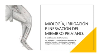
  • 1. MIOLOGÍA, IRRIGACIÓN E INERVACIÓN DEL MIEMBRO PELVIANO. IP. MVZ. Sebastián Ordóñez Ramírez. Esp. Pedagogía | Est. MSc Medicina veterinaria de pequeños animales | Mtr Medicina y cirugía de aves y animales exóticos | Mtr Neurología clínica.
  • 3. M. Glúteo medio. M. Glúteo superficial. M. Coccígeo. M. Bíceps femoral. M. Tensor de la fascia lata. Fascia lata.
  • 4. M. Gracilis. M. Bíceps femoral. M. Semitendinoso. M. Gastrocnemio lateral. Tendón calcáneo común. Fascia de la pierna. Fascia lata. M. Flexor digital superficial. M. Flexor digital lateral. Ligamento rotuliano.
  • 5. MÚSCULO. ORIGEN. INSERCIÓN. FUNCIÓN. INERVACIÓN. M. biceps femoral. Hueso sacro y pelvis. Rótula. Fascia de la pierna. Flexor de la articulación de la rodilla. Extensor del tarso. N. glúteo caudal. M. biceps femoral cabeza isquiática. Hueso sacro y pelvis Tendón calcáneo común. Abducción del miembro. N. tibial. M. separador caudal de la pierna. Ligamento sacrotuberoso. Fascia de la pierna. Abducción del miembro. Supinación del miembro. N. peroneo o fibular.
  • 6. M. Separador caudal de la pierna. M. Glúteo medio. M. Tensor de la fascia lata. M. Sartorio parte craneal. M. Semitendinoso. M. Semimembranoso. M. Aductor. Fascia lata. Ligamento rotuliano. M. Gastrocnemio.
  • 7. MÚSCULO. ORIGEN. INSERCIÓN. FUNCIÓN. INERVACIÓN. M. semitendinoso. Cabeza vertebral y cabeza pelviana. Borde craneal de la tibia. Tendón calcáneo común. Flexor de la rodilla. Extensor de la articulación coxal. N. glúteo caudal. N. tibial. Ms. semimembranoso. Cabeza pelviana. Cóndilos mediales del fémur y de la tibia. Extensor de la articulación de la rodilla. Aductor del miembro pelviano. N. glúteo caudal. N. tibial.
  • 8. M. Glúteo profundo. M. Piriforme. M. Sartorio parte caudal. M. Sartorio parte craneal. M. Gemelo craneal. M. Gemelo caudal. M. Cuadrado femoral. M. Aductor. M. Vasto lateral.
  • 9. M. Recto femoral. M. Vasto intermedio. M. Obturador interno. M. Obturador externo. M. Gracilis. M. Iliopsoas.
  • 10. MÚSCULO. ORIGEN. INSERCIÓN. FUNCIÓN. INERVACIÓN. M. obturador interno. Cara interna del agujero obturador. Fosa trocantérica. Supinación. N. isquiático. M. obturador externo. Cara externa del agujero obturador. Fosa trocantérica. Supinación. N. obturador. Ms. gemelos. Cabeza vertebral y cabeza pelviana. Borde craneal de la tibia. Tendón calcáneo común. Flexor de la rodilla. Extensor de la articulación coxal. N. isquiático. M. articular de la cadera. Cabeza pelviana. Cóndilos mediales del fémur y la tibia. Extensor de la articulación de la rodilla. Adductor del miembro pelviano. N. isquiático.
  • 11. MÚSCULO. ORIGEN. INSERCIÓN. FUNCIÓN. INERVACIÓN. M. pectíneo. Eminencia iliopubiana. Cresta troclear medial del fémur. Aducción. N. obturador. N. femoral. M. sartorio. Tuberosidad coxal. Cuerpo del ilion o tendón del M. psoas menor. Fascia de la pierna. Adelanta y aduce el miembro. N. femoral. M. gracilis. Aponeurosis en la sínfisis pelviana. Fascia de la pierna. Aducción. N. obturador. Ms. aductores. Cara ventral de la pelvis y en el tendón del M. gracilis. Borde medial del fémur. Aducción. N. obturador.
  • 12. M. Recto femoral. M. Vasto medial. M. Sartorio parte caudal. M. Sartorio parte craneal.
  • 13. MÚSCULO. ORIGEN. INSERCIÓN. FUNCIÓN. INERVACIÓN. M. cuadrado femoral. Hueso isquion. Fosa troncantérica. Supinación N. isquiatico. M. cuadriceps femoral. M. recto femoral. Cuerpo del ilion. Rótula. Tuberosidad de la tibia. Extensor de la articulación de la rodilla. N. femoral. M. vasto lateral. Cara lateral del fémur. M. vasto medial. Cara medial del fémur. M. vasto intermedio. Cara craneal del fémur.
  • 14. M. Sartorio parte caudal. M. Sartorio parte craneal. M. Pectíneo. M. Iliopsoas. M. Aductor. M. Gracilis. M. Obturador interno. M. Piriforme.
  • 15. M. Sartorio parte caudal. M. Sartorio parte craneal. M. Recto femoral (M. Cuadriceps femoral). M. Vasto medial (M. Cuadriceps femoral). M. Pectíneo.
  • 16. M. Tensor de la fascia lata. M. Pectíneo. M. Recto femoral (M. Cuadriceps femoral). M. Aductor. M. Semimembranoso. M. Semitendinoso. M. Vasto medial (M. Cuadriceps femoral).
  • 17. M. Vasto medial (M. Cuadriceps femoral). M. Semimembranoso. M. Semitendinoso. M. Gastrocnemio. M. Flexor digital superficial. M. Tibial craneal. M. Flexor digital profundo. M. Poplíteo.
  • 18. MÚSCULO. ORIGEN. INSERCIÓN. FUNCIÓN. INERVACIÓN. M. gastrocnemio. Parte distal del fémur. Tuberosidad del calcáneo (tendón de Aquiles). Extensor del tarso. Flexor de la rodilla. N. tibial. M. soleo. Peroné o fíbula. Tendón de Aquiles. Extensor del tarso. N. tibial. M. flexor digital superficial. Fosa supracondílea. Tuberosidad supracondílea lateral. Falange media. Flexor del tarso y de los dedos. Extensor del tarso. Aducción. N. tibial. Ms. flexor digital profundo. N. tibial.
  • 19.
  • 20.
  • 21. M. Recto femoral (M. Cuadriceps femoral). M. Vasto medial (M. Cuadriceps femoral). M. Poplìteo. M. Vasto lateral. (M. Cuadriceps femoral).
  • 22. M. Glúteo medio. M. Glúteo superficial. M. Biceps femoral. M. Semimembranoso. M. Semitendinoso. M. Gracilis. M. Gastrocnemio.
  • 23. M. Obturador interno. M. Obturador externo. M. Semimembranoso. M. Semitendinoso. M. Aductor. M. Gracilis.
  • 24. M. Obturador interno. M. Obturador externo.
  • 25. M. Peroneo largo. M. Flexor digital medial. M. Tibial craneal. M. Extensor digital largo. M. Gastrocnemio (cabeza lateral). M. Flexor digital lateral.
  • 26. M. Extensor digital corto. M. Flexor digital superficial. M. Peroneo largo. M. Extensor digital largo. M. Flexor digital lateral.
  • 27. MÚSCULO. ORIGEN. INSERCIÓN. FUNCIÓN. INERVACIÓN. M. tibial craneal. Cóndilo lateral de la tibia. Medialmente en el taso y en la parte proximal del metatarso. Flexor de la articulación del tarso. N. peroneo. M. flexor digital medial. Tibia Tendón común del flexor profundo. Falange distal. Flexor de los dedos. N. tibial. M. flexor digital lateral. Peroné y tibia. Tendón común del flexor profundo. Falange distal. Flexor de los dedos. N. tibial. Ms. peroneo largo. Peroné y cóndilo lateral de la tibia. Medialmente en el tarso y en la parte proximal del metatarso. Flexor de la articulación del tarso. N. peroneo.
  • 28. M. Extensor digital largo. M. Extensor digital corto. Tendón del M. Extensor digital lateral. M. Extensor digital largo del dedo I.
  • 29. M. Extensor digital largo del dedo I. M. Extensor digital lateral. M. Peroneo corto. M. Extensor digital corto.
  • 30. M. Poplíteo. M. Gastrocnemio (cabeza medial). M. Tibial craneal. M. Flexores digitales profundos.
  • 31. M. Gastrocnemio (cabeza lateral). M. Gastrocnemio (cabeza medial). M. Flexores digitales profundos. Tendón del M. Flexor digital superficial.
  • 32. M. Flexor digital lateral. M. Flexor digital medial. M. Poplíteo. Tendón del M. Flexor digital superficial. Tendón del M. Flexor digital profundo.
  • 33.
  • 34. M. Flexor digital lateral. M. Flexor digital medial.
  • 35. MÚSCULO. ORIGEN. INSERCIÓN. FUNCIÓN. INERVACIÓN. M. poplíteo. Cóndilo lateral del fémur. Borde medial de la tibia. Flexor de la articulación de la rodilla. Prondor. N. tibial. M. peroneo corto. Peroné o fíbula. Metatarsiano V Flexor del tarso. N. peroneo. M. tercer peroneo. Fosa extensora del fémur. Huesos del tarso y metatarsianos. Flexor del tarso. Extensor de la articulación de la rodilla N. peroneo. M. tibial caudal. Peroné y tibia. Tendón común del flexor profundo. Falange distal. Flexor de los dedos.. N. tibial.
  • 36. MÚSCULO. ORIGEN. INSERCIÓN. FUNCIÓN. INERVACIÓN. M. extensor digital largo. Fosa extensora del fémur. Apófisis extensora de la falange III. Extensor de los dedos. Extensor de la articulación de la rodilla. N. peroneo. M. extensor digital lateral. Peroné y cóndilo lateral de la tibia. Falange II del dedo V o IV, y apófisis extensora de la falange distal. Extensor de los dedos. N. peroneo. M. extensor largo del dedo I. Peroné o fíbula. Dedo II. Extensor del dedo II. N. peroneo.
  • 37. Tendón del M. Extensor digital largo. Tendón del M. Peroneo largo. Tendón del M. Extensor digital lateral. M. Interóseo del dedo V Tendón del M. Extensor digital superficial.
  • 38. Tendón del M. Extensor digital lateral. M. Extensor digital corto. Tendón del M. Extensor digital largo. Tendón del M. Tibial craneal.
  • 40. M. Flexor digital profundo. M. Flexor digital medial. M. Flexor digital superficial.
  • 41.
  • 42.
  • 43.
  • 45. L1 = N. Iliohipogástrico craneal. • M. Cuadrado lumbar. • M. Psoas menor. • Ms. Abdominales. L2 = N. Iliohipogástrico caudal. L3 = N. Ilioinguinal. • Región inguinal. • Cara cráneo lateral región femoral. L4 = N. Cutáneo femoral lateral. • Cara lateral región femoral y de la rodilla. L3 – L4 = N. Genitofemoral. • M. Cremaster. • Región femoral caudo medial. • Prepucio/ Gs Mamarias caudales. L4 – L5 – L6 = N. Femoral. • M. Cuadriceps femoral. • M. Sartorio. • M. Safeno. • Caras mediales región femoral, tibial y pie. L5 – L6 = N. Obturador. • M. Obturador externo. • Ms. Aductores. • M. Pectíneo y Gracilis.
  • 46.
  • 47.
  • 48.
  • 49. A. ILIACA CIRCUNFLEJA PROFUNDA. A. ILIACA EXTERNA. A. FEMORAL PROFUNDA. A. FEMORAL CIRCUNFLEJA MEDIAL. A. EPIGASTRICA CAUDAL. A. PUDENDA EXTERNA. A. MAMARIAS / ESCROTALES. A. CRANEAL DEL PENE. A. EPIGASTRICA CUADAL SUPERFICIAL. TRONCO PUDENDO EPIGASTRICO. A. POPLÍTEA. A. FEMORAL. A. TIBIAL CRANEAL. A. TIBIAL CAUDAL.
  • 50. A. FEMORAL. A. FEMORAL CAUDAL. A. SAFENA. A. GENICULAR DESCENDENTE. RAMA CRANEAL RAMA CAUDAL (A. TIBIAL RECURRENTE). A. PLANTAR MEDIAL. A. PLANTAR LATERAL. RAMA PROFUNDA. RAMA SUPERFICIAL (A. DIGITAL PLANTAR MEDIAL)
  • 51.
  • 52.
  • 53.
  • 54.
  • 55. BIBLIOGRAFIA. • Budras, K. D., Habel, R. E., Mülling, C. K. W., Greenough, P. R., Jahrmärker, G., Richter, R., & Starke, D. (2011). Bovine Anatomy. In Bovine Anatomy. https://doi.org/10.1201/9783842683594 • Budras, K. D., Habel, R. E., Mülling, C. K. W., Greenough, P. R., Jahrmärker, G., Richter, R., & Starke, D. (2012). Anatomy of the horse. In Veterinary Record (Vol. 170, Issue 1). https://doi.org/10.1136/vr.e16 • König, E., & Liebich, H. (2005). Anatomía de los animales domésticos: texto y atlas. In Panamericana (Vol. 2, p. 11). http://bit.ly/3jYovk6 • Mccarthy, K. B. P. H., Horowitz, A., Berg, R., & Fricke, W. (1891). The anatomy of the dog. In Nature (Vol. 45, Issue 1149). https://doi.org/10.1038/045016a0