2. • There is no lovelierway to thank god for u’r sight than by
giving a helpinghand to those in the dark……
Helen Keller
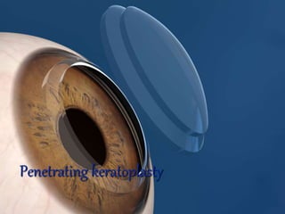
3. history
• F.Reisinger:term keratoplasty
• First succesful pkp was performed by
edward konrad zirm in 1906.
• V.p philatov:father of modern eye banking.
• Ramon castroviejo was another pioneer
• McCarrey& H.Kaufman :storage media
4. aims
• Ability to see with acceptable optic
correction
• Restore binocularity
• Elimination of corneal disease
• Improvement in function& life style
• Relieve pain
8. • Chemical injuries
• Mechanical trauma,non surgical
• Regraft due to allograft rejection
• Regraft non related to allograft
tissue failure
vitreo corneal touch
rec stromal dystrophy
pseudophakic corneal oedema
• Other causes
silicon oil keratopathy
uveitis
9. PROGNOSIS
• Grade 1:excellent prognosis
90% success
central corneal disease.normal
periphery,sensation&tear film
normal,limbus normal
• Grade 11:very good prognosis[80-90]
.involve part or whole of corneal
periphery with minimal vascularisation,<2
quadrants
10. • Grade 111 :fair prognosis{50-80}
extremes of corneal disease involving large
part of limbus
• Grade 1V:poor prognosis{,50%}
absence of nl limbal stem cells&nl corneal
epithelial maturation
11. CONTRA INDICATION
• Only absolute CI is no pl
• Relative
.ambulatory vision in the eye with better
vision
.no tear film,bad ocular surface
.multiple graft failure
.inacccurate PR with underlying RD
12. PRE OP EVALUATION
• General history
• Ocular history
• Ocular examination
• s/l examination
• Iop
• Fundus{if possible}
• Vision& refraction
• Tear film status
• Keratometry
• Specular microscopy
• Usg
• Gonio{optional}
13. Contra indication for donor use
• Death of unknown cause
• Death from cns disease of unexplained
diagnosis
• Viral:CJD,HIV,rabies etc
• Active septiceamia
• Active bact or fungal endocarditis
• Leukemia,lymphoma
• Intrinsic eye disease
• Prior intraocular surgery
14. Donor tissue evaluation
• Gross examination
• s/l
• Specular
Criteria to consider cornea unsatisfactory
• Very low endothelial cell count <1500
• Extreme polymegathism or polymorphism
• Presence of significant cornea guttae
• Very severe oedema
• Presence of infl cells on endothelium
15. Surgical instruments
• For globe exposure
• To cut recipient& donor corneas
• To secure donor &to remove &replace lens
implants
• For maintanence& re construction of AC
16. Globe exposure
• eye speculum
• globe supporting rings
used to maintain corneal architecture after
removing corneal button as in aphakic,paediatric
eyes.measured to size slightly less than inter
palpebral opening.suutured with 7.0 vicryl to 50%
scleral thickness.ex;filieringa’s ring
18. • Corneal trephine: used to create circular
corneal incision
• Criteria for a good trephine
should be available in various sizes
should be comfortable to use &stable
during cutting
should have a clear view of the
cornea within and around the trephine
while cutting
should be able 2 rotate in a direction
perpendicular 2 axis of trephine
19. • 5 types
hand held
mechanized
suction fixation type
special purpose
skin biopsy punches
non contact trephination
20. Hand held
• Varying sizes—3to17mm
• Some cases there is a central
obturator which can be
adjusted to select depth of corneal cut.
disadv:central view is obstructed.
21. 2. mechanized trephine
cutting blade is driven
by motor present in the main body
&hav a circular trephine at the motor
shaft end.used to trephine recipient cornea
Disadv:corkscrew edge effect
Adv:rapid breaking within 0.1 sec,less stromal
disruption&smoother interface
22. • 3.suction fixation trephine
outer corneal suction
ring for fixation&
Inner circular cutting
blade which is sharp
ex: hassburg baron vacuum trephine.
used to cut recipient cornea
Adv:low cost,consistency of cut,control of
depth
Disadv:;tendency to undercut&increased
endothelial damage
23. • Olson calibrated corneal trephine
used to cut both recipient&
donor cornea.
4.Special purpose trephine
used in case of optical zone laceration in recipient
cornea
5.Skin biopsy punches :used in small
Patch graft.size 2-2.5mm.used 2 cut both donor
&recipient cornea
• Single point cutting trephine
fixate at limbus ,so less corneal
distortion
24. • Combination corneal trephine
Salient characteristics of above
Ex;hannah trephine system
2 parts:limbal suction ring system&
A mechanical trephine fitted with
suction ring which fixates on cornea.
• Non contact trephination.
using laser
adv:better visualisation,centration&less
topographic distortion
Disadv:cost&endothelial injury
25. • Globe fixation
gauze piece
tudor thomas stand
• Cutting blocks
paraffin,teflon,polycarbonated nylon
block
• Corneal endothelial punch
to cut cornea from endothelial
side.ex ..cottingham corneal punch
Troutmann,liebermann gravity action punch
Adv:sharp vertical cuts without bevelling
26. • Cutting instruments
• Grasping
• Holding
• Sutures:
nylon is the suture of choice b’coz of low
tissue reactivity.tensile strength>I yr
• Needles:
full curve
mini curve
bicurve
compound curve
27. techniques
Harvesting donor cornea
donor button is cut b’fore recipient
Size of graft
1. if diameter of recipient bed is>9 or
<7mm—graft larger by 1mm
2,btw 7-9mm-
aphakic….0.5mm
pseudophakic or
phakic….0.25mm
28. Harvesting donor tissue from whole globe
using hand held or suction fixaton trephine
donor graft cut from epithelial side.globe is
held in non dominant hand&trephine in dominant
hand,,using counter pressure with one hand
,trephine is firmly placed on the centre,rotate with
fingers &exert downward pressure
. release of pressure is
noted when AC is entered.
.finish cut with scissors
29. • Trephination from preserved corneo scleral
button
cut from endothelial side.--- with hand
held trephine or with endothelial punch.
Hand held trephine …donor button is
kept over a cutting block.cut by
punching¬ rotation.audidle click
indicates
complete cut
Using endothelial punch
30. • Trephination using artificial AC maintainer
a drop of visco is applied on the
endothelial side& then endothelium is kept
on AC maintainer.air is used 2 create AC.
Suction ring is placed over donor cornea&
then activate by releasing the syringe.
Following placement of trephine on the
suction ring,the lever
of suction ring is pressed
to lower blade onto donor
tissue&trephine is turned
to complete the cut
31. • Non mechanical laser trephination
. from epithelial side.
..using 193 nm excimer laser
…app 11000 laser pulses used 2
perforate cornea
….avoids mechanical disruption
during trephination&giv smooth
perpendicular edges
32. recipient
• Anaesthesia
• Paint &drape
• Exposure
• Placement of scleral fixation ring if needed
• Marking the host cornea
centering of graft is imp as decentration
can lead to graft rejection
& high post op astig.suture
marker stained with gentian
violet for exact suture placement
33. Trephination of recipient cornea
• Size of graft depends on dimeter of pt’s
cornea,extent of corneal disease ,etc…
• Too small graft{<6.5mm} –high post op
astigmatism
• Large graft:risk of immunologic reaction more
• Routinely uses 7-8mm
• Larger graft in infectiouskeratitis, keratoconus,
fuch’s &bullous keratopathy
• Conventional hand held or suction and automatic
trephines can be used
34. With hand held trephine
trephine held perpendicular to cornea,
align the centration mark on the cornea with
centre of the blade.trephine is rotated btw
thumb& forefinger maintaining a
downward pressure. Escape of aqueous is
noted 2 ensure full thickness incision.visco
elastics injected 2 deepen AC. Corneal
scissors used 2 complete the cut.
Stabilise the cornea with forceps
once half is cut
.edge is trimmed
35. With suction trephines
this fix the cornea with suction during
trephination.so useful in perforated corneas as less
AC collapse &corneal distortion.
Keep the trephine in zero position. blade is
then retracted by 0.18mm by turning the spoke 3
timesplunger is pressed,cross hairs of trephine
centered,plunger released abruptly.if suction was
there plunger stops at 4ml mark
.stabilise the trephine Blade
returned to 0 position.then no of spokes
depending on desired depth is turned.
then release plunger.
36. Non mechanical trephination
• using 193 nm excimer laser
• 7000 pulses required for focal corneal
perforation.
• femto second laser can be used.set at 850
µm.for safety 70µm of post stroma is
retained un cut.ac entered.then complete the
cut.
37.
38. • Suturing:
place donor cornea on recipient.first place
inf edge.ac is maintained with visco.
Placement of cardinal sutures.
1st at 12’o clock
11nd at 6’o clock.
1mm on both sides.
3rd&4th --90° from first two
in donor graft passed just ant to DM
suture tied with a triple throw followed by 2
single throw.
tension on the cardinal suture—a diamond
shaped bow appears after placement
39. • Placement of other sutures
.interrupted
..combined interrupted& continuous
…single continuous
….double continuous
40. • Interrupted
in infants&children
in highly vascularised cornea
in therapeutic keratoplasty
Total 16 sutures with 2nd four equidistant
btw first four.
Second eight equidistant btw the first eight
• Combined continuous
bites of continuous
placed btw each of the
interrupted.90-95% depth
41. • Single continuous
after 4 cardinal suture,24 bite
continuous suture.then put a
temporary knot at 12’0 c lock.
cardinal sutures are removed.
put permanent knot
• Double continuous
12 bite continuous suture
with knot at 12‘0 clock.second
continuous suture of 50-60%
depth btw earlier bites
42. • Check for wound leak
• Intra op suture adjustment 2 reduce
astigmatism
intra op keratometer
alternative; safety pin
• S/C genta&dexa
• P&B for 24 hrs
43. Triple procedure
Pkp& cataract extraction
.
Cataract removed when VA is<20/50
Nuclear sclerosis>gr11
PSC
ADVANTAGE: needs only one procedure
Offers imm visual improvement
Less cost
Less risk 2 transplanted corneal
endo due to 2nd surgery
44. • Dis advantage
iol power calculation difficult
prolonged surgery time
Specific indications
fuch’s
if endo cell count<1000/mm²
corneal thickness >0.62mm
obvious corneal changes or s/l like
guttae,stromal oedema
cornea of fellow eye decompensated
45. Herpetic keratitis
Bacterial kerayitis
Chemical burns
Corneolenticular trauma
Corneal opacity in eelderly patient
contraindication
Perforated corneal ulcers
Interstitial keratitis with meltingcornea
Ocular cicatrical pemphigoid
Rec mod to severe uveitis
46. Surgical techniques
• Simultaneous ecce with pkp
• Phaco followed by pkp
• Temp graft for closed system cat surgery
• Temp keratoprosthesis to perform sics
48. • Phaco followed by pkp
adv:less pc rent&choroidal hemorrrhage
scleral tunnel preferred
Nucleotomy done by 4 quqdrqnt technique
• Temporary graft for closed system cat
surgery
• Temporary keratoprsthesis to perform sics
49. Pkp for bullous
keratopathy&iol exchange
• Detaied history&examination
• i/v mannitol prior to surgery
• Insert scleral ring
• use relatively large graft
• In aphakia can put aciol
• In pseudophakia decide onexplanation
/exchange of iol
50. Indications for iol exchange
• Poorly controlled glaucoma
• Rec hypheama
• Metal clips or loops on iol
• Any closed loop AC iol
• Iris supported iol with optic infront of iris
iol retained
• When correct diopric power
• Stable¬ touching corneal endothelium
• Not asso with inflammation or pas or cme or
vitreous in ac
51. Technique of exchange
• Remove recipient cornea
aciol
• Amputation of haptic
• Haptic rotated&then removed
• If any vitreous adhesion snip it off
Pciol
• Identify haptic
• Using sinsky’s hook iol is rotated to release
adhesions
52. Anterior segment reconstruction
• Gonioplasty:
viscodissection
using sinsky’s hook
iridotomy
• Iridoplasty ; to pull iris from angle
• Iol implantation
53. Paediatric keratoplasty
Indications
• Congenital
cong sclerocornea
cong hereditary endothelial dystrophy
peter’s anomaly
glaucoma with corneal oedema
muco polysaccharidosis
• Acquired traumatic
• Acquired non traumatic
herpes,bacterial,fungal keratitis
steven johnson
ophthalmia neonatorum
interstitial keratitis
54. • Pre-op eveluation
• EUA—3-6wks
• Pre op conselling
• Timing of surgery-
in neonatal glaucoma—not before 2 mths
usually at 8-12wks
2nd____2-3 mths after 1st
• Preop mannitol
• Donor age__4-19yrs
• Donor conea 1/2mm larger
55. • Take care that no adhesion btw iris&cornea
• If adhesions remove with cyclodialysis
spatula
• Visco separation also done
• Cornea trephined
• Always expect high positive vitreous
pressure
• So remove& replace cornea fast.
56. • If rapid &immediate bulge;each quadrant
after removal ,suture back with 7’0
silk.after3,0 quad removal,put visco on
surface.donor cornea placed over visco.two
9.0 sutures are passed 180° apart.last quad
is cut.7’0 removed.6& 12’oclock sutures
tightened.host cornea slowly removed.instill
atropine.put rmaining sutures.
• If delayed bulge: app 270° of cornea
excised.put visco over it.donor slid on top of
pt’s cornea.sutured with 10’0 at 6& 12.host
cornea then cut& slid out
57. keratoconus
• Penetrating keratoplasty
• Mushroom/hat/doublepunc keratoplasty
• Total 9mm graft with central 5-6mm full
thickness &periphery lamellar 200µm
thickness.
• femto sec laser assisted surgery
• Adv;less post op astigmatism
improved wound healing
faster visual recovery
less rejection
58. Mushroom shaped penetrating
Keratoplasty
Indication and Basic operation method
healthy recipient’s peripheral endothelium with
large stromal disease
Prepared
Donor cornea
Stepwise
Trephinized
Recipient
Cornea