
Recommended
More Related Content
Similar to Lecture 3- Anatomy of the Pleura and Lung
Similar to Lecture 3- Anatomy of the Pleura and Lung (20)
Recently uploaded
Recently uploaded (20)
Lecture 3- Anatomy of the Pleura and Lung
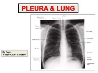
- 1. By Prof. Saeed Abuel Makarem
- 2. Pleura الرئوي الغشاء • Double-layered serous membrane enclosing the lung. • Has two layers: – Parietal layer, which lines the thoracic walls. – Visceral layer, which covers the surfaces of the lung. • The two layers continue with each other around the root of the lung الصوره انظر, where it forms a loose cuff hanging down called the pulmonary ligament. • The space between the two layers, the pleural cavity, contains a thin film جدا دقيق of pleural fluid ( 5-10 ml.). Root of the lung: واالوعيه الشرايين دخول منطقه المنطقه هذه وفي للرئه والقصبات الرئوي الغشاء طبقتي التحم يتم واحده بطبقه Pulmonary ligament
- 3. Parietal Pleura • Divided according to the region in which it lies and the surfaces it covers, into: 1- Cervical عنقي 2- Costal 3- Mediastinal 4- Diaphragmatic القادمه بالشرائح مفصال تشرح
- 4. Parietal Pleura • Cervical Pleura: • Projects up تبرز into the neck about 1-1.5 inches above the medial1/3rd of clavicle. • It lines the under surface of the suprapleural membrane الرئوي الغشاء فوق غشاء. • Costal pleura: • lines, the back of the: • 1- sternum, • 2- Ribs & costal cartilages, • 3- Intercostal spaces & • 4- Sides of vertebral bodies
- 5. Parietal Pleura • Mediastinal pleura: covers the mediastinum الصوره انظر. • At the hilum دخول منطقه االوعيه ( شرحها سبق ) , it is reflected on to the vessels and bronchi, to become continuous with the visceral pleura • Diaphragmatic pleura: covers the thoracic (upper) surface of the diaphragm
- 6. Pleural Recesses : تجويفات • 1- Costodiaphragmatic: • Slit عميق دقيق تجويف like space between costal and diaphragmatic pleurae. The inferior border الحقا ياتي of the lung descends ينزل into it during deep inspiration • 2- Costomediastinal: • Slit like space between costal and mediastinal pleurae, is filled by the anterior border of the lung during deep inspiration
- 7. Costophrenic حاله الصوره الزايه الن مرضيه هواء يصلها ال
- 8. Pleura: Nerve Supply • Parietal pleura: • is sensitive to pain, temperature, touch & pressure and is supplied as follows: Costal pleura segmentally supplied by the intercostal nerves. Mediastinal pleura supplied by phrenic nerves. Diaphragmatic pleura supplied over the domes by phrenic nerves, around the periphery by lower six intercostal nerves. • Visceral pleura sensitive to stretch only and is supplied by the autonomic fibers from the pulmonary plexus
- 9. Lungs
- 10. Lungs • Located in the thoracic cavity, one on each side of the mediastinum • Each lung is: Conical in shape. Covered by the visceral pleura. Suspended معلقه free in its own pleural cavity. Attached to the mediastinum only by its root.
- 11. Borders حدود معناها يعني الرئه اطرافها • Each lung has: 1- A thin anterior امامي border that overlaps the heart. The left lung shows cardiac notch along this border. 2- A thick posterior خلفي border that lies beside the vertebral column. 3- A thin inferior سفلي border, that is related to diaphragm cardiac notch
- 12. Surfaces يعني الرئه سطوح الحدود غير الباقيه االجزاء Each lung has: • An apex قمه, which projects upward into the neck for about 1 inch يتمد الغالف تذكر ونصف النش above the clavicle. • A concave base, which rests on the diaphragm. • A convex costal surface, which corresponds to the concave chest wall.
- 13. • A mediastinal surface, which is molded تطبع عليها ( واللي الصوره مثل بالعملي ورونا ) to the mediastinal structures. • At bout مرض - نوبه the middle of this surface is a depression تنضغط, the hilum, where the structures enter the lung: • (bronchi, bronchial & pulmonary arteries) or leave (bronchial & pulmonary veins, nerves & lymphatics)
- 14. Mediastinal surface االجزاء للرئه المجاوره والتي اليمنى كم عليها تطبع ا المربعات في المرقمه of the right lung 1 2 3 Right atrum 4 تحفظ مهمه فصوص ثالث بها اليمنى الرئه المرقمه الدوائر في كما 1 2 3
- 15. Mediastinal surface of the left lung 1 2 Left ventricle اللسان : يوجد فقط اليسرى بالرئه القلب وجود بسبب نوتش كارديك : يوجد فقط اليسرى بالرئه القلب وجود بسبب 3 لها اليسرى الرئه فقط فصين 2 1
- 16. Fissures الفاصل الخط & Lobes الفصوص • Right Lung: • Divided by two fissures, the oblique & horizontal, into: • 1- Superior, • 2-Middle and • 3- Inferior • Left lung: • Divided by only one oblique fissure into: • 1- Superior and • 2- Inferior
- 17. Fissures • Oblique fissure: • Runs from the inferior border upward and backward across the medial and costal surfaces until cuts the posterior border about 2½ inches below the apex • Horizontal fissure: runs horizontally across the costal surface at the level of right 4th costal cartilage to meet the oblique fissure in the midaxillary الخط االبطي الفاصل line
- 18. Blood Supply للرئه • Arterial supply: • By bronchial arteries, branches of the descending thoracic aorta. • Venous Drainage االورده: • By bronchial veins, which drain into azygos & hemiazygos veins.
- 19. Nerve Supply • Through pulmonary plexuses located at the root of each lung, and composed of: – Sympathetic fibers from the sympathetic trunk – Parasympathetic fibers from the vagus nerve
- 20. Root of the Lung • Formed by the structures entering or leaving the lung: bronchi, pulmonary vessels, lymphatics, bronchial vessels and nerves. • Surrounded by a tubular sheath of pleura which hangs down to form pulmonary ligament. • The pulmonary ligament provides a potential space for the movement of pulmonary vessels and large bronchi
- 21. Surface Anatomy of Pleurae & Lungs للرئه الكتاب اقرا صفحه 68 اقرا الرئوي وللغشاء صفحه 72
- 24. توضح صوره النفس سماع اماكن والصورة للرئه نفسها تشرح مهمه
- 25. تابع
- 26. Bronchopulmonary segments: للرئه الشعب تدخل عندما ش سيقمنت لكل ان فائدتها السيقمينت هذه تكون بالرئه ترتبط عندما بالرئه ترتبط حتى تتفرع ريان الرئه عمل على تؤثر وال ازالتها يمكن به مشكله حدوث فعند به خاص وعصب • Definition: Are the smallest anatomic, surgical, and functional units of the lung. Each segment is pyramidal in shape with its apex directed medially toward the lung root, and its base toward the lung surface. Each segment receives segmental bronchus, branch of pulmonary artery, its own lymphatic vessels, and autonomic nerve. The branch of pulmonary vein lie in the connective tissue between the segment.
- 29. Clinical Notes • Pleural effusion • Pleuritis, pleural rub, pleural adhesions • Pneumothorax • Empyema • الدرس باشرحها الدكتور قال الجاي