
Recommended
Recommended
More Related Content
Similar to Intro to Soft x ray vector ptycho tomography.pptx
Similar to Intro to Soft x ray vector ptycho tomography.pptx (20)
Recently uploaded
Recently uploaded (20)
Intro to Soft x ray vector ptycho tomography.pptx
- 1. Rana†, Liao†, et al. Nature Nanotechnology 18, 227 (2023). Soft x-ray vector ptycho-tomography: A quantitative vector nanoimaging method for spin textures in 3D
- 2. Soft x-ray vector ptycho-tomography: a new quantitative vector nanoimaging method for spin textures in 3D Chen-Ting Liao1,2,*, Arjun Rana2,3, Ezio Iacocca4,5, Ji Zou3, Minh Pham2,6, Xingyuan Lu3,7, Emma-Elizabeth Cating Subramanian1,2, Yuan Hung Lo2,3, Sinéad A. Ryan1,2, Charles S. Bevis1,2, Robert M. Karl Jr1,2, Andrew J. Glaid8, Jeffrey Rable8, Pratibha Mahale8,9, Joel Hirst10, Thomas Ostler10,11, William Liu2,3, Colum M. O’Leary2,3, Young-Sang Yu12, Karen Bustillo13, Hendrik Ohldag12, David A. Shapiro12, Sadegh Yazdi14, Thomas E. Mallouk8,9, Stanley J. Osher2,6, Henry C. Kapteyn1,2, Vincent H. Crespi8, John V. Badding8, Yaroslav Tserkovnyak3, Margaret M. Murnane1,2, and Jianwei Miao2,3 1JILA and Department of Physics, University of Colorado and NIST, 440 UCB, Boulder, Colorado 80309, USA; 2STROBE Science and Technology Center, USA; 3Department of Physics & Astronomy and California NanoSystems Institute, University of California, Los Angeles, CA 90095, USA; 4Department of Mathematics, Physics, and Electrical Engineering, Northumbria University, Newcastle upon Tyne, NE1 8ST, U.K.; 5Center for Magnetism and Magnetic Materials, University of Colorado, Colorado Springs, CO 80918, USA; 6Department of Mathematics, University of California, Los Angeles, CA 90095, USA; 7School of Physical Science and Technology, Soochow University, Suzhou 215006, China; 8Departments of Chemistry, Physics, Materials Science and Engineering and Materials Research Institute, Penn State University, University Park, PA 16802, USA; 9Department of Chemistry, University of Pennsylvania, Philadelphia PA 19104, USA; 10Materials and Engineering Research Institute, Sheffield Hallam University, Howard Street, Sheffield S1 1WB, U.K.; 11Department of Physics and Mathematics, University of Hull, Cottingham Road, Hull, HU67RX, U.K.; 12Advanced Light Source, Lawrence Berkeley National Laboratory, Berkeley, CA 94720, USA; 13National Center for Electron Microscopy, Molecular Foundry, Lawrence Berkeley National Laboratory, Berkeley, CA, 94720, USA; 14Renewable and Sustainable Energy Institute, University of Colorado, Boulder, CO 80309, USA.
- 3. ABSTRACT Understanding topological spin textures is important because of scientific interests and technological applications. However, observing nanoscale magnetization and mapping out their interactions in 3D have been challenging – due to the lack of nondestructive vector nanoimaging techniques that penetrate thick samples. Recently, we developed a new characterization technique, soft x-ray vector ptycho-tomography, to image spin textures with a 3D vector spatial resolution of 10 nm. Using 3D magnetic metamaterial as an example, we demonstrated the creation and observation of topological magnetic monopoles and their interactions. We expect this method to be applied broadly to image vector fields in magnetic samples and beyond. Keywords: topological spin texture, Skyrmions, soft x-ray, 3D vector imaging, ptychography, tomography, coherent diffractive imaging, computational imaging
- 4. INTRODUCTION Probing and mapping topological spin textures such as magnetic Skyrmions and hedgehogs is of paramount importance. In fact, topological magnetic whirls not only reveal new physics, but they have also risen and become leading candidates for next-generation information encoding and storage platforms. Within the last decade, it has been demonstrated that nanoscale topological spin textures in various materials at room temperature can be experimentally created, manipulated, and erased1,2. However, observing the magnetization directly and mapping out their interactions at 10s of nanometer scales has been challenging. This is due to the lack of high-resolution (<20 nm), nondestructive 3D imaging techniques that penetrate relatively thick (>100nm) samples.
- 5. METHODOLOGY Recently, we developed a new imaging technique, soft x-ray vector ptycho- tomography, to characterize spin textures with a 3D vector spatial resolution of 10 nm3. This new coherent x-ray imaging method combines x-ray magnetic circular dichroism (XMCD), ptychography (a scanning coherent diffractive imaging method), scalar tomography based on a new algorithm called RESIRE4, and vector tomography, to quantitatively image and reconstruct spin textures. This new method contrasts with the 3D vector imaging methods using Maxwell's equations as a constraint5,6. By measuring diffraction patterns with high differential magnetic contrast at the L3-edge resonance of transition metals, we improved the spatial resolution close to the magnetic exchange length of transition metals, representing a significant advance of the resolution over previous soft and hard x-ray vector tomography methods7,8.
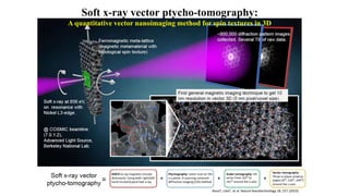
- 6. Figure 1. Schematic experiment setup for soft x-ray vector ptycho-tomography and its results. Circularly polarized soft x-ray (purple) at 856 eV (λ ~ 1.45 nm) is on resonance with nickel L3-edge and illuminates the sample (gray). The topological magnetic metamaterial sample is a ferromagnetic meta-lattice, which is made of fcc-packed 60nm silica nanospheres with infiltrated nickel in-between the nanospheres. An x-ray camera collects coherent diffractive images (~800,000 diffraction patterns in total, several TB of raw data). Bottom insets: after reconstructions, 3D scalar images (gray, representing nickel) and spin textures (red and blue arrows, representing magnetization fields in nickel) can be observed. Image credit: the artistic rendering is done by Steven Burrows at JILA, based on experimentally measured and reconstructed data.
- 8. RESULTS To showcase the method, we demonstrated the creation and observation of topological magnetic monopoles (TMMs) and their interactions in a nanoscale magnetic metamaterial3,9,10. More than 100 stable TMMs are created in a frustrated ferromagnetic meta-lattice. These field-free, room-temperature TMMs do not possess strong anisotropy or the Dzyaloshinskii-Moriya interaction (DMI). The complex 3D curved surfaces of the nanospheres in the meta-lattice create a magnetically frustrated configuration harboring topological spin texture.
- 9. CONCLUSIONS Our work demonstrates that magnetic meta-lattices could be used as a new platform to create and investigate the interactions of TMMs. Furthermore, we expect soft x-ray vector ptycho-tomography to be applied broadly to quantitatively image 3D vector fields in magnetic and anisotropic nanomaterials based on facility-scale (synchrotrons)11,12 and table-top (laser-driven high harmonic generation) 13,14 coherent soft x- ray sources. ACKNOWLEDGEMENTS This work was primarily supported by STROBE: a U.S. National Science Foundation Science and Technology Center under award DMR1548924. Soft X-ray ptycho-tomography experiments were performed at COSMIC and used resources of the Advanced Light Source, which is a U.S. Department of Energy Office of Science User Facility under contract number DE-AC02-05CH11231.
- 10. REFERENCES 1. Everschor-Sitte, K. et al. "Perspective: Magnetic skyrmions—Overview of recent progress in an active research field." Journal of Applied Physics 124.24 (2018). 2. Göbel, B. et al. "Beyond skyrmions: Review and perspectives of alternative magnetic quasiparticles." Physics Reports 895, 1-28 (2021). 3. Rana†, A., Liao†, C.-T. et al. "Three-dimensional topological magnetic monopoles and their interactions in a ferromagnetic meta-lattice." Nature Nanotechnology 18, 227 (2023). https://doi.org/10.1038/s41565-022-01311-0 4. Pham, M. et al. "Accurate real space iterative reconstruction (RESIRE) algorithm for tomography." Sci. Rep. 13, 5624 (2023). 5. Phatak, C. et al. "Three-dimensional study of the vector potential of magnetic structures." Phys. Rev. Lett. 104, 253901 (2010). 6. Davis, Timothy J. et al. "Ultrafast vector imaging of plasmonic skyrmion dynamics with deep subwavelength resolution." Science 368.6489, eaba6415 (2020). 7. Donnelly, C. et al. "Three-dimensional magnetization structures revealed with X-ray vector nanotomography." Nature 547, 328 (2017). 8. Donnelly, C. et al. "Time-resolved imaging of three-dimensional nanoscale magnetization dynamics." Nature Nanotechnology 15, 356 (2020). 9. Han, J. E. and Crespi, V. H. "Abrupt Topological Transitions in the Hysteresis Curves of Ferromagnetic Metalattices." Phys. Rev. Lett. 89, 197203 (2002). 10. Liu, Y. et al. “Confined chemical fluid deposition of ferromagnetic metalattices.” Nano Lett. 18, 301 546 (2018). 11. Shapiro, D., et al. "The COSMIC Imaging Beamline at the Advanced Light Source: A new facility for spectro-microscopy of nano-materials." Microscopy and Microanalysis, 24, 8-11 (2018). 12. Lo, Y. H., Liao, C.-T. et al. "Multimodal x-ray and electron microscopy of the Allende meteorite." Science Advances, 5(9), eaax3009 (2019). 13. Shi, X., Liao, C.-T. et al. "Attosecond light science and its applications for probing quantum materials." Journal of Physics B: Atomic, Molecular and Optical Physics 53, 184008 (2020). 14. Loetgering, L. et al. "Advances in laboratory-scale ptychography using high harmonic sources." Optics Express 30, 4133 (2022).