HCSPHARMA Importance of microenvironment in cerebral in vitro models for phenotypic screening

Aim: About 90% of drug-candidates failed in clinical trials, in particular in neurology, due to a lack of efficacy. That highlights a lack of relevance in preclinical models, including in vitro models, which do not take into account the microenvironment, composed by glial cells and the Extracellular Matrix (ECM). The objective was to study the influence of the microenvironment in cerebral in vitro models, in the frame of Parkinson’s Disease (PD). Methods: First, we analyzed the influence of astrocytes on Luhmes cell sensitivity, a dopaminergic neuronal cell line, in 2D culture. Then, we developed a hyaluronic acid-based hydroscaffold for 3D cell culture, which mimics the ECM, and study the sensitivity of Luhmes cells in this model. Thirdly, we performed a co-culture of Luhmes cells and astrocytes in this matrix, to form a complex model including both the glial and the matricial microenvironments. Results: We observed a protective effect of astrocytes in 2D culture. In the hydroscaffold, Luhmes cells displayed a lower sensitivity compared to 2D culture, that was explained by a partial retention of toxic molecules in the matrix, and differences in neuronal protein expression. In the co-culture, we observed spheroids containing both neurons and astrocytes. Conclusions: This work highlighted that the microenvironment of neurons can modify the neuronal response in vitro, and should thus be considered carefully in academic research and in drug discovery. This model can be now used to study the microenvironment modifications in pathological conditions, and to develop innovative drugs targeting the microenvironment.
Recommended

Recommended
More Related Content
Similar to HCSPHARMA Importance of microenvironment in cerebral in vitro models for phenotypic screening
Similar to HCSPHARMA Importance of microenvironment in cerebral in vitro models for phenotypic screening (20)
More from HCS Pharma
More from HCS Pharma (20)
Recently uploaded
Recently uploaded (20)
HCSPHARMA Importance of microenvironment in cerebral in vitro models for phenotypic screening
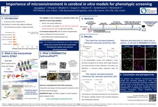
- 1. 1. Introduction • Low success rate in drug discovery: 90% of failure in clinical trials (safety and efficacy) • The Central Nervous System is highly concerned: This highlights a lack of relevance in preclinical models used upstream to select drug candidates: • Animal: inter-species differences • In vitro models: too simple, do not take into account the microenvironment of neurons (glial cells and ECM). Ex in Parkinson disease (PD), with the death of dopaminergic neurons involving the microenvironment: • Neuroinflammation mediated by glial cells • ECM modifications Importance of microenvironment in cerebral in vitro models for phenotypic screening De Conto V.1*, Cheung V.1, Maubon G.1, Souguir Z.1, Maubon N.1, Vandenhaute E.1, Bérézowski V.2 . 1HCS Pharma, Loos, France ; 2Lille Neuroscience & Cognition, Univ. Lille, Inserm, CHU-Lille, Lille, France 2. What is the extracellular matrix (ECM)? The ECM has diverse and crucial roles, in health and disease *GAG: Glycosaminoglycans Ratio GAG*/Collagen = 10/1 Low stifness Goal : To improve the predictivity of in vitro models • Taking into account the 3D cell organization • Including the cellular and matricial microenvironments 3. What is BIOMIMESYS® hydroscaffoldTM? Hydroscaffold Hydrogels: Biohydric medium Cells encapsulated Pathological or irrelevant ECM Solid Scaffolds: Porous, No ECM Decellularized organs Complete ECM Difficult tissue access, not reproducible (Scanning electron microscopy) Hyaluronic acid grafted with other ECM components: Composition and physico-chemical properties tailored to mimic the ECM of each tissue/organ BIOMIMESYS® Brain: HA, RGDS, Collagen IV, cationic polymer Elastic modulus 0.1 kPa. Porous, physiological, reproducible matrix 4. Methods 5. Results For more information, go to: hcs-pharma.com or scan this QR-code. * : veronique.deconto@hcs-pharma.com Neurons and astocytes in mono and co- culture, in 2D and in BIOMIMESYS® Brain Scale bar = 200µm GFAP TUBB3 Reference : Barnes, J.M., Przybyla, L., and Weaver, V.M. (2017). Tissue mechanics regulate brain development, homeostasis and disease. J Cell Sci 130, 71–82 ; Morgan, P., Brown, D.G., Lennard, S., Anderton, M.J., Barrett, J.C., Eriksson, U., Fidock, M., Hamrén, B., Johnson, A., March, R.E., et al. (2018). Impact of a five-dimensional framework on R&D productivity at AstraZeneca. Nat Rev Drug Discov 17, 167–181 ; Lu, P., Weaver, V.M., and Werb, Z. (2012). The extracellular matrix: A dynamic niche in cancer progression. The Journal of Cell Biology 196, 395. The cellular microenvironment influences the neuronal sensitivity (2D) The presence of astrocytes decreased the cell sensitivity to PD inducers, probably linked to their secretion properties, like antioxydative molecules. The matricial microenvironment influences neuronal sensitivity (3D) In the hydroscaffold, Luhmes cells displayed a lower sensitivity compared to 2D culture, which might explained by a partial retention of toxic molecules in the matrix, and differences in neuronal protein expression (data not shown). Source : CMR (2010-2017) Morgan et al., 2018 Barnes et al., 2017 Adapted from Lu et al. 2021 Brain ECM: 20% of cerebral volume 6. Conclusion and perspectives The cellular and matricial microenvironments modify the neuronal response in vitro, and should thus be considered carefully in academic research and in drug discovery. This model can now be used to study the microenvironment modifications in pathological conditions, and to develop innovative drugs targeting the microenvironment.