_ETC and Oxidative phosphorylation.pptx
•
0 likes•3 views

The electrons generated from different metabolic pathways of the cell are channeled to the electron transport chain by electron acceptors. The electrons then contributes for the synthesis of ATP.

Recommended
Recommended
More Related Content
Similar to _ETC and Oxidative phosphorylation.pptx
Similar to _ETC and Oxidative phosphorylation.pptx (20)
More from Alisha Shaikh
More from Alisha Shaikh (16)
Recently uploaded
Recently uploaded (20)
_ETC and Oxidative phosphorylation.pptx
- 1. ETC (Electron Transport Chain) and Oxidative phosphorylation
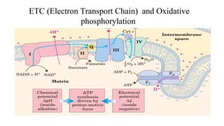
- 2. Introduction ● Oxidative phosphorylation is the culmination of energy yielding metabolism in aerobic organisms. ● All oxidative steps in the degradation of carbohydrates, fats, and amino acids converge at this final stage of cellular respiration, in which the energy of oxidation drives the synthesis of ATP. ● In eukaryotes, oxidative phosphorylation occurs in mitochondria. Oxidative phosphorylation involves the reduction of O2 to H2O with electrons donated by NADH and FADH2.
- 3. Biochemical anatomy of a mitochondrion. ● The convolutions (cristae) of the inner membrane provide a very large surface area. ● The inner membrane of a single liver mitochondrion may have more than 10,000 sets of electron- transfer systems (respiratory chains) and ATP synthase molecules, distributed over the membrane surface. Heart mitochondria, which have more profuse cristae and thus a much larger area of inner membrane, contain more than three times as many sets of electron-transfer systems as liver mitochondria. ● The mitochondrial pool of coenzymes and intermediates is functionally separate from the cytosolic pool.
- 4. Electron transport chain ● Oxidative phosphorylation begins with the entry of electrons into the respiratory chain. ● Most of these electrons arise from the action of dehydrogenases that collect electrons from catabolic pathways and funnel them into universal electron acceptors—nicotinamide nucleotides (NAD or NADP) or flavin nucleotides (FMN or FAD).
- 5. Complex I: NADH Dehydrogenase, NADH:ubiquinone oxidoreductase Complex II: Succinate dehydrogenase Complex III:cytochrome bc1 complex or ubiquinone:cytochrome c oxidoreductase Complex IV: Cytochrome oxidase Complexes of Electron Transport Chain
- 6. Complex I: NADH dehydrogenase/NADH:ubiquinone oxidoreductase ● It is a large enzyme composed of 42 different polypeptide chains, including an FMN-containing flavoprotein and at least six iron sulfur centers. ● High-resolution electron microscopy shows Complex I to be L-shaped, with one arm of the L in the membrane and the other extending into the matrix.
- 7. ● Complex I catalyzes the transfer of a hydride ion from NADH to FMN, from which two electrons pass through a series of Fe-S centers to the iron sulfur protein N-2 in the matrix arm of the complex. ● Electron transfer from N-2 to ubiquinone on the membrane arm forms QH2, which diffuses into the lipid bilayer. ● This electron transfer also drives the expulsion from the matrix of four protons per pair of electrons. ● Proton flux produces an electrochemical potential across the inner mitochondrial membrane (N side negative, P side positive), which conserves some of the energy released by the electron-transfer reactions. This electrochemical potential drives ATP synthesis.
- 8. ● Complex I catalyzes two simultaneous and obligately coupled processes: (1) the exergonic transfer to ubiquinone of a hydride ion from NADH and a proton from the matrix, expressed by Ubiquinol (QH2, the fully reduced form) diffuses in the inner mitochondrial membrane from Complex I to Complex III, where it is oxidized to Q in a process that also involves the outward movement of H. (2) the endergonic transfer of four protons from the matrix to the intermembrane space.
- 9. ● Succinate dehydrogenase,is the only membrane-bound enzyme in the citric acid cycle ● It has four different protein subunits (A,B,C,D) and contain a heme group, heme b,and a binding site for ubiquinone, the final electron acceptor in the reaction catalyzed by Complex II. ● Electrons move from succinate to FAD, then through the three Fe-S centers to ubiquinone.The heme b is not on the main path of electron transfer but protects against the formation of reactive oxygen species (ROS) by electrons that go astray. Complex II: Succinate dehydrogenase
- 10. Structure of complex II: ● The enzyme has two transmembrane subunits, C (green) and D (blue); the cytoplasmic extensions contain subunits B (orange) and A (purple). ● Just behind the FAD in subunit A (gold) is the binding site for succinate ● Subunit B has three sets of Fe-S centers (yellow and red) ● ubiquinone (yellow) is bound to subunit C ● and heme b (purple) is sandwiched between subunits C and D. ● A cardiolipin molecule is so tightly bound to subunit C that it shows up in the crystal structure (gray spacefilling).
- 11. ● It couples the transfer of electrons from ubiquinol (QH2) to cytochrome c with the vectorial transport of protons from the matrix to the intermembrane space. ● The complex has two distinct binding sites for ubiquinone, QN and QP. Complex III: cytochrome bc1 complex or ubiquinone:cytochrome c oxidoreductase
- 13. Electron transport by complex III: The path of electrons through Complex III is shown by blue arrows. On the P side of the membrane, two molecules of QH2 are oxidized to Q near the P side, releasing two protons per Q (four protons in all) into the intermembrane space. Each QH2 donates one electron (via the Rieske Fe-S center) to cytochrome c1, and one electron (via cytochrome b) to a molecule of Q near the N side, reducing it in two steps to QH2. This reduction also uses two protons per Q, which are taken up from the matrix.
- 14. ● In the final step of the respiratory chain, Complex IV, also called cytochrome oxidase,carries electrons from cytochrome c to molecular oxygen, reducing it to H2O. ● Complex IV is a large enzyme (13 subunits; Mr204,000) of the inner mitochondrial membrane. Complex IV: cytochrome oxidase
- 15. ● The three proteins critical to electron flow are subunits I, II, and III. ● The larger green structure includes the other ten proteins in the complex. ● Electron transfer through Complex IV begins when two molecules of reduced cytochrome c (top) each donate an electron to the binuclear center CuA. From here electrons pass through heme a to the Fe-Cu center (cytochrome a3 and Cu B). ● Oxygen now binds to heme a3 and is reduced to its peroxy derivative (O2 2- ) by two electrons from the Fe-Cu center. Delivery of two more electrons from cytochrome c (making four electrons in all) converts the O2 2- to two molecules of water, with consumption of four “substrate” protons from the matrix. At the same time, four more protons are pumped from the matrix by an as yet unknown mechanism.
- 16. Summary For each pair of electrons transferred to O2, four protons are pumped out by Complex I, four by Complex III, and two by Complex IV. The vectorial equation for the process is therefore ● The electrochemical energy inherent in this difference in proton concentration and separation of charge represents a temporary conservation of much of the energy of electron transfer. ● The energy stored in such a gradient, termed the proton-motive force, has two components: 1. The chemical potential energy due to the difference in concentration of a chemical species (H) in the two regions separated by the membrane 2. The electrical potential energy that results from the separation of charge when a proton moves across the membrane without a counterion
- 17. ATP Synthesis ● How is a concentration gradient of protons transformed into ATP? ● We have seen that electron transfer releases, and the proton-motive force conserves, more than enough free energy (about 200 kJ) per “mole” of electron pairs to drive the formation of a mole of ATP, which requires about 50 kJ. ● But what is the chemical mechanism that couples proton flux with phosphorylation?
- 18. The chemiosmotic model ● The chemiosmotic model, proposed by Peter Mitchell, gives insights into the mechanism. ● According to the model, the electrochemical energy inherent in the difference in proton concentration and separation of charge across the inner mitochondrial membrane—the proton-motive force—drives the synthesis of ATP as protons flow passively back into the matrix through a proton pore associated with ATP synthase. ● To emphasize this crucial role of the proton motive force, the equation for ATP synthesis is sometimes written
- 20. ● In this simple representation of the chemiosmotic theory applied to mitochondria, electrons from NADH and other oxidizable substrates pass through a chain of carriers arranged asymmetrically in the inner membrane. ● Electron flow is accompanied by proton transfer across the membrane, producing both a chemical gradient (Δ pH) and an electrical gradient (Δψ ). ● The inner mitochondrial membrane is impermeable to protons; protons can re-enter the matrix only through proton-specific channels (Fo). ● The proton-motive force that drives protons back into the matrix provides the energy for ATP synthesis, catalyzed by the F1 complex associated with Fo.
- 21. Complex V: ATP Synthase ● ATP synthase, also called Complex V, has two distinct components: F1, a peripheral membrane protein, and Fo (o denoting oligomycin-sensitive), which is integral to the membrane. ● Fo has a proton pore through which protons leak as fast as they are pumped by electron transfer, and without a proton gradient the F1-depleted vesicles cannot make ATP. ● Mitochondrial ATP synthase is an F-type ATPase. This large enzyme complex of the inner mitochondrial membrane catalyzes the formation of ATP from ADP and Pi, accompanied by the flow of protons from the P to the N side of the membrane.
- 22. ● Mitochondrial F1 has nine subunits of five different types, with the composition α3 β3γδε. Each of the three subunits has one catalytic site for ATP synthesis. The corresponding subunit conformations are designated β-ATP, β-ADP, and β-empty. ● The Fo complex making up the proton pore is composed of three subunits, a, b, and c, in the proportion ab2c10–12. Subunit c is a small (Mr8,000), very hydrophobic polypeptide, consisting almost entirely of two transmembrane helices, with a small loop extending from the matrix side of the membrane.
- 23. Binding-change model for ATP synthase (rotational catalysis)