BURN and its related anaesthesia complication
•Download as PPTX, PDF•
0 likes•11 views

BURN and its related anaesthesia complication

Recommended
More Related Content
Similar to BURN and its related anaesthesia complication
Similar to BURN and its related anaesthesia complication (20)
More from ZIKRULLAH MALLICK
More from ZIKRULLAH MALLICK (20)
Recently uploaded
Recently uploaded (20)
BURN and its related anaesthesia complication
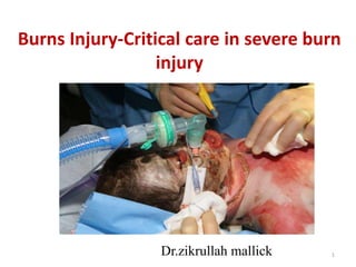
- 1. 1 Burns Injury-Critical care in severe burn injury Dr.zikrullah mallick
- 2. Definition of Burn • Damage to body tissues caused by heat, chemicals, electricity, sunlight, or radiation. • Scalds from hot liquids & steam, building fires &, flammable liquids & gases are the most common causes.
- 3. The global burden • Burn injuries are among the most devastating of all injuries and a major global public health crisis. • Burns are the 4th most common type of trauma worldwide, following traffic accidents, falls, and interpersonal violence. [The Global Burden of Disease: World Health Organization, Geneva 2008] • Approximately 90% of burns occur in low to middle income countries, regions that generally lack the necessary infrastructure to reduce the incidence and severity of burns. [Peck M, Pressman MA. The correlation between burn mortality rates from fire and flame and economic status of countries. Burns 2013; 39:1054] 3
- 4. 4 • The WHO latest figures says nearly 1.95 lakh deaths every year are caused by burns. Women in the south- east Asian region have the highest rate of burns, accounting for 27% of global burn deaths and nearly 70% of fatalities in the region. • Burns are the 11th leading cause of death of children aged between 1-9 years and are also the 5th most common cause of non-fatal childhood injuries.
- 5. 5 "Non-fatal burn injuries are a leading cause of morbidity even though burns are preventable. Burns are a global public health problem accounting for an estimated 195, 000 deaths annually. In many high- income countries, burn death rates have been decreasing, and the rate of child deaths from burns is currently over 7 times higher in low and middle- income countries than in high-income countries.“ - World Health Organization's (WHO)
- 6. Anatomy of The Skin • Skin is the largest, most important organ • 16% of total body weight • 1.73 sq meter – surface area Functions of the Skin • Protection • Sensation • Temperature regulation • Insulation from trauma • Appearance 6
- 7. Anatomy of The Skin • Layers – Epidermis – Dermis – Subcutaneous – Underlying Structures • Fascia • Nerves • Tendons • Ligaments • Muscles • Organs 7
- 8. Thermal Injuries / Burns • Tissue destruction caused by application of heat in any form to the external or internal surface of the body. Causes of Burn Injuries Fire burn – flash/flame Scald burn - liquids or steam, Contact burn - hot solid materials Chemical burn - noxious chemicals /corrosives Electrical burn - electrical current, lightning Radiation burn - X ray, UV rays, laser, Radioactive materials 8
- 9. Burn Classification • 1st degree - localized to the epidermis • 2nd degree (Superficial) - epidermis, superficial dermis • 2nd degree (deep) - epidermis and deep dermis • 3rd degree - epidermis, dermis, subcutaneous fat • 4th degree - epidermis, dermis, subcutaneous fat, underlying muscle, bone or organ 9
- 10. Superficial (1st degree) • Least destructive • Painful, erythematous, blanch to the touch. • Sunburn or a minor scald from a kitchen accident. • Do not result in scarring. 10
- 11. Partial-thickness (2nd degree) • Epidermis destroyed • Involvement of varying depth of dermis. • Two types: superficial and deep. • distinction is based on the depth of injury into this structure 11
- 12. 12 Superficial dermal burns: • Erythematous, painful, blanch to touch, blister. • Spontaneously re-epithelialize in 7 to 14 days. • After healing, slight skin discoloration. Deep dermal burns: • Painful, do not blanch to touch, pale. • Heal in 14 to 35 days by re-epithelialization. • Severe scarring as a result of the loss of dermis.
- 13. Full-thickness (3rd degree) • Epidermis and dermis • ESCHAR - Painless, hard, leathery dead skin that is black, white, or cherry red. • Compartment syndrome • Chest restriction • Patient will need grafting 13
- 14. Deep Full-thickness (4th degree) 14 • Extend beyond skin into underlying fascia and tissues • Muscle, bone and tendon damage with exposure to surface • Blackened and depressed, little or no sensation • Early excision and grafting beneficial.
- 15. 15
- 16. 16
- 17. 17
- 18. 18
- 19. Jackson’s Theory of Thermal Wounds • 3-D model showing burn depth and TBSA burned 19
- 20. 20 Zone of Coagulation • Inner area nearest the burn • The necrotic area of a burn where cells have been disrupted • Ruptured cell memb. clotted blood and thrombosed vessels Zone of Stasis • area immediately surrounding the necrotic zone. • moderate degree of insult with decreased tissue perfusion • Potentially salvable; susceptible to additional injury Zone of Hyperemia • Peripheral area of burn • vasodilation from inflammation surrounding the burn wound. • Area of least cellular injury & increased blood flow • Viable tissue from which the healing process begins.
- 21. American Burn Association (ABA) Major Burn - >25% of TBSA • Functionally significant involvement of hands, face, feet, or perineum. • Electrical or inhalation injury Moderate Burn – 15% to 25% of TBSA • No complications of involvement of hands, face, feet, or perineum • No electrical or inhalation injury Minor Burn - <15% of TBSA • No involvement of hands, face, feet, or perineum • No electrical or inhalation injury 21
- 22. Rule of Nines 22
- 23. Infant Rule of nines • 18 Head • 9 Each upper limb • 18 Front trunk • 18 Back trunk • 13.5 Each lower limb • 1 perineum ADULT Rule of Nines • 9 Head • 9 Each upper limb • 18 Front trunk • 18 Back trunk • 18 Each lower limb • 1 perineum 23
- 24. Palm Rule • Area of the open hand (including the palm and extended fingers) of the patient to be approximately 1% of TBSA. • An alternate system for approximating the extent of the burn. • An accurate assessment of size can be made by using the patient's palmar hand surface, including the digits, • This method is helpful when evaluating splash burns and other burns of mixed distribution as well as smaller burns. 24
- 25. Lund and Browder Chart • Children have much larger heads and smaller thighs in proportion to total body size than do adults. • child's body proportions do not fully reach adult percentages until adolescence • More complicated and time consuming method to estimate BSA burned. • BUT MORE ACCURATE!! 25
- 26. Laser Doppler Flowmeter • Currently, burn depth is most accurately assessed by the judgment of experienced practitioners. Accurate depth determination is critical because wounds that will heal with local treatment are treated differently from those requiring operative intervention. • Several recent reports claim superiority of this method over clinical judgment in the determination of wounds requiring skin grafting for timely healing 26
- 27. Laser Doppler Flowmeter • The sensor is placed on the skin in question, which returns a value of perfusion units. • A value of 0 is necrotic, whereas values of 80 indicate viable skin. 27
- 29. • Burn are caused by a transfer of energy from a heat source to the body. • Tissue destruction results from coagulation, protein denaturation, or ionization of cellular content. • Disruption of the skin can lead to increased fluid loss, infection, hypothermia, scarring, compromised immunity, and changes in function, appearance and body image. • The depth of the injury depends on the temp of the burning agent and the duration of contact with the agent • For example in the case of scald burns in adults, 15 seconds of contact with hot tap water at 56 degree C may result in a burn that destroys both the epidermis and the dermis, causing a full thickness (third degree) injury. 29
- 30. Inflammatory Response to Burn Prostaglandins, Catecholamines, Bradykinin, Vasoactive amines, Leukotrienes, and activated Complement, Histamine, Serotonin, Thromboxane A2 • Vasoconstriction and Vasodilation, • Increased capillary permeability, and • Edema • Mesenteric vasoconstriction and decreased blood flow to the gut that compromise gut mucosal integrity and immune function. 30
- 31. Burn Shock • Combination of hypovolaemic, and distributive shock, refractory to i.v. resuscitation. • Causes are: • ↑ extracellular fluid • ↓ Intravascular volume • ↓ plasma volume • Characterized by specific hemodynamic changes: • ↑ systemic vascular resistance, and • ↓ pulmonary artery occlusion pressures, • ↓ cardiac output . • ↓ contractility 31
- 32. Fluid/Electrolyte Changes Fluid Shift • ↑ capillary hydrostatic pressure and ↑ capillary permeability • Leak of plasma from intravascular into interstitial space. Fluid remobilization • Capillary leak ceases and fluid shifts back into the circulation • Restores fluid balance and renal perfusion • Increased urine formation & diuresis Electrolyte derangements: • Initially tissue destruction - Hyperkalemia • Later, Renal wasting & Gastric losses - Hypokalemia • ↑ extracellular volume - Hyponatremia 32
- 33. Metabolic Response to Burn Hypermetabolism • Resting energy expenditure (REE) after burn injury increases as much as 100%. • Increase in oxygen consumption because of increased production of catecholamines, cortisol, and glucagon. • Increased heat loss from the burn wound secondary to increased blood flow and increased beta-adrenergic stimulation are probably primary factors. • REE helps in assessing the nutritional status. 33
- 34. Glucose Metabolism • ↑ Gluconeogenesis • ↑ glycogenolysis • ↑ basal rate of glucose production • Hyperglycemia complicates the acute management, related to poor outcomes, specifically increased risk of infection & decreased graft take. • Exogenous insulin administration to achieve euglycemia has been shown to decrease donor site healing time and decrease length of stay. 34
- 35. Lipid Metabolism • Lipolysis occurs at a rate more than the requirements for fatty acids as an energy source in burn patients resulting in ↑ FFA. • The majority of released fatty acids are not oxidized, rather re-esterified into triglycerides, resulting in fat accumulation in the liver and hepatic steatosis. • Beta-blockade using propranolol appears helpful in manipulating peripheral lipolysis and in preventing hepatic steatosis. 35
- 36. Protein Metabolism • Muscle protein breakdown is ↑ and is excreted in the urine as urea. This results in an ↑ efflux of amino acids from the skeletal muscle and hypoproteinemia. • Wound healing & immunologic activity require enhanced protein synthesis. • Protein intake 1 g/kg/day has been recommended for all burn patients, and for patients with normal renal function, the intake is 2 g/kg/day. 36
- 37. Neuroendocrine Response • Catecholamines ↑ & Glucagon ↑ and appear to be the major endocrine mediators of the hypermetabolic response in burn patients. • Burn injuries abolish the normal diurnal variation in glucocorticoid secretion, producing persistent hypercortisolemia. • growth hormone (GH) ↓ • T3 ↓ & T4 ↓ and reverse T3 ↑ 37
- 38. Systemic Changes • Cardiac: ↓ CO, ↓ BP • Pulmonary: FRC ↓ lung & chest wall compliance ↓ • Renal: RBF ↓ GFR ↓ • Gastrointestinal: ↓ or absent motility, Curling’s ulcer • Immune System: • ↓ production of immunoglobulin • ↓ opsonic activity • ↓ bactericidal activity • Impairment in host defense causes the burned patient to become especially prone to infection. 38
- 40. At the Site • Remove to safe area, if possible • Stop the burning process • Extinguish fire • Remove clothing and jewellery • Cut around areas where clothing is stuck to skin 40
- 41. Emergency Care • The initial management of a severely burnt patient is similar to that of any trauma patient. • Primary and secondary survey should follow ATLS principles. • Do not get distracted by the burn • Burn injury must not distract from this sequential assessment, otherwise serious associated injuries may be missed. 41
- 42. Initial care based on ATLS Guidelines • Many of the components are being assessed simultaneously, which is essential for immediately identifying life-threatening situations and its management. • If initial interaction reveals that the patient is alert, talking, oriented, and moving all extremities… • Patient has an adequate airway • Is breathing adequately • Oxygen is being circulated to the brain, and • There is no major neurologic injury. 42
- 43. Airway Control • Early tracheal intubation should be considered in the presence of any of the following features: • Stridor, • Hypoxaemia or Hypercapnia, • GCS ≤ 8 • Facial burns • Inhalational injury / CO poisoning • Upper airway / oropharyngeal oedema • Full-thickness neck burns and • Chest wall restriction 43
- 44. • Delay in performing the intubation can result in total obstruction of the upper airway. • In presence of facial burns and when the fasting status of the patient is unknown, RSI should be performed. • Succinylcholine is safe in the first 24 h after a burn. After this time, its use is contraindicated due to the risk of hyperkalaemia leading to cardiac arrest, thought to be due to release of potassium from extrajunctional acetylcholine receptors. This can persist up to 1 year post-burn. • The intubation can be done by simple laryngoscopy, intubation stylet, FOB or LMA-Fastrach(ILMA). 44
- 45. 45 • In patient with 3rd to 4th degree burns of the neck, laryngoscopy may be impossible because of the rigidity of the cervical tissue, an incision in neck can facilitate laryngoscopy. • Use of modern technique which involve less extension like lightwand, airtraq or FOB. • Extensive or circumferential 3rd to 4th degree burns to the chest wall can cause severe restriction and may require immediate escharotomy of the anterior chest wall.
- 46. Tube fixation • The fixation of the ET is a challenge in the burned patient. • Secure the tube safely without additional injury to the tissue of the face and is flexible enough to adjust to edema formation. • Adhesive tape are not effective in the burn patient because they do not adhere adequately. Usually, a soft sling ribbon is used. It is tied at the back of the head (not the neck). • Suturing the tube to the gums, wiring the tube around a tooth. 46
- 47. Inhalational Injury • Any pulmonary insult associated with a burn injury Three types: • Heat injury to the airways • Inhalation of toxic product of combustion like CO • Chemical burn with deposition of carbon particles in the lower airways. • Leading cause of death from fires. • Higher mortality than burn patients without inhalation injury. 47
- 48. 48
- 49. Warning Signs of Airway Burn Suspect airway burn if: • Enclosed space • Stridor, hoarseness, or cough • Burns to face, lips, mouth, pharynx, or nasal mucosa • Soot in sputum, nose, or mouth • Dyspnoea, ↓ level of consciousness, or confusion • Low O2 saturation on pulse oximetry • Increased CO2( > 2%) 49
- 50. CO Poisoning • When carboxy Hb >15% in blood. • Causes tissue hypoxia due to: • Binds to Hb 200-300 times more readily than O2 in alveoli • Rate of dissociation of CO Hb slow. • Creates a left shift of O2 - Hb dissociation curve • Impairs O2 unloading at tissue level. • Signs and Symptoms: • Headache, nausea/vomiting, dyspnea/tachypnea • Cherry red appearance of mucous membranes • Persistent metabolic acidosis with adequate volume resuscitation. 50
- 51. Treatment • The clinical manifestations are often delayed but are usually apparent by 24 hours. • There is no specific treatment for airway burns other than ensuring adequate oxygenation and minimising iatrogenic lung insult. • Prophylactic corticosteroids or antibiotics have no role in treatment. • All patients suspected of having CO Poisoning should be given 100% O2 . 51
- 52. 52 • The half life of CO-Hb breathing room air is 90 minutes, whereas, when breathing 90 to 100% oxygen is 30 minutes, i.e., the concentration of carboxyhemoglobin is reduced by approximately 50% every 30 minutes if an oxygen concentration of 90 to 100% is used. • Hyperbaric oxygen produces more rapid displacement and is useful in cases of prolonged exposure. • ET intubation and use of 100% oxygen with mechanical ventilation is indicated for those patients with impaired neurologic function and a high carboxyhemoglobin.
- 53. Breathing • Breathing, chest movement, and tracheal position should be assessed clinically. • All burn patients should receive 100% O2 through a non-rebreathing mask on presentation. • Oxygen administration is required for all major burns until carbon monoxide toxicity has been ruled out. • The oxygen saturation is most readily assessed by the use of a pulse oximeter, as the clinical determination of adequate oxygenation is virtually impossible by any other noninvasive means. • Artificial ventilation if required. • If in doubt intubate!! 53
- 54. Circulation • Two large-bore IV catheters are inserted into the peripheral veins, Traditional sites for i.v. access may be unavailable and unusual peripheral venous sites or central venous access may be required. • Intravenous fluid if : • >15 - 20% burn in adults • >10% burn in children • Age >65yrs or <2yrs • Ringers lactate solution preferred. • Pulse rate and character, color and temperature of the skin, and mental status are rapidly evaluated to assess perfusion. • BP is determined at the onset of resuscitation and then every 5 to 10 minutes until the patient's vital signs have stabilized. 54
- 55. Disability: neurologic status • This should consist of determining the GCS score and examining the pupils for size, symmetry, and reaction to light. • The GCS is the sum of scores for three areas of assessment, including (a) eye opening, (b) verbal response, and (c) best motor response. • Pupillary size, symmetry, and reaction to light are important diagnostic tools that aid in determining if there is a lateralizing brain injury. 55
- 56. Exposure and Estimation • Expose and ensure all jewellery and watches are removed from burnt limbs. • Burn patients become hypothermic easily, so should be covered and warmed as soon as possible. • The patient should be examined (including the back - log roll if appropriate) to get an accurate estimate of the burn area and to check for any concomitant injuries. • A standard Lund–Browder chart is used for a quick assessment of BSA burnt. • If this is not available, the ‘Rule of Nines’ is fairly accurate in adult patients. 56
- 57. Intensive Care Burn Units • Severely burned patients are cared in specialized intensive wards which have semi-sterile laminar flow boxes, their own operating rooms, and special bathrooms for patients. • Intensive care therapy is a basis for further plastic surgery therapy and plays an important role for the survival of the patient. • Controlled fluid and electrolyte management with continuous monitoring of various laboratory parameters are the basis of intensive care and decreases the risk of common complications. 57
- 58. Intensive Care Management • Fluid resuscitation and electrolyte management • Airway burn/Inhalational injury • Mechanical ventilation • Nutrition & Metabolism • Prevention of Infection • Prevention of multiorgan failure • Compartment syndrome, Hypothermia, DVT • Bed sores • Other supportive measures 58
- 59. ABA Burn Center Referral Criteria • Burns >10% TBSA in patients <10 or >50 years of age • Burns >20% TBSA in other age groups • Full-thickness burns >5% TBSA in any age group • Burns involving the face, hands, feet, genitalia, perineum, or major joints. • Electrical burns/Chemical burns/Inhalation injury • Burn injury in patients with preexisting medical disorders. • Burn with concomitant trauma (e.g., fractures) in which the burn poses the greatest risk of morbidity or mortality. • Burn injury in children admitted to a hospital without qualified personnel or equipment for pediatric care. 59
- 60. Fluid Management • TBSA <20% is associated with minimal fluid shifts and can generally be resuscitated with oral hydration, except in cases of facial, hand and genital burns, as well as burns in children and the elderly. • TBSA >20% is associated with massive fluid shifts, which result in burn oedema and burn shock. OBJECTIVES: • HR < 110/minute • Normal sensorium (awake, alert, oriented) • Urine output – 0.5 ml/kg/hr (adult); 0.5-1 ml/kg/hr (paed) • Resuscitation formulae provide estimates, • Adjust to individual patient responses. 60
- 61. Choice of Fluid • Isotonic crystalloids, hypertonic solutions and colloids have been used for this purpose, but every solution has its advantages and disadvantages. • Plasma proteins generate the inward oncotic force that counteracts the outward intravascular hydrostatic force. Without protein, intravascular volume could not be maintained. • The sodium shift into the cell results in cellular oedema and hypo-osmolar intravascular fluid volume. Rapid infusion of hypertonic sodium solutions has proven to increase the plasma osmolality and limit cellular oedema. 61
- 62. • Normal saline should be avoided, as the large volumes required for resuscitation invariably lead to hyperchloremic metabolic acidosis • None of them is ideal, and none is superior to any of the others. Combination of fluids can be used to achieve the desired goal of end-organ perfusion. • Crystalloid, in particular LR is the most extensively used resuscitation fluid. 62
- 63. Fluid Resuscitation Formula • Parkland formula • Brooke formula • Modified Brooke formula • Evan’s formula • Monafo formula Formulas developed for children • Shriner’s cincinnati • Galveston 63
- 64. Parkland formula Initial 24 hours: • RL @ 4 ml/kg/% burn for adults • RL @ 3 ml/kg/% burn for children. • Half in first 8 hrs, remaining half in next 16 hrs • no colloid in the initial 24 hours. • Next 24 hours: Colloids given as 20–60% of calculated plasma volume. No crystalloids. • maintain a urinary output of 0.5–1 ml/kg/hour in adults and 1 ml/kg/hour in children. 64
- 65. Brooke formula • Initial 24 hours: RL @ 1.5 ml/kg/% burn plus colloids @ 0.5 ml/kg/% burn plus 2000 ml free water with glucose • Next 24 hours: RL @ 0.5 ml/kg/% burn, colloids @ 0.25 ml/kg/% burn and the same amount of glucose in water as in the first 24 hours Modified Brooke • Initial 24 hours: No colloids. RL @ 2 ml/kg/% burn in adults and 3 ml/kg/% burn in children • Next 24 hours: Colloids @ 0.3–0.5 ml/kg/% burn and no crystalloids are given. 65
- 66. Evans formula • First 24 hours: Crystalloids 1 ml/kg/% burn plus colloids at 1 ml/kg/% burn plus 2000 ml glucose in water • Next 24 hours: Crystalloids at 0.5 ml/kg/% burn, colloids at 0.5 ml/kg/% burn and the same amount of glucose in water as in the first 24 hours. Monafo formula • Monafo recommends using a solution containing 250 mEq Na, 150 mEq lactate and 100 mEq Cl. • The amount is adjusted according to the urine output. 66
- 67. Galveston • Initial 24 hours: RL 5000 ml/m² burn area + 1500 ml/m² total area • (1/2 of total volume over 8 hours, rest of the total volume in 16 hours) 67
- 68. Nutrition and metabolism • Burn injury is associated with a considerable hypermetabolic response, mediated by the systemic response to the burn and related to the extent of the burn injury. • Even small burns can be associated with hyperpyrexia directly due to hypermetabolism. • Close attention to nutritional needs is critical to prevent protein breakdown, decrease infection, increase wound healing and improve immune status. 68
- 69. • In view of the greatly increased nutritional requirements, appropriate nutrition must be initiated rapidly through enteral route. • Enteral food supply should be targeted as early as possible, in order to avoid regression of intestinal villi. • The capillary leak, which is responsible for the massive displacement of fluids, spontaneously ceases after 24 hours. Till then, intensive fluid therapy must be continued, in order to counteract the decreased cardiac output, the reduced perfusion of the kidney, the liver and the intestine. 69
- 70. Routes Oral Enteral: Energy requirements are proportional to the size of the burn and should be met by enteral nutrition, and this should be established as soon as possible after the burn injury. i.e. duodenal / gastrostomy / jejunaostomy tube Parenteral i.e. TPN and PPN Total parenteral nutrition is associated with immunosuppression, an increase in infective complications, and reduced survival. 70
- 71. • High-protein & high-calorie diet • Often requiring various supplements- vitamin A & C. • Glutamine, arginine, and omega 3 fatty acid supplementation may improve immunity and gut function. 71
- 72. Management of the hypermetabolic response • Reduce heat loss - environmental conditioning • Excision and closure of burn wound • Early enteral feeding • Recognition and treatment of infection 72
- 73. Infection in Burns Patients • After the initial resuscitation, up to 75% of mortality in burns patients is related to infection. • Infective pulmonary complications are now the commonest types of infection seen in burns patients. Several factors contribute to the high frequency and severity of infection: Destruction of the skin or mucosal surface barrier Presence of necrotic tissue and serosanguinous exudate provides a medium to support growth of microorganisms Invasive monitoring provides portals for bacterial entry. Impaired immune function allows microbial proliferation 73
- 74. Signs of Wound Infection • Change in wound appearance: a) Discoloration of surrounding skin b) Offensive exudate • Delayed healing • Graft failure • Conversion of partial thickness wound to full thickness 74
- 75. Causative Agents Bacteria • Haemolytic streptococci— Streptococcus pyogenes. • Staphylococci— MRSA • Gram negative bacteria— Pseudomonas aeruginosa, Acinetobacter, Proteus species. Fungi • Candida—Most common fungal isolate. • Filamentous fungi— Aspergillus, Fusarium, and phycomycetes Viruses • Herpes simplex—Characterised by vesicular lesions 75
- 76. Treatment • Use of topical antimicrobial agents are effective. • Silver sulfadiazine, Silver nitrate and Mafenide are most frequently used. • Early closure of the burn wound by surgical techniques then lessens the surface area available for further microbial colonisation and subsequent infection. • Prophylactic use of systemic antibiotics is controversial and is not indicated, in patients with burns covering less than 40% of TBSA. 76
- 77. Prevention of Multiorgan damage Pulmonary complications • Aggressive airway toilet is essential. • Diluted heparin and acetyl cystine nebulisation may be helpful. • Early surgical debridement, enteral feeding, mobilisation of the patient, and early extubation are desirable. • Antibiotics should be reserved for established infections and guided by regular microbiological surveillance. 77
- 78. Heart Failure • Myocardial dysfunction is a potential consequence of major burn injury, attributed to a circulating myocardial depressant factor. • Administration of an inotropic agent is preferable to overloading a failing myocardium with large volumes of fluid. • However, the inotropic drug can produce vasoconstriction in the burn wound, reducing the viability of critically injured tissue. • Inotropic drugs should not be used until adequate fluid resuscitation has been ensured. • Inotropic drugs that do not produce vasoconstriction (such as dopexamine or dobutamine) will preserve wound viability. 78
- 79. Renal Failure • Early renal failure after burn injury is usually due to delayed or inadequate fluid resuscitation or from substantial muscle break down or haemolysis. • Delayed renal failure is usually the consequence of sepsis and is often associated with other organ failure. • A reduced urine output, despite adequate fluid administration, is usually the first sign of ARF. • This will be followed by a rise in serum creatinine and urea concentrations. • Early renal support (CRRT) will control serum electrolytes and accommodate the large volumes of nutritional supplementation required in a major burn. 79
- 80. Cerebral Injury • Hypoxic cerebral insults and closed head injuries are not uncommon in burn injuries. • Fluid administration for the burn injury will increase cerebral oedema and intracranial pressure. • Monitoring intracranial pressure may help in minimising the adverse effects of trying to achieve two contradictory treatment goals. 80
- 81. Compartment Syndromes • A lifethreatening complication caused by high volume resuscitation is abdominal compartment syndrome (ACS). • Defined as intra-abdominal pressure >20 mm Hg plus at least one new organ dysfunction. • ACS has been associated with renal impairment, gut ischemia, and cardiac and pulmonary hypoperfusion. • Clinical manifestations include tense abdomen, decreased pulmonary compliance, hypercapnia, and oliguria. 81
- 82. • Appropriate intravascular volume, appropriate body positioning, pain management, sedation, nasogastric decompression, muscle relaxants if required, and torso escharotomy are all interventions to increase abdominal wall compliance and decrease intra-abdominal pressures. Extremity compartment syndromes can also result from extensive edema formation. • Patients may require escharotomies, fasciotomies, or both for the release of extremity compartment syndrome. 82
- 83. • Hypothermia - Strategies to vigorously prevent hypothermia include a warmed room, warmed inspired air, warming blankets, and countercurrent heat exchangers for infused fluids. • Deep Venous Thrombosis - The incidence of DVT in burn patients is estimated to be 1% to 23% • Prevention of BED SORES - These patients may require a prolong bed rest, so care should be taken concerning development of BED SORES. • Stress Ulcers - Patients with major burn injuries are at risk so should receive routine prophylaxis beginning at admission. 83
- 84. • Neutropenia - Transient leukopenia is common, primarily due to a decreased neutrophil count. Maximal WBC depression occurs several days after admission with rebound to normal a few days later. Use of silver sulfadiazine has been associated with this transient leukopenia. • Adrenal Insufficiency - Those with massive burns have higher cortisol levels but may be resistant to serum cortisol increases in response to stimulation. Absolute adrenal insufficiency occurs in up to 36% of patients with major burns . 84
- 85. Other Supportive Measures • Appropriate analgesia: - Avoid IM injections!!! - Intravenous: Ketamine 0.5mg/kg Morphine 0.2mg/kg every 5 mins • All patients with burns > 10% 0.5ml Tetanus toxoid. • Gastric decompression with NGT in all major burns. • Antibiotic use is controversial. • Neck, oral & joint splints can prevent deformities. 85
- 86. Key Points • Fluid resuscitation must be based on frequent reassessment. Formulas are only a guide. • Pulse oximetry readings may be normal in carbon monoxide toxicity • Unnecessary intubation is preferable to systemic hypoxia. • Early enteral nutrition in major burns may improve survival by increasing immune status and reducing risk of infection. • Antibiotics should be used wisely to limit emergence of multiresistant organisms: close liaison with a clinical microbiologist is crucial. • Failure to respond to treatment should trigger an escalation in the invasiveness of the monitoring 86
- 87. Deaths are only part of the problem, for every person who dies as a result of their burns; many more are left with lifelong disabilities and disfigurements. For some this means living with the stigma and rejection that all too often comes with disability and disfigurement. [Krug E. A WHO plan for burn prevention and care, Geneva, Switzerland, 2008] 87
- 88. Follow-up and Rehabilitation • Rehabilitation measures should be implemented as soon as possible. • Early respiration training deepens inhalation and therefore prevents pulmonary infections. • Edema prophylaxis and therapy, scar care, compression garments and the specific prophylaxis of scarred contractures in critical locations (throat, face, hands, and joints) are the fundamental pillars of multimodal rehabilitation • After completion of the intensive care period, the patients are transferred to a follow-up ward where further wound care, physiotherapy and psychiatric care help to maximize the patient’s autonomy. . 88
- 89. • A burn injury is a frightening & potentially life-changing event for patients and families. Because of this loss/change in their lives, they can often face many difficult emotions at varying stages after the injury. • The impact of the burn can result in permanent disability and disfigurement. In the long term, many patients suffer from their changed appearance. • Burns of the face and hands are felt as especially disturbing, as they are continuously visible to other people. 89
- 90. • Given the nature of burn injury and its treatment there are many stressors that may trigger psychological problems, particularly those associated with anxiety and depression • Over 60% of all severely burned patients develop psychological problems like PTSD, depression and sleep disturbances. • A multidisciplinary team of physiotherapists, psychologists, nurses, and councillors are vital to aid rehabilitation and reduce long-term impairment. 90
- 91. Indian Scenario • According to WHO, in India, over 10 lakh people are moderately or severely burnt every year. • The MOHFW says India records 70 lakh burn injury cases annually of which 1.4 lakh people die of burn every year. Around 70% of all burn injuries occur in most productive age group (15-35 years). Around four out of five burnt cases are women and children. As many as 80% of cases admitted are a result of accidents at home (kitchen-related incidents). [The Times of India. New Delhi. June 7, 2012] 91
- 92. • Of all burn death cases, 80.8% were females, 82.4% married ones, 71.9% belonged to the young age group of 21-40 years and 75.0% came from the rural parts of the district. • In all female suicides, burns was the commonest method adopted by over 60% females. Torture by in- laws (32.1%) was the commonest reason for committing suicide by burns in married women. [Batra AK. Burn mortality: recent trends and sociocultural determinants in rural India. Burns. 2003 May;29(3):270-5.] 92
- 93. National Programme for Prevention of Burn Injuries • The high incidence is attributed to illiteracy, poverty and low level safety consciousness in the population. • The situation becomes further grim due to the absence of organized burn care at primary and secondary health care level. 93
- 94. • The GOAL of National programme for prevention of burn injuries (NPPBI) would be to ensure prevention and capacity building of infrastructure and manpower at all levels of health care delivery system in order to reduce incidence, provide timely and adequate treatment to burn patients to reduce mortality, complications and provide effective rehabilitation to the survivors. 94
- 95. The programme note says… "Among all traumas, burn cases have highest duration of hospital bed occupancy. Cost of hospitalized burn injury case management is extremely high which may cost enormous financial burden to the country. The rehabilitation of the individual may be a challenging and daunting task.“ National programme for prevention of burn injuries (NPPBI) 95
- 96. THANK YOU 96