unit 1 cytoskeletal structures ECM docx.pdf sh.pdf
•
0 likes•350 views

this notes provides text on cell organelles like ECM , cytoskeletal structures

Recommended
More Related Content
What's hot
What's hot (20)
Similar to unit 1 cytoskeletal structures ECM docx.pdf sh.pdf
Similar to unit 1 cytoskeletal structures ECM docx.pdf sh.pdf (20)
More from MSCW Mysore
More from MSCW Mysore (20)
Recently uploaded
Recently uploaded (20)
unit 1 cytoskeletal structures ECM docx.pdf sh.pdf
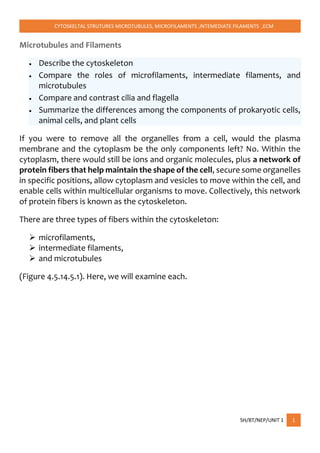
- 1. CYTOSKELTAL STRUTURES MICROTUBULES, MICROFILAMENTS ,INTEMEDIATE FILAMENTS ,ECM SH/BT/NEP/UNIT 1 1 Microtubules and Filaments Describe the cytoskeleton Compare the roles of microfilaments, intermediate filaments, and microtubules Compare and contrast cilia and flagella Summarize the differences among the components of prokaryotic cells, animal cells, and plant cells If you were to remove all the organelles from a cell, would the plasma membrane and the cytoplasm be the only components left? No. Within the cytoplasm, there would still be ions and organic molecules, plus a network of protein fibers that help maintain the shape of the cell, secure some organelles in specific positions, allow cytoplasm and vesicles to move within the cell, and enable cells within multicellular organisms to move. Collectively, this network of protein fibers is known as the cytoskeleton. There are three types of fibers within the cytoskeleton: microfilaments, intermediate filaments, and microtubules (Figure 4.5.14.5.1). Here, we will examine each.
- 2. CYTOSKELTAL STRUTURES MICROTUBULES, MICROFILAMENTS ,INTEMEDIATE FILAMENTS ,ECM SH/BT/NEP/UNIT 1 2 Figure 4.5.14.5.1: Microfilaments thicken the cortex around the inner edge of a cell; like rubber bands, they resist tension. Microtubules are found in the interior of the cell where they maintain cell shape by resisting compressive forces. Intermediate filaments are found throughout the cell and hold organelles in place. Microfilaments Are Of the three types of protein fibers in the cytoskeleton, microfilaments are the narrowest. They function in cellular movement, have a diameter of about 7 nm, and are made of two intertwined strands of a globular protein called actin (Figure 4.5.24.5.2). For this reason, microfilaments are also known as actin filaments.
- 3. CYTOSKELTAL STRUTURES MICROTUBULES, MICROFILAMENTS ,INTEMEDIATE FILAMENTS ,ECM SH/BT/NEP/UNIT 1 3 Figure 4.5.24.5.2: Microfilaments are made of two intertwined strands of actin. Actin is powered by ATP to assemble its filamentous form, which serves as a track for the movement of a motor protein called myosin. This enables actin to engage in cellular events requiring motion, such as cell division in animal cells and cytoplasmic streaming, which is the circular movement of the cell cytoplasm in plant cells. Actin and myosin are plentiful in muscle cells. When your actin and myosin filaments slide past each other, your muscles contract. Microfilaments also provide some rigidity and shape to the cell. They can depolymerize (disassemble) and reform quickly, thus enabling a cell to change its shape and move. White blood cells (your body’s infection-fighting cells) make good use of this ability. They can move to the site of an infection and phagocytize the pathogen.
- 4. CYTOSKELTAL STRUTURES MICROTUBULES, MICROFILAMENTS ,INTEMEDIATE FILAMENTS ,ECM SH/BT/NEP/UNIT 1 4 Intermediate Filaments Intermediate filaments are made of several strands of fibrous proteins that are wound together (Figure 4.5.34.5.3). These elements of the cytoskeleton get their name from the fact that their diameter, 8 to 10 nm, is between those of microfilaments and microtubules. Figure 4.5.34.5.3: Intermediate filaments consist of several intertwined strands of fibrous proteins. Intermediate filaments have no role in cell movement. Their function is purely structural. They bear tension, thus maintaining the shape of the cell, and anchor the nucleus and other organelles in place. Figure 4.5.14.5.1 shows how intermediate filaments create a supportive scaffolding inside the cell. The intermediate filaments are the most diverse group of cytoskeletal elements. Several types of fibrous proteins are found in the intermediate filaments. You are probably most familiar with keratin, the fibrous protein that strengthens your hair, nails, and the epidermis of the skin. Microtubules As their name implies, microtubules are small hollow tubes. The walls of the microtubule are made of polymerized dimers of α-tubulin and β-tubulin, two globular proteins (Figure 4.5.44.5.4). With a diameter of about 25 nm, microtubules are the widest components of the cytoskeleton. They help the cell resist compression, provide a track along which vesicles move through
- 5. CYTOSKELTAL STRUTURES MICROTUBULES, MICROFILAMENTS ,INTEMEDIATE FILAMENTS ,ECM SH/BT/NEP/UNIT 1 5 the cell, and pull replicated chromosomes to opposite ends of a dividing cell. Like microfilaments, microtubules can dissolve and reform quickly. Figure 4.5.44.5.4: Microtubules are hollow. Their walls consist of 13 polymerized dimers of α-tubulin and β-tubulin (right image). The left image shows the molecular structure of the tube. Microtubules are also the structural elements of flagella, cilia, and centrioles (the latter are the two perpendicular bodies of the centrosome). In fact, in animal cells, the centrosome is the microtubule-organizing center. In eukaryotic cells, flagella and cilia are quite different structurally from their counterparts in prokaryotes. EXTRA-INFORMATION The cytoskeleton is a structure that helps cells maintain their shape and internal organization, and it also provides mechanical support that enables cells to carry out essential functions like division and movement. There is no single cytoskeletal component. Rather, several different components work together to form the cytoskeleton. What Is the Cytoskeleton Made Of?
- 6. CYTOSKELTAL STRUTURES MICROTUBULES, MICROFILAMENTS ,INTEMEDIATE FILAMENTS ,ECM SH/BT/NEP/UNIT 1 6 The cytoskeleton of eukaryotic cells is made of filamentous proteins, and it provides mechanical support to the cell and its cytoplasmic constituents. All cytoskeletons consist of three major classes of elements that differ in size and in protein composition. Microtubules are the largest type of filament, with a diameter of about 25 nanometers (nm), and they are composed of a protein called tubulin. Actin filaments are the smallest type, with a diameter of only about 6 nm, and they are made of a protein called actin. Intermediate filaments, as their name suggests, are mid-sized, with a diameter of about 10 nm. Unlike actin filaments and microtubules, intermediate filaments are constructed from a number of different subunit proteins. What Do Microtubules Do? Tubulin contains two polypeptide subunits, and dimers of these subunits string together to make long strands called protofilaments. Thirteen protofilaments then come together to form the hollow, straw-shaped filaments of microtubules. Microtubules are ever-changing, with reactions constantly adding and subtracting tubulin dimers at both ends of the filament (Figure 1). The rates of change at either end are not balanced — one end grows more rapidly and is called the plus end, whereas the other end is known as the minus end. In cells, the minus ends of microtubules are anchored in structures called microtubule organizing centers (MTOCs). The primary MTOC in a cell is called the centrosome, and it is usually located adjacent to the nucleus. Microtubules tend to grow out from the centrosome to the plasma membrane. In nondividing cells, microtubule networks radiate out from the centrosome to
- 7. CYTOSKELTAL STRUTURES MICROTUBULES, MICROFILAMENTS ,INTEMEDIATE FILAMENTS ,ECM SH/BT/NEP/UNIT 1 7 provide the basic organization of the cytoplasm, including the positioning of organelles. What Do Actin Filaments Do? Figure 2 Figure Detail The protein actin is abundant in all eukaryotic cells. It was first discovered in skeletal muscle, where actin filaments slide along filaments of another protein called myosin to make the cells contract. (In nonmuscle cells, actin filaments are less organized and myosin is much less prominent.) Actin filaments are made up of identical actin proteins arranged in a long spiral chain. Like microtubules, actin filaments have plus and minus ends, with more ATP- powered growth occurring at a filament's plus end (Figure 2). In many types of cells, networks of actin filaments are found beneath the cell cortex, which is the meshwork of membrane-associated proteins that supports and strengthens the plasma membrane. Such networks allow cells to hold — and move — specialized shapes, such as the brush border of microvilli. Actin filaments are also involved in cytokinesis and cell movement (Figure 3).
- 8. CYTOSKELTAL STRUTURES MICROTUBULES, MICROFILAMENTS ,INTEMEDIATE FILAMENTS ,ECM SH/BT/NEP/UNIT 1 8 Figure 3: Actin filaments support a variety of structures in a cell. What Do Intermediate Filaments Do? Intermediate filaments come in several types, but they are generally strong and ropelike. Their functions are primarily mechanical and, as a class, intermediate filaments are less dynamic than actin filaments or microtubules. Intermediate filaments commonly work in tandem with microtubules, providing strength and support for the fragile tubulin structures. All cells have intermediate filaments, but the protein subunits of these structures vary. Some cells have multiple types of intermediate filaments, and some intermediate filaments are associated with specific cell types. For example, neurofilaments are found specifically in neurons (most prominently in the long axons of these cells), desmin filaments are found specifically in muscle cells, and keratins are found specifically in epithelial cells. Other intermediate filaments are distributed more widely. For example, vimentin filaments are found in a broad range of cell types and frequently colocalize with microtubules. Similarly, lamins are found in all cell types, where they form a meshwork that reinforces the inside of the nuclear membrane. Note that intermediate filaments are not polar in the way that actin or tubulin are (Figure 4).
- 9. CYTOSKELTAL STRUTURES MICROTUBULES, MICROFILAMENTS ,INTEMEDIATE FILAMENTS ,ECM SH/BT/NEP/UNIT 1 9 Figure 4: The structure of intermediate filaments Intermediate filaments are composed of smaller strands in the shape of rods. Eight rods are aligned in a staggered array with another eight rods, and these components all twist together to form the rope-like conformation of an intermediate filament. How Do Cells Move? Cytoskeletal filaments provide the basis for cell movement. For instance, cilia and (eukaryotic) flagella move as a result of microtubules sliding along each other. In fact, cross sections of these tail-like cellular extensions show organized arrays of microtubules. Other cell movements, such as the pinching off of the cell membrane in the final step of cell division (also known as cytokinesis) are produced by the contractile capacity of actin filament networks. Actin filaments are extremely dynamic and can rapidly form and disassemble. In fact, this dynamic action underlies the crawling behavior of cells such as amoebae. At the leading edge of a moving cell, actin filaments are rapidly polymerizing; at its rear edge, they are quickly depolymerizing (Figure 5). A large number of other proteins participate in actin assembly and disassembly as well.
- 10. CYTOSKELTAL STRUTURES MICROTUBULES, MICROFILAMENTS ,INTEMEDIATE FILAMENTS ,ECM SH/BT/NEP/UNIT 1 10 Figure 5: Cell migration is dependent on different actin filament structures. (A) In a cell, motility is initiated by an actin-dependent protrusion of the cell’s leading edge, which is composed of armlike structures called lamellipodia and filopodia. These protrusive structures contain actin filaments, with elongating barbed ends orientated toward the plasma membrane. (B) During cellular arm extension, the plasma membrane sticks to the surface at the leading edge. (C) Next, the nucleus and the cell body are pushed forward through intracellular contraction forces mediated by stress fibers. (D) Then, retraction fibers pull the rear of the cell forward. Conclusion The cytoskeleton of a cell is made up of microtubules, actin filaments, and intermediate filaments. These structures give the cell its shape and help organize the cell's parts. In addition, they provide a basis for movement and cell division. Refrence
- 11. CYTOSKELTAL STRUTURES MICROTUBULES, MICROFILAMENTS ,INTEMEDIATE FILAMENTS ,ECM SH/BT/NEP/UNIT 1 11 1.© 2008 Nature Publishing Group Mattila, P.K. & Lappalainen, P. Filopodia: molecular architecture and cellular functions. Nature Reviews Molecular Cell Biology 9, 446-454 (2008). All rights reserved. The extracellular matrix and cell wall The extracellular matrix and cell wall. Collagen, integrins, fibronectin, cellulose, and pectin. Introduction We’ve spent a lot of time looking at what’s inside a cell. What, then, is on the outside? It depends a lot on what kind of cell you’re looking at. Plants and fungi have a tough cell wall for protection and support, while animal cells can secrete materials into their surroundings to form a meshwork of macromolecules called the extracellular matrix. Here, we’ll look in more detail at these external structures and the roles they play in different cell types. Extracellular matrix of animal cells Most animal cells release materials into the extracellular space, creating a complex meshwork of proteins and carbohydrates called the extracellular matrix (ECM). A major component of the extracellular matrix is the protein collagen. Collagen proteins are modified with carbohydrates, and once they're released from the cell, they assemble into long fibers called collagen fibrils^{1}1start superscript, 1, end superscript. Collagen plays a key role in giving tissues strength and structural integrity. Human genetic disorders that affect collagen, such as Ehlers-Danlos syndrome, result in fragile tissues that stretch and tear too easily. In the extracellular matrix, collagen fibers are interwoven with a class of carbohydrate-bearing proteoglycans, which may be attached to a long
- 12. CYTOSKELTAL STRUTURES MICROTUBULES, MICROFILAMENTS ,INTEMEDIATE FILAMENTS ,ECM SH/BT/NEP/UNIT 1 12 polysaccharide backbone as shown in the picture below. The extracellular matrix also contains many other types of proteins and carbohydrates. Diagram showing the extracellular matrix and its connections to the cell. A network of collagen fibers and proteoglycans is found outside of the cell. Collagen connects to integrin proteins in the plasma membrane via fibronectin. On the inside of the cell, the integrins link up to the microfilaments of the cytoskeleton. Image credit: OpenStax Biology.
- 13. CYTOSKELTAL STRUTURES MICROTUBULES, MICROFILAMENTS ,INTEMEDIATE FILAMENTS ,ECM SH/BT/NEP/UNIT 1 13 The extracellular matrix is directly connected to the cells it surrounds. Some of the key connectors are proteins called integrins, which are embedded in the plasma membrane. Proteins in the extracellular matrix, like the fibronectin molecules shown in green in the diagram above, can act as bridges between integrins and other extracellular matrix proteins such as collagen. On the inner side of the membrane, the integrins are linked to the cytoskeleton. Integrins anchor the cell to the extracellular matrix. In addition, they help it sense its environment. They can detect both chemical and mechanical cues from the extracellular matrix and trigger signaling pathways in response. Blood clotting provides another example of communication between cells and the extracellular matrix. When the cells lining a blood vessel are damaged, they display a protein receptor called tissue factor. When tissue factor binds to a molecule present in the extracellular matrix, it triggers a range of responses that reduce blood loss. For instance, it causes platelets to stick to the wall of the damaged blood vessel and stimulates them to produce clotting factors. The cell wall Though plants don't make collagen, they have their own type of supportive extracellular structure: the cell wall. The cell wall is a rigid covering that surrounds the cell, protecting it and giving it support and shape. Have you ever noticed that when you bite into a raw vegetable, like celery, it crunches? A big part of that crunch is the rigidity of celery’s cell walls. Fungi also have cell walls, as do some protists (a group of mostly unicellular eukaryotes) and most prokaryotes—though I don't recommend biting into any of those to see if they crunch! Like the animal extracellular matrix, the plant cell wall is made up of molecules secreted by the cell. The major organic molecule of the plant cell wall
- 14. CYTOSKELTAL STRUTURES MICROTUBULES, MICROFILAMENTS ,INTEMEDIATE FILAMENTS ,ECM SH/BT/NEP/UNIT 1 14 is cellulose, a polysaccharide composed of glucose units. Cellulose assembles into fibers called microfibrils, as shown in the diagram below. Most plant cell walls contain a variety of different polysaccharides and proteins. In addition to cellulose, other polysaccharides commonly found in the plant cell wall include hemicellulose and pectin, shown in the diagram above. The middle lamella, shown along the top of the diagram, is a sticky layer that helps hold the cell walls of adjacent plant cells together. Refrence Urry, L. A., Cain, M. L. 1., Wasserman, S. A., Minorsky, P. V., Reece, J. B., & Campbell, N. A. (2017). Campbell biology. Eleventh edition. New York, NY, Pearson Education, Inc. www.nptel.com www.khanacademy.com openstaxbiology