Approach to ABG Analysis
•Download as PPTX, PDF•
14 likes•382 views

stepwise Systematic Approach and Practice interpreting ABG

Recommended
More Related Content
What's hot
What's hot (20)
Similar to Approach to ABG Analysis
Similar to Approach to ABG Analysis (20)
Recently uploaded
Recently uploaded (20)
Approach to ABG Analysis
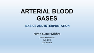
- 1. ARTERIAL BLOOD GASES Navin Kumar Mishra Junior Resident-III IMS-BHU 23-07-2018 BASICS AND INTERPRETATION
- 2. OBJECTIVES • Understand correct sample handling • Outline an interpretation strategy • Describe causes for acid-base disturbances • Delineate a workup strategy for common disorders @ NKMishra23/07/18 2
- 3. ABG • ABG is a very useful diagnostic tool in our day to day practice. • The first arterial puncture was performed in 1912 by Hurter, a German physician. • Drawn from artery- Radial, Brachial, Femoral, Dorsalis pedis, Posterior tibial • It is an invasive procedure. @ NKMishra23/07/18 3
- 4. Acid-Base Physiology • pH is the negative logarithm to the base 10 of the hydrogen ion concentration in mmol/L • pH = - log10[H+] • An increase in pH indicates a proportionate decrease in the [H+] and a decrease in the pH indicates a proportionate increase in the [H+]. • H2CO3 generates 12,500 mmol H+ per day. • Normal metabolism of proteins and nucleotides generates about 100 mmol H+ per day in the form of sulphuric and phosphoric acids.@ NKMishra23/07/18 4
- 5. Calculation of pH • pH is calculated from Henderson-Hasselbalch equation . • pH = pK + log acid/bas • pH = 6.1 + log HCO3- H2CO3 Kassirer and Bliech modified equation • H+ = 24 x PCO2/HCO3- @ NKMishra23/07/18 5
- 6. Regulation of pH pH is maintained in narrow range by • 1) In seconds: buffer systems • 2)In minutes: CO2 excretion by the lungs • 3)In hours to days: renal excretion of H+, reabsorption of HCO3 @ NKMishra23/07/18 6
- 7. Regulation of arterial pH • 1.BUFFERS –Buffer systems minimize the change in pH resulting from production of acid . • Main buffer system in humans is HCO3- in ECF and protein and phosphate buffers in ICF. 2.ROLE OF THE RESPIRATORY SYSTEM–Elimination of volatile acid CO2. • a. Respiratory centers in the brain respond to changes in pH of CSF and blood to affect ventilatory rate. • b. Ventilation directly controls the elimination of CO2. @ NKMishra23/07/18 7
- 8. ROLE OF KIDNEY It retains and regenerate HCO3- thereby regenerating the body buffer with the net effect of eliminating the non- volatile acid load a. H+ secretion 1. Free urinary H+ - minimal contribution 2. Ammonia 3. Phosphorus b. HCO3- reabsorption 1. Proximal tubule – 90% 2. Distal tubule -10% @ NKMishra23/07/18 8
- 9. Applications of ABG • To document respiratory failure and assess its severity. • To monitor patients on ventilators and assist in weaning. • To assess acid base imbalance in critical illness. • To assess response to therapeutic interventions and mechanical ventilation. • To assess pre-op patients. @ NKMishra23/07/18 9
- 10. VENTILATION PaCO2 = VCO2 x K VA Hypercapnea > 45 mm Hg (Hypoventilation) Respiratory Acidosis Hypocapnea < 35 mm Hg (Hyperventilation) Respiratory Alkalosis VA=Portion of total ventilation participate in gas exchange with pulmonary blood @ NKMishra23/07/18 10
- 11. OXYGENATION • P(A-a)O2 • O2 content • PaO2 / FiO2 ratio • arterial-Alveolar O2 tension ratio @ NKMishra23/07/18 11
- 12. P(A-a)O2= PAO2- PaO2 • PAO2- partial pressure of oxygen in alveolar gas pAO2 = pIO2 – (paCO2 / R) PAO2= (PB-P h2o) x FiO2- (paCO2/R) Eg.: =(760-47) x .21- (40/0.8) =100 PAO2-Pao2= 100-80=20 N= <15 PAO2 = partial pressure of oxygen in alveolar gas, PB = barometric pressure (760mmHg), Ph2o = water vapor pressure (47 mm Hg), FiO2 = fraction of inspired oxygen, PCO2 = partial pressure of CO2 in the ABG, R = respiratory quotient (0.8) @ NKMishra23/07/18 12
- 13. P(A-a)O2= PAO2- PaO2 • PaO2- partial pressure of oxygen in blood PaO2 = FiO2 × 5 PaO2 = 109 - 0.4 (Age) PaO2 is dependant upon Age, FiO2, Patm HYPOXEMIA Mild (60-80) mmHg Moderate(40-60) mmHg Severe <40 mmHg @ NKMishra23/07/18 13
- 15. Arterial Blood pH 7.4 Neutral Academia Acidosis? Alkalemia Alkalosis? 7.35~7.45 6.8 7.8 @ NKMishra23/07/18 15
- 16. TECHNIQUES of Sampling @ NKMishra23/07/18 16
- 17. SAMPLE MANAGEMENT • Sample needs to be iced • At room temperature sample lasts 15 – 20 min • PaCO2 increases • PaO2 decreases • pH decreases Red cell glycolysis @ NKMishra23/07/18 17
- 18. CAUTION pH PaCO2 PaO2 Heparin ↓ ↓ Air Bubbles ↓ ↑ @ NKMishra23/07/18 18
- 20. Technical Errors Risk of alteration of results with: 1)size of syringe/needle 2)vol of sample Syringes must have > 50% blood Use only 3ml or less syringe 25% lower values if 1 ml sample taken in 10 ml syringe (0.25 ml heparin in needle) @ NKMishra23/07/18 20
- 21. Parameters Excessive Heparin Air bubbles pH ↓ or remain the same ↑ PCO2 ↓ ↓ PO2 May altered May altered HbO2% sat May altered May altered HbCO2% sat Will not altered Will not altered Hb content ↓ Is not altered HCO3 ↓ ↓ Base Excess ↓ ↓ Oxygen content May be altered Maybe altered @ NKMishra23/07/18 21
- 22. Technical Errors WBC Counts 0.01 ml O2 consumed/dL/min Marked increase in high TLC/plt counts : dec.pO2 Chilling / immediate analysis ABG Syringe must be transported earliest via COLD CHAIN Change/10 min Uniced 370C Iced 40C pH 0.01 0.001 pCO2 1 mm Hg 0.1 mm Hg pO2 0.1% 0.01% @ NKMishra23/07/18 22
- 23. Site Selection • Radial Artery – 45. insertion angle • Brachial Artery – 60-90 .insertion angle • Femoral Artery – 90 insertion angle • Dorsalis Pedis Artery • Posterior Tibial artery @ NKMishra23/07/18 24
- 29. Contraindication • No absolute contraindications • Dialysis shunt – choose another site • Patient on anticoagulant/aspirin therapy – may have to hold pressure on puncture site longer than normal @ NKMishra23/07/18 30
- 30. Site specific contraindication Radial : Buergers disease Raynauds Absent Ulnar collateral circulation AV dialysis shunt Femoral: Local infection @ NKMishra23/07/18 31
- 32. @ NKMishra Only the person who collect the sample can tell if he has drawn a pulsating blood Partly mixed sample- Difficult to recognize Arterial Venous PH 7.35 - 7.45 7.36- 7.39 Pco2 35- 45 44 - 48 Po2 80 - 100 38 - 42 Hco3 24- 26 20 - 24 Sao2 95- 100 % 75% 23/07/18 33
- 34. Step 1: Assess the internal consistency of the values using the Henderseon-Hasselbach equation: • [H+] = 24(PaCO2) [HCO3-] @ NKMishra23/07/18 35
- 35. pH Approximate [H+] (mmol/L) 7.00 100 7.05 89 7.10 79 7.15 71 7.20 63 7.25 56 7.30 50 7.35 45 7.40 40 7.45 35 7.50 32 7.55 28 7.60 25 7.65 22 If the pH and the [H+] are inconsistent, the ABG is probably not valid. @ NKMishra23/07/18 36
- 36. Step 2: Is there alkalemia or acidemia present? • pH < 7.35 acidemia pH > 7.45 alkalemia • This is usually the primary disorder • Remember: an acidosis or alkalosis may be present even if the pH is in the normal range (7.35 – 7.45) • You will need to check the PaCO2, HCO3- and anion gap @ NKMishra23/07/18 37
- 37. • Step 3: Is the disturbance respiratory or metabolic? • What is the relationship between the direction of change in the pH and the direction of change in the PaCO2? • In primary respiratory disorders, the pH and PaCO2 change in opposite directions; in metabolic disorders the pH and PaCO2 change in the same direction. Acidosis Respiratory pH ↓ PaCO2 ↑ ROME Acidosis Metabolic& pH ↓ PaCO2 ↓ ROME Alkalosis Respiratory pH ↑ PaCO2 ↓ ROME Alkalosis Metabolic pH ↑ PaCO2 ↑ ROME @ NKMishra23/07/18 38
- 38. • Step 4: Is there appropriate compensation for the primary disturbance? • Usually, compensation does not return the pH to normal (7.35 – 7.45) except….? Disorder Expected compensation Correction factor Metabolic acidosis PaCO2 = (1.5 x [HCO3-]) +8 ± 2 Acute respiratory acidosis Increase in [HCO3-]= ∆ PaCO2/10 ± 3 Chronic respiratory acidosis (3-5 days) Increase in [HCO3-]= 3.5(∆ PaCO2/10) Metabolic alkalosis Increase in PaCO2 = 40 + 0.6(∆HCO3-) Acute respiratory alkalosis Decrease in [HCO3-]= 2(∆ PaCO2/10) Chronic respiratory alkalosis Decrease in [HCO3-] = 5(∆ PaCO2/10) to 7(∆ PaCO2/10) If the observed compensation is not the expected compensation, it is likely that more than one acid-base disorder is present. @ NKMishra23/07/18 39
- 39. • Step 5: Calculate the anion gap (if a metabolic acidosis exists): • AG= [Na+]-( [Cl-] + [HCO3-] )=12 ± 2 • A normal anion gap is approximately 12 meq/L. • In patients with hypoalbuminemia, the normal anion gap is lower than 12 meq/L; the “normal” anion gap in patients with hypoalbuminemia is about 2.5 meq/L lower for each 1 gm/dL decrease in the plasma albumin concentration (for example, a patient with a plasma albumin of 2.0 gm/dL would be approximately 7 meq/L.) • If the anion gap is elevated, consider calculating the osmolal gap in compatible clinical situations. • Elevation in AG is not explained by an obvious case (DKA, lactic acidosis, renal failure • Toxic ingestion is suspected • OSM gap = measured OSM – (2[Na+] - glucose/18 – BUN/2.8 • The OSM gap should be < 10 @ NKMishra23/07/18 40
- 40. • Step 6: If an increased anion gap is present, assess the relationship between the increase in the anion gap and the decrease in [HCO3-]. • Assess the ratio of the change in the anion gap (∆AG ) to the change in [HCO3- ] (∆[HCO3-]): ∆AG/∆[HCO3-] • This ratio should be between 1.0 and 2.0 if an uncomplicated anion gap metabolic acidosis is present. • If this ratio falls outside of this range, then another metabolic disorder is present: • If ∆AG/∆[HCO3-] < 1.0, then a concurrent non-anion gap metabolic acidosis is likely to be present. • If ∆AG/∆[HCO3-] > 2.0, then a concurrent metabolic alkalosis is likely to be present. • It is important to remember what the expected “normal” anion gap for your patient should be, by adjusting for hypoalbuminemia (see Step 5, above.) @ NKMishra23/07/18 41
- 41. Disorder pH Primary problem Compensation Metabolic acidosis ↓ ↓ in HCO3- ↓ in PaCO2 Metabolic alkalosis ↑ ↑ in HCO3- ↑ in PaCO2 Respiratory acidosis ↓ ↑ in PaCO2 ↑ in [HCO3-] Respiratory alkalosis ↑ ↓ in PaCO2 ↓ in [HCO3-] Table 1: Characteristics of acid-base disturbances @ NKMishra23/07/18 42
- 42. Table 2: Selected etiologies of respiratory acidosis oAirway obstruction - Upper - Lower o COPD o asthma o other obstructive lung disease oCNS depression oSleep disordered breathing (OSA or OHS) oNeuromuscular impairment oVentilatory restriction oIncreased CO2 production: shivering, rigors, seizures, malignant hyperthermia, hypermetabolism, increased intake of carbohydrates oIncorrect mechanical ventilation settings @ NKMishra23/07/18 43
- 43. Table 3: Selected etiologies of respiratory alkalosis oCNS stimulation: fever, pain, fear, anxiety, CVA, cerebral edema, brain trauma, brain tumor, CNS infection oHypoxemia or hypoxia: lung disease, profound anemia, low FiO2 oStimulation of chest receptors: pulmonary edema, pleural effusion, pneumonia, pneumothorax, pulmonary embolus oDrugs, hormones: salicylates, catecholamines, medroxyprogesterone, progestins oPregnancy, liver disease, sepsis, hyperthyroidism oIncorrect mechanical ventilation settings @ NKMishra23/07/18 44
- 44. Table 4: Selected causes of metabolic alkalosis oHypovolemia with Cl- depletion o GI loss of H+ o Vomiting, gastric suction, villous adenoma, diarrhea with chloride-rich fluid o Renal loss H+ o Loop and thiazide diuretics, post-hypercapnia (especially after institution of mechanical ventilation) oHypervolemia, Cl- expansion o Renal loss of H+: edematous states (heart failure, cirrhosis, nephrotic syndrome), hyperaldosteronism, hypercortisolism, excess ACTH, exogenous steroids, hyperreninemia, severe hypokalemia, renal artery stenosis, bicarbonate administration @ NKMishra23/07/18 45
- 45. Table 5: Selected etiologies of metabolic acidosis oElevated anion gap: o Methanol intoxication o Uremia o Diabetic ketoacidosisa, alcoholic ketoacidosis, starvation ketoacidosis o Paraldehyde toxicity o Isoniazid o Lactic acidosisa o Type A: tissue ischemia o Type B: Altered cellular metabolism o Ethanolb or ethylene glycolb intoxication o Salicylate intoxication a Most common causes of metabolic acidosis with an elevated anion gap b Frequently associated with an osmolal gap oNormal anion gap: will have increase in [Cl-] o GI loss of HCO3- o Diarrhea, ileostomy, proximal colostomy, ureteral diversion o Renal loss of HCO3- o proximal RTA o carbonic anhydrase inhibitor (acetazolamide) o Renal tubular disease o ATN o Chronic renal disease o Distal RTA o Aldosterone inhibitors or absence o NaCl infusion, TPN, NH4+ administration @ NKMishra23/07/18 46
- 46. Disorder Characteristics Selected situations Respiratory acidosis with metabolic acidosis ↓in pH ↓ in HCO3 ↑ in PaCO2 •Cardiac arrest •Intoxications •Multi-organ failure Respiratory alkalosis with metabolic alkalosis ↑in pH ↑ in HCO3- ↓ in PaCO2 •Cirrhosis with diuretics •Pregnancy with vomiting •Over ventilation of COPD Respiratory acidosis with metabolic alkalosis pH in normal range ↑ in PaCO2, ↑ in HCO3- •COPD with diuretics, vomiting, NG suction •Severe hypokalemia Respiratory alkalosis with metabolic acidosis pH in normal range ↓ in PaCO2 ↓ in HCO3 •Sepsis •Salicylate toxicity •Renal failure with CHF or pneumonia •Advanced liver disease Metabolic acidosis with metabolic alkalosis pH in normal range HCO3- normal •Uremia or ketoacidosis with vomiting, NG suction, diuretics, etc. Table 6: Selected mixed and complex acid-base disturbances @ NKMishra23/07/18 47
- 47. ARTERIAL BLOOD GAS MEASURES • PaCO2 • PaO2 • pH CALCULATES • Bicarbonate • SaO2 @ NKMishra23/07/18 48
- 49. Normal Values • pH - 7.35 - 7.45 • PaCO2 - 35-45 mmHg • PaO2 - 80-100 mmHg • HCO3 - 22-26 • O2sat - 95-100% • Base Excess - +/-2 m Eq/L @ NKMishra23/07/18 50
- 50. Base excess and deficit • The BE (or base deficit) is defined as the amount of acid (or base) required to be added to whole blood to achieve a pH of 7.4 at 37˚C and paCO2 of 40mmHg. If the base is in excess • may be due to decrease in metabolic acids • may be due to increase in buffers (e.g. HCO3-) If the base is in deficit • may be due to excess metabolic acids @ NKMishra23/07/18 51
- 51. Step 1: is it reliable?? STEP -2 : Comprehensive history and physical examination. STEP -3 : Acidosis or alkalosis..??? See the pH (<7.35 or >7.45) STEP -4 : Identify the primary disorder See the change in PCo2 & HCO3 STEP -5 : Calculate the compensatory response Is adequately compensated??? @ NKMishra23/07/18 52
- 52. STEP -6 : Calculate anion gap STEP -7 : Calculate the delta gap (unmask hidden mixed disorders) STEP -8 : Calculate the osmolar gap (for high AG acidosis) STEP -9 : Calculate the urinary anion gap (Non AG metabolic acidosis) STEP -10 : Formulate differential diagnosis @ NKMishra23/07/18 53
- 53. INTERPRETATION STEPS 1. pH 2. PaCO2 3. Anion Gap - Delta ratio 4. Compensation pH PaCO2 Disorder ↓ ↓ Metabolic Acidosis ↑ ↑ Metabolic Alkalosis ↓ ↑ Respiratory Acidosis ↑ ↓ Respiratory Alkalosis @ NKMishra23/07/18 54
- 54. ANION GAP (AG) • Normal value 8 – 12 • Primarily determined by negatively charged plasma proteins • Albumin ↓ 1 g/dL (below 4/4.5) AG ↓ 2.5 • AG needs correction for hypoalbuminemia @ NKMishra23/07/18 55
- 55. UNADJUSTED AG ADJUSTED AG Decreased 26.7 % Decreased 4.7 % Normal 22.0 % Normal 62.6 % Normal 36.4 % Increased 26.3 % Increased 10.7 % Increased 10.7 % J Lab Clin Med 2005;146:317–20 @ NKMishra23/07/18 56
- 56. Some quick clues: • 1. if CO2 and HCO3 in opposite direction , it indicates mixed d/o • 2. if pH and Co2 same direction, it indicates Metabolic d/o=ROME • 3. if pH and Co2 same direction, it indicates Respiratory d/o= ROME • If the difference between digits after decimal in PH and CO2 is • <15= metabolic • > 15= Respiratory @ NKMishra23/07/18 57
- 58. Case 0 • pH 7.28 • PaCO2 27 mm Hg • PaO2 105 mm Hg • H+ 70 mmhg • Na+ 134 mmol/L • K+ 3.7 mmol/L • Cl- 109 mmol/L • HCO3 - 13 mmol/L • Albumin 4.0 g/dL • AG 12 @ NKMishra23/07/18 59
- 59. • H+ = 24 X 27/ 13 • = 49.8 • Not Reliable test. @ NKMishra23/07/18 60
- 60. CASE 1 @ NKMishra23/07/18 61
- 61. CASE 1 • pH 7.28 • PaCO2 27 mm Hg • PaO2 105 mm Hg • H+ 50 mmhg • Na+ 134 mmol/L • K+ 3.7 mmol/L • Cl- 109 mmol/L • HCO3 - 13 mmol/L • Albumin 4.0 g/dL ANION GAP = 12 @ NKMishra23/07/18 62
- 62. 1. pH: ↓ 2. PCO2: ↓ 3. Anion Gap: - Delta ratio 4. Compensation CASE 1 METABOLIC ACIDOSIS NON-ANION GAP pH PaCO2 Disorder ↓ ↓ Metabolic Acidosis ↑ ↑ Metabolic Alkalosis ↓ ↑ Respiratory Acidosis ↑ ↓ Respiratory Alkalosis Normal @ NKMishra23/07/18 63
- 63. COMPENSATION PaCO2 = 1.5 x HCO3 - + 8 ± 2 PaCO2 = 1.5 x 13 + 8 ± 2 PaCO2 = 19.5 + 8 ± 2 PaCO2 = 27.5 ± 2 PaCO2 = 25.5 - 29.5 PaCO2 = 27 @ NKMishra23/07/18 64
- 64. 1. pH: ↓ 2. PCO2: ↓ 3. Anion Gap: Normal - Delta ratio 4. Compensation CASE 1 METABOLIC ACIDOSIS NON-ANION GAP ADEQUATE COMPENSATION @ NKMishra23/07/18 65
- 65. NON-ANION GAP ACIDOSIS • Hyperchloremic: Net Bicarbonate loss • Two main different routes • GI: diarrhea, fistulas • Renal: RTAs, drugs • What is used to differentiate? Urine AG = Na+ + K+ - Cl- UAG = Negative normally bcz CL- content is high in urine. • NAG Acidosis: Diarrhea NAG M.Acidosis UAG= Neg K+= dec RTA NAG M.Acidosis UAG= Positive K+= Inc @ NKMishra23/07/18 66
- 67. URINE ANION GAP [Na+] + [K+] + [NH4 +] + [UC] ≈ [Cl-] + [HCO3-] + [UA] [Na+] + [K+] - [Cl-] ≈ [UA] - [UC] UAG ≈ [Na+] + [K+] - [Cl-] ? @ NKMishra23/07/18 68
- 68. HOW TO USE IT NORMAL RESPONSE TO ACIDOSIS UAG ≈ [Na+] + [K+] - [Cl-] UAG < 0 IF KIDNEYS UNABLE TO EXCRETE H+ UAG ≈ [Na+] + [K+] - [Cl-] UAG > 0 NH4 + Cl- NH4 + Cl - @ NKMishra23/07/18 69
- 69. NON ANION GAP ACIDOSIS GASTROINTESTINAL - Diarrhea - External pancreatic or bowel drainage - Ureterosigmoidostomy - Drugs * Calcium Chloride * Magnesium Sulfate * Cholestyramine RENAL - Hypokalemic * Proximal RTA (type 2) * Distal RTA (type 1) * Drugs: Acetazolamide, Amphotericin B - Hyperkalemic * Type 4 RTA * Mineralocorticoid deficiency * Tubulointerstitial disease * Ammonium excretion defect * Drugs: K+ sparing diuretics, ACEi ARBs, Trimethoprim, Pentamidine, NSAIDs, Cyclosprine, tacrolimus, etc @ NKMishra23/07/18 70
- 70. URINE ANION GAP NEGATIVE Gastrointestinal Diarrhea Small bowel / pancreatic drainage IATROGENIC Parenteral nutrition Saline Anion exchange resins POSITIVE URINE pH > 5.5 Type I RTA URINE pH < 5.5 Low K+ Type II RTA High K+ Aldosterone deficiency Type IV RTA @ NKMishra23/07/18 71
- 71. CASE 2 @ NKMishra23/07/18 72
- 72. CASE 2 • pH 7.30 • PaCO2 34 mm Hg • PaO2 235 mm Hg • H+ 48 mmhg • Na+ 144 mmol/L • K+ 4.5 mmol/L • Cl- 105 mmol/L • HCO3 - 17 mmol/L • Albumin 4.0 g/dL ANION GAP = 22 @ NKMishra23/07/18 73
- 73. 1. pH: ↓ 2. PCO2: ↓ 3. Anion Gap: ↑ - Delta Ratio 4. Compensation CASE 2 METABOLIC ACIDOSIS ANION GAP pH PaCO2 Disorder ↓ ↓ Metabolic Acidosis ↑ ↑ Metabolic Alkalosis ↓ ↑ Respiratory Acidosis ↑ ↓ Respiratory Alkalosis @ NKMishra23/07/18 74
- 74. DELTA RATIO • Delta ratio = Δ anion gap / Δ bicarbonate Actual AG – 12 Delta Ratio = 24 – HCO3 - 22 – 12 12 Delta Ratio = = = 1.7 24 – 17 7 @ NKMishra23/07/18 75
- 75. NON-ANION GAP ACIDOSIS (↑↑ AG – 12) Delta Ratio = ----------------------- (24-↓↓↓HCO3) • Delta Ratio < 0.8 METABOLIC ALKALOSIS (↑↑ AG – 12) Delta Ratio = ----------------------- (24 - ≈↑HCO3) • Delta Ratio > 1.5 Δ AG (↑AG-12) Delta Ratio = ------------- = ----------------- Δ HCO3- (24-↓HCO3) (0.8-1.5)1 @ NKMishra23/07/18 76
- 76. DELTA RATIO Δ RATIO < 0.8 0.8 - 1.5 > 1.5 Anion Gap metabolic acidosis Anion Gap metabolic acidosis Anion Gap metabolic acidosis Non anion gap acidosis Metabolic alkalosis @ NKMishra23/07/18 77
- 77. 1. pH: ↓ 2. PCO2: ↓ 3. Anion Gap: ↑ - Delta Ratio = 1.7 4. Compensation CASE 2 METABOLIC ACIDOSIS ANION GAP METABOLIC ALKALOSIS @ NKMishra23/07/18 78
- 78. COMPENSATION PaCO2 = 1.5 x HCO3 - + 8 ± 2 PaCO2 = 1.5 x 17 + 8 ± 2 PaCO2 = 25.5 + 8 ± 2 PaCO2 = 33.5 ± 2 PaCO2 = 31.5 - 35.5 PaCO2 = 34 @ NKMishra23/07/18 79
- 79. 1. pH: ↓ 2. PCO2: ↓ 3. Anion Gap: ↑ - Delta Ratio = 1.7 4. Compensation CASE 2 METABOLIC ACIDOSIS ANION GAP METABOLIC ALKALOSIS ADEQUATE COMPENSATION @ NKMishra23/07/18 80
- 80. HIGH ANION GAP M ethanol U remia D iabetic Ketoacidosis: alcohol, starvation P araldehyde, paracetamol I ron, Isoniazid, inborn errors of metabolism L actic Acidosis E thylene glycol, Ethanol S alicylates @ NKMishra23/07/18 81
- 81. CASE 3 @ NKMishra23/07/18 82
- 82. CASE 3 • pH 7.24 • PaCO2 60 mm Hg • PaO2 158 mm Hg • Na+ 140 mmol/L • K+ 4.4 mmol/L • Cl- 103 mmol/L • HCO3 - 25 mmol/L • Albumin 4.0 g/dL ANION GAP = 12 @ NKMishra23/07/18 83
- 83. 1. pH: ↓ 2. PCO2: ↑ CASE 3 RESPIRATORY ACIDOSIS pH PaCO2 Disorder ↓ ↓ Metabolic Acidosis ↑ ↑ Metabolic Alkalosis ↓ ↑ Respiratory Acidosis ↑ ↓ Respiratory Alkalosis ACUTE OR CHRONIC? @ NKMishra23/07/18 84
- 84. RESPIRATORY DISORDERS Δ10 PaCO2 ACUTE CHRONIC Δ pH 0.08 0.03 @ NKMishra23/07/18 85
- 85. ACUTE OR CHRONIC? • PaCO2 = 60 • pH = 7.24 Δ10 PaCO2 ACUTE CHRONIC Δ pH 0.08 0.03 Δ 2x10 = 20 Δ 2x0.08 =0.16 @ NKMishra23/07/18 86
- 86. 1. pH: ↓ 2. PCO2: ↑ 3. Anion Gap: Normal - Delta Ratio 4. Compensation CASE 3 RESPIRATORY ACIDOSIS ACUTE @ NKMishra23/07/18 87
- 87. HCO3 - ??? 1 2 4 5 Δ 10 PaCO2 ACUTE CHRONIC ACIDOSIS ALKALOSIS @ NKMishra23/07/18 88
- 88. CASE 3 • pH 7.24 • PaCO2 60 mm Hg • HCO3 - 25 mmol/L 1 2 4 5 Δ 10 PaCO2 ACUTE CHRONIC ACIDOSIS ALKALOSIS Δ 2x10=20 Δ 1 @ NKMishra23/07/18 89
- 89. 1. pH: ↓ 2. PCO2: ↑ 3. Anion Gap: Normal - Delta Ratio 4. Compensation CASE 3 RESPIRATORY ACIDOSIS ACUTE ADEQUATE COMPENSATION @ NKMishra23/07/18 90
- 90. CASE 4 @ NKMishra23/07/18 91
- 91. CASE 4 • pH 7.33 • PaCO2 60 mm Hg • PaO2 158 mm Hg • Na+ 146 mmol/L • K+ 4.4 mmol/L • Cl- 102 mmol/L • HCO3 - 32 mmol/L • Albumin 4.0 g/dL ANION GAP = 12 @ NKMishra23/07/18 92
- 92. 1. pH: ↓ 2. PCO2: ↑ CASE 4 RESPIRATORY ACIDOSIS pH PaCO2 Disorder ↓ ↓ Metabolic Acidosis ↑ ↑ Metabolic Alkalosis ↓ ↑ Respiratory Acidosis ↑ ↓ Respiratory Alkalosis ACUTE OR CHRONIC? @ NKMishra23/07/18 93
- 93. RESPIRATORY DISORDERS Δ10 PaCO2 ACUTE CHRONIC Δ pH 0.08 0.03 @ NKMishra23/07/18 94
- 94. ACUTE OR CHRONIC? • PaCO2 = 60 • pH = 7.33 Δ10 PaCO2 ACUTE CHRONIC Δ pH 0.08 0.03 Δ 2x10=20 Δ2x0.03= 0.06 @ NKMishra23/07/18 95
- 95. 1. pH: ↓ 2. PCO2: ↑ 3. Anion Gap: Normal - Delta Ratio 4. Compensation CASE 4 RESPIRATORY ACIDOSIS CHRONIC @ NKMishra23/07/18 96
- 96. CASE 4 • pH 7.33 • PaCO2 60 mm Hg • HCO3 - 32 mmol/L 1 2 4 5 Δ 10 PaCO2 ACUTE CHRONIC ACIDOSIS ALKALOSIS ↑2 x 10 = 20 ↑ 2 x 4 = 8 @ NKMishra23/07/18 97
- 97. 1. pH: ↓ 2. PCO2: ↑ 3. Anion Gap: Normal - Delta Ratio 4. Compensation CASE 4 RESPIRATORY ACIDOSIS CHRONIC ADEQUATE COMPENSATION @ NKMishra23/07/18 98
- 98. CASE 5 @ NKMishra23/07/18 99
- 99. CASE 5 • pH 7.47 • PaCO2 45 mm Hg • PaO2 146 mm Hg • Na+ 143 mmol/L • K+ 4.2 mmol/L • Cl- 101 mmol/L • HCO3 - 33 mmol/L • Albumin 3.0 g/dL ANION GAP = 9 @ NKMishra23/07/18 100
- 100. 1. pH: ↑ 2. PCO2: ↑ 3. Anion Gap: Normal - Delta Ratio 4. Compensation CASE 5 METABOLIC ALKALOSIS pH PaCO2 Disorder ↓ ↓ Metabolic Acidosis ↑ ↑ Metabolic Alkalosis ↓ ↑ Respiratory Acidosis ↑ ↓ Respiratory Alkalosis @ NKMishra23/07/18 101
- 101. COMPENSATION PaCO2 = 0.7 x HCO3 - + 21 ± 2 PaCO2 = 0.7 x 33 + 21 ± 2 PaCO2 = 23.1 + 21 ± 2 PaCO2 = 44.1 ± 2 PaCO2 = 42.1 - 46.1 PaCO2 = 45 @ NKMishra23/07/18 102
- 102. 1. pH: ↑ 2. PCO2: ↑ 3. Anion Gap: Normal - Delta Ratio 4. Compensation CASE 5 METABOLIC ALKALOSIS ADEQUATE COMPENSATION @ NKMishra23/07/18 103
- 103. METABOLIC ALKALOSIS • Decreased H+ concentration • Chloride responsive • Chloride resistant • Measure Urine Cl- • IF Urine CL <20MEQ = Cl responsive M.Alk • IF Urine CL >20MEQ = Cl resistant M.Alk @ NKMishra23/07/18 104
- 104. URINE CHLORIDE CHLORIDE RESPONSIVE Gastric fluid loss Diuretics Post hypercapnia Villous adenoma Congenital chloridorrhea CHLORIDE RESISTANT Laxative abuse Severe K+ depletion Diuretic abuse Bartter or Gitelman Syndrome Primary aldosteronism Adrenal Hyperplasia Cushing syndrome < 20 mEq/L > 20 mEq/L @ NKMishra23/07/18 105
- 105. METABOLIC ALKALOSIS RENAL HYDROGEN LOSS - Primary mineralocorticoid excess - Mineralocorticoid excess-like states * Licorice ingestion * Liddle syndrome - Loop or thiazide diuretics - Bartter or Gitelman syndrome - Post-hypercapnic alkalosis - Hypercalcemia and the milk-alkali syndrome INTRACELLULAR shift of H+ - Severe hypokalemia * Villous Adenoma * Laxative abuse GASTROINTESTINAL H+ LOSS - Vomiting or nasogastric suction - Congenital chloride diarrhea ALKALI ADMINISTRATION WITH REDUCED RENAL FUNCTION CONTRACTION ALKALOSIS @ NKMishra23/07/18 106
- 106. CASE 6 @ NKMishra23/07/18 107
- 107. CASE 6 • pH 7.40 • PaCO2 38 mm Hg • PaO2 106 mm Hg • Na+ 141 mmol/L • K+ 4.8 mmol/L • Cl- 97 mmol/L • HCO3 - 23 mmol/L • Albumin 4.0 g/dL ANION GAP = 21 @ NKMishra23/07/18 108
- 108. 1. pH: Normal 2. PCO2: Normal 3. Anion Gap: ↑ - Delta Ratio 4. Compensation CASE 6 METABOLIC ACIDOSIS ANION GAP pH PaCO2 Disorder ↓ ↓ Metabolic Acidosis ↑ ↑ Metabolic Alkalosis ↓ ↑ Respiratory Acidosis ↑ ↓ Respiratory Alkalosis @ NKMishra23/07/18 109
- 109. DELTA RATIO • Delta ratio = Δ anion gap / Δ bicarbonate Actual AG – 12 Delta Ratio = 24 – HCO3 - 21 – 12 9 Delta Ratio = = = 9 24 – 23 1 @ NKMishra23/07/18 110
- 110. DELTA RATIO Δ RATIO < 0.8 0.8 - 1.5 > 1.5 Anion Gap metabolic acidosis Anion Gap metabolic acidosis Anion Gap metabolic acidosis Non anion gap acidosis Metabolic alkalosis @ NKMishra23/07/18 111
- 111. 1. pH: Normal 2. PCO2: Normal 3. Anion Gap: ↑ - Delta Ratio = 9 4. Compensation CASE 6 METABOLIC ACIDOSIS ANION GAP METABOLIC ALKALOSIS @ NKMishra23/07/18 112
- 112. COMPENSATION PaCO2 = 1.5 x HCO3 - + 8 ± 2 PaCO2 = 1.5 x 23 + 8 ± 2 PaCO2 = 34.5 + 8 ± 2 PaCO2 = 42.5 ± 2 PaCO2 = 40.5 - 44.5 PaCO2 = 38 @ NKMishra23/07/18 113
- 113. 1. pH: Normal 2. PCO2: Normal 3. Anion Gap: ↑ - Delta Ratio = 9 4. Compensation CASE 6 METABOLIC ACIDOSIS ANION GAP METABOLIC ALKALOSIS RESPIRATORY ALKALOSIS @ NKMishra23/07/18 114
- 114. Case 7 • pH: 7.4 • PCO2: 40MM/HG • HCO3: 25MMOL/L • AG: 30 • M.AC with M.Alk with R.Alk @ NKMishra23/07/18 115
- 115. • Case 8. A 54y/o male, Alcoholic presented to ER with c/o vomiting and increased respiratory rate. • PH: 7.4 • PCO2: 30 • HCO3:25 • AG: 30 • M. AC(Alcoholic Ketoacidosis) • with M.Alk(Vomiting) • with R. Alk( Hyperventilation due to hepatic dysfxn or alcohol withdrawal) @ NKMishra23/07/18 116
- 118. SUMMARY 1. pH 2. PaCO2 3. Anion Gap - Delta ratio 4. Compensation DISORDER EXPECTED PCO2 Metabolic Acidosis PaCO2 = 1.5 x HCO3 + 8 ± 2 Metabolic Alkalosis PaCO2 = 0.7 x HCO3 + 21 ± 2 Δ RATIO < 0.8 0.8 - 1.5 > 1.5 Anion Gap metabolic acidosis Anion Gap metabolic acidosis Anion Gap metabolic acidosis Non anion gap acidosis Metabolic alkalosis pH PaCO2 Disorder ↓ ↓ Metabolic Acidosis ↑ ↑ Metabolic Alkalosis ↓ ↑ Respiratory Acidosis ↑ ↓ Respiratory Alkalosis @ NKMishra23/07/18 119
- 119. SUMMARY 1. pH 2. PaCO2 3. Anion Gap - Delta ratio 4. Compensation Δ10 PaCO2 ACUTE CHRONIC Δ pH 0.08 0.03 1 2 4 5 Δ 10 PaCO2 ACUTE CHRONIC ACIDOSIS ALKALOSIS @ NKMishra23/07/18 120
- 120. Treat the patient not the ABG. Thank you!! Have a Nice Day!! @ NKMishra23/07/18 121
- 121. Case-1 • 60 years old M, presents to the ED with rapid breathing and less responsive than usual. No other history available. ABG results pH 7.31 PCO₂ 10 HCO₃ 5 Na 123 K 5 Cl 99 @ NKMishra23/07/18 122
- 122. Stepwise interpretation 1. At pH 7.3 H+ conc. Should be ≈50nmol/L • Calculated H+ = 24 × 10/5 = 24 × 2 = 48 • Both values corroborate, hence result is valid. 2. pH is 7.3, i.e Acidosis 3. HCO₃ value has gone down, primary process is metabolic 4. Respiratory compensation: • Calculated PCO₂ = (1.5 × 5)+8 ± 2 = 13.5 to 17.5 • Partially compensated M.Acidosis a/w respiratory alkalosis :- Mixed disorder @ NKMishra23/07/18 123
- 123. 5. Anion gap: (123+5) – (99 + 5) = 24 • High anion gap metabolic acidosis Finally:- Mixed acid base disorder, with presence of high AG metabolic acidosis and respiratory alkalosis. @ NKMishra23/07/18 124
- 124. Case-2 • A k/c/o COPD with cor pulmonale on treatment presented with progressive breathlessness. ABG results pH 7.42 PCO₂ 67 HCO₃ 42 Na 140 K 3.5 Cl 88 @ NKMishra23/07/18 125
- 125. • pH is normal; but PCO₂ & HCO₃ both are increased. • Change in PCO₂ is 67-40 = 27 • Expected rise in HCO₃ should be 27 × 0.4 = 10.8 • Expected HCO₃ = 24+10.8 ≈ 35 • Actual HCO₃ = 42 • AG = 12 (N) • Mixed disorder, both respiratory acidosis & metabolic alkalosis. @ NKMishra23/07/18 126
- 126. Case -3 • A known case of chronic kidney disease, discontinued dialysis & presented to the emergency in an altered state of sensorium. Attendants gave history of repeated episodes of vomiting at home. ABG results pH 7.42 PCO₂ 40 HCO₃ 25 Na 140 K 3.0 Cl 95@ NKMishra23/07/18 127
- 127. • pH, PCO₂, HCO₃ all WNL • AG = 23 (↑) • Delta gap = 13 – 1 = 12 (↑) • AG >> HCO3 – • Mixed disorder with presence of both high AG metabolic acidosis and metabolic alkalosis. @ NKMishra23/07/18 128
- 128. Case-4 • 65 yrs old M, past h/o Acute MI on medication, presented with high grade fever with, cough & yellowish expectoration for 5 days. Acute increase in shortness of breath. ABG results pH 7.3 PCO₂ 38 HCO₃ 16 Na 136 K 4 Cl 102 @ NKMishra23/07/18 129
- 129. • pH 7.3 = Acidosis • HCO₃ is low ; primary disorder is metabolic acidosis • Expected PCO₂ = (1.5 × 16) + 8 = 32 • Calculated PCO₂ < estimated PCO₂ • AG = 22 • Delta gap = (10-8) = 2 • Mixed disorder with metabolic acidosis & respiratory acidosis @ NKMishra23/07/18 130
- 133. Case-6 • ABHG parameters are following:- PH = 7.39 PaCO2 = 24 HCO3 = 14 Na = 140 K = 4 Cl = 106 AG = 20 @ NKMishra23/07/18 134
- 134. • Result:- High anion gap Metabolic Acidosis with Respiratory Alkalosis • Examples- Lactic acidosis,Sepsis @ NKMishra23/07/18 135
- 135. Case-7 • ABHG parameters are following:- PH = 7.42 PaCO2 = 67 HCO3 = 42 Na = 140 K = 3.5 Cl = 88 AG = 10 @ NKMishra23/07/18 136
- 136. • Result:- Metabolic Alkalosis with Respiratory Acidosis • Examples- COPD Patients @ NKMishra23/07/18 137
- 137. Case-8 • ABHG parameters are following:- PH = 7.3 PaCO2 = 38 HCO3 = 18 Na = 140 K = 4 Cl = 102 AG = 20 @ NKMishra23/07/18 138
- 138. • Result:- Metabolic Acidosis with Respiratory Acidosis • Examples- Pneumonia, Pulmonary Edema @ NKMishra23/07/18 139
- 139. Case-9 • ABHG parameters are following:- PH = 7.42 PaCO2 = 40 HCO3 = 25 Na = 140 K = 3 Cl = 95 AG = 23 @ NKMishra23/07/18 140
- 140. • Result:- Mixed Disorder-Metabolic Acidosis with Metabolic Alkalosis • Examples- Uremia with Vomiting @ NKMishra23/07/18 141
- 141. Case-10 • ABG parameters are following:- PH = 7.2 PaCO2 = 25 HCO3 = 10 Na = 132 K = 3 Cl = 110 AG = 15 @ NKMishra23/07/18 142
- 142. • Result:- Normal AG Hyperchloremic Metabolic Acidosis • Examples- DKA @ NKMishra23/07/18 143
- 144. Causes of High AG Met Acidosis 1. Ketoacidosis: Diabetic Alcoholic Starvation 2. Lactic Acidosis: Type A (Inadequate O2 Delivery to Cells) Type B (Inability of Cells to utilise O2) Type D (Abnormal bowel anatomy) 3. Toxicity: Salicylates Paraldehyde Methanol Toluene Ethylene Glycol 4. Renal Failure 5. Rhabdomyolsis @ NKMishra23/07/18 145
- 145. CAUSES OF NORMAL ANION GAP METABOLIC ACIDOSIS 1.HCO3 loss: GIT Diarrhoea Pancreatic or biliary drainage Urinary diversions (ureterosigmoidostomy) Renal Proximal (type 2) RTA Ketoacidosis (during therapy) Post-chronic hypocapnia @ NKMishra23/07/18 146
- 146. 2. Impaired renal acid excretion: Distal (type 1) RTA Hyperkalemia (type 4) RTA Hypoaldosteronism 3. Misc: Acid Administration (NH4Cl) Hyperalimentation Cholestyramine Cl HCl therapy (Rx of severe met alkalosis) @ NKMishra23/07/18 147
- 148. CAUSES OF METABOLIC ALKALOSIS 1. EXOGENOUS HCO3- LOADS Acute alkali administration Milk – alkali syndrome 2. Effective ECFV contraction, normotention, K+ deficiency and secondary Hyperreninemic hyperaldosteronism: GI LOSS: Vomiting Gastric Aspiration Villous adenoma @ NKMishra23/07/18 149
- 149. RENAL LOSS : Diuretics Post hypercapnic state Hypercalcaemia Recovery from LA/KA Mg2+ deficiency Bartters/Gitelmans syndr Nonreabs anions – penicill 3. ECFV expansion, hypertension,K+ deficiency, and mineralocorticoid excess: HIGH RENIN : RAS Accelerated hypertension Renin sec tumor @ NKMishra23/07/18 150
- 150. LOW RENIN : PRIMARY ALDOSTERONISM – Adenoma, hyperplasia , carcinoma ADRENAL ENZYME DEFECTS – 11 b Hydroxylase CUSHINGS SYNDROME OR DIS. 4. Gain of function mutation of renal sodium channel with ECF expansion , hypertension , K+ deficiency and hyporeninemic hypoaldosteronism : called as LIDDLES SYNDROME @ NKMishra23/07/18 151
- 152. Causes of Respiratory Acidosis 1. CENTRAL : Drugs( anesthetics, morphine , sedatives) Stroke Infection 2. AIRWAY : Obstruction Asthma 3. PARENCHYMA : Emphysema Pneumoconiosis Bronchitis ARDS Barotrauma @ NKMishra23/07/18 153
- 153. • 4. NEUROMUSCULAR : • Poliomyelitis • Kyphoscoliosis • Myasthenia • Muscular dystrophies 5. MISCELLANEOUS • Obesity • Hypoventilation • Permissive Hypercapnia @ NKMishra23/07/18 154
- 155. Causes of Respiratory Alkalosis 1.CENTRAL NERVOUS SYSTEM STIMULATION Structural Causes Non Structural Causes Head trauma Pain Brain tumor Anxiety CVA Fever Meningitis, encephalitis Psychosis 2. HYPOXEMIA OR TISSUE HYPOXIA Pneumonia, pulm oedema Aspiration High Altitude @ NKMishra23/07/18 156
- 156. 3. STIMULATION OF CHEST RECEPTORS : • Hemothorax • Flail chest • Cardiac failure • Pulmonary embolism 4. MIXED/UNKNOWN MECHANISMS: Drugs – Salicylates Nicotine Progesterone Thyroid hormone Catecholamines Xanthines (Aminophylline & related compounds) Cirrhosis Gram –ve Sepsis Pregnancy Heat exposure Mechanical Ventilation @ NKMishra23/07/18 157
- 157. • ABG is a very useful diagnostic tool for our day to day practice. • Approach to interpret should be step wise & in a systematic manner. • Any abnormal result should be analyzed cautiously in light of clinical context. • Appropriate use of this tool using clinical judgment is of paramount importance @ NKMishra23/07/18 158