HCS PHARMA - (ECM 2022) Development of innovative hiPSC-based model including an innovative 3D modified hyaluronic acid hydroscaffold for phenotypic screening

<!-- wp:paragraph --> <p>We previously showed that human pluripotent stem cells (hiPSCs) provide a suitable model to study<br>metabolic diseases upon hepatocyte-like cell (HLC) differentiation. With a non-invasive approach, hiPSCs can be generated from urine samples of patients and HLCs have been used to model cholesterol metabolism regulation, by the study of LDLR- and PCSK9-mediated autosomal dominant hypercholesterolemia (ADH) as well as PCSK9-mediated familial hypobetalipoproteinemia (FHBL). This model provides promising advantages with a direct link to the patient and with an unlimited source of HLCs. But like all models, there are limitations, mainly by the neonatal characteristic of HLCs lead to difficulties for pharmacological investigations.</p> <!-- /wp:paragraph --> <!-- wp:paragraph --> <p>Therefore, to overcome these burdens, we chose to 1. Differentiate hiPSCs into HLCs in an innovative<br>3D <a href="https://hcs-pharma.com/biomimesys/">hyaluronic acid-based hydroscaffold</a>, BIOMIMESYS® produces by HCS Pharma to enhance their maturation. 2. Adapt our 3D differentiation process to a 96-well format to make it compatible for drug screening. 3. Characterization of the 3D HLCs model by metabolism tests and compare to primary human hepatocyte (PHH).</p> <!-- /wp:paragraph --> <!-- wp:paragraph --> <p>We gathered 3’ SRP data all along the differentiation process and RNAseq has been performed by comparing 2D and 3D differentiation conditions to characterize hiPSCs differentiation into liver organoids. We observed an enhanced expression of most hepatic genes and genes expressed by non-parenchymal cells such as stellate cells. Immunofluorescence data confirmed the co-localization of albumin-positive<br>hepatocytes, desmin-positive stellate cells and LYVE1-positive endothelial cells in liver organoids. Finally, at a functional level, several CYP activities including CYP3A4 were detected at the basal level and successfully induced. Liver organoids responded to pharmacological treatments as shown by their ability to accumulate lipids upon amiodarone treatment or uptake LDL-bodipy upon statin treatment.</p> <!-- /wp:paragraph --> <!-- wp:paragraph --> <p>Altogether, our development gave rise to functional liver organoids generated with a unique and common procedure, in a process of automating for future high throughput screening.</p> <!-- /wp:paragraph -->
Recommended

Recommended
More Related Content
More from HCS Pharma
More from HCS Pharma (20)
HCS PHARMA - (ECM 2022) Development of innovative hiPSC-based model including an innovative 3D modified hyaluronic acid hydroscaffold for phenotypic screening
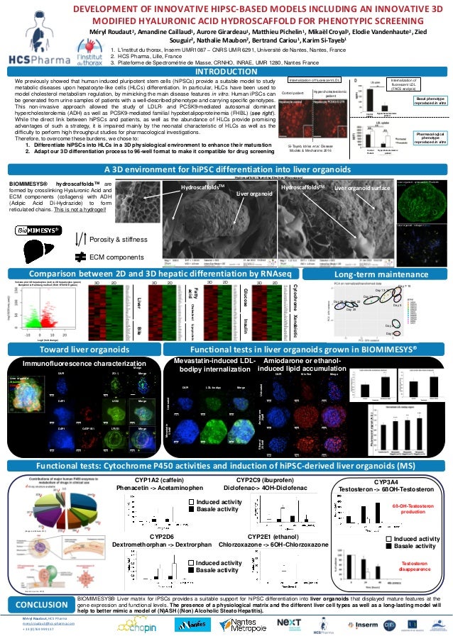
- 1. 1. L'institut du thorax, Inserm UMR1087 – CNRS UMR 6291, Université de Nantes, Nantes, France 2. HCS Pharma, Lille, France 3. Plateforme de Spectrométrie de Masse, CRNHO, INRAE, UMR 1280, Nantes France DEVELOPMENT OF INNOVATIVE HIPSC-BASED MODELS INCLUDING AN INNOVATIVE 3D MODIFIED HYALURONIC ACID HYDROSCAFFOLD FOR PHENOTYPIC SCREENING Méryl Roudaut2, Amandine Caillaud1, Aurore Girardeau1, Matthieu Pichelin1, Mikaël Croyal3, Elodie Vandenhaute2, Zied Souguir2, Nathalie Maubon2, Bertrand Cariou1, Karim Si-Tayeb1 INTRODUCTION We previously showed that human induced pluripotent stem cells (hiPSCs) provide a suitable model to study metabolic diseases upon hepatocyte-like cells (HLCs) differentiation. In particular, HLCs have been used to model cholesterol metabolism regulation, by mimicking the main disease features in vitro. Human iPSCs can be generated from urine samples of patients with a well-described phenotype and carrying specific genotypes. This non-invasive approach allowed the study of LDLR- and PCSK9-mediated autosomal dominant hypercholesterolemia (ADH) as well as PCSK9-mediated familial hypobetalipoproteinemia (FHBL) (see right). While the direct link between hiPSCs and patients, as well as the abundance of HLCs provide promising advantages of such a strategy, it is impaired mainly by the neonatal characteristic of HLCs as well as the difficulty to perform high throughput studies for pharmacological investigations. Therefore, to overcome these burdens, we chose to: 1. Differentiate hiPSCs into HLCs in a 3D physiological environment to enhance their maturation 2. Adapt our 3D differentiation process to 96-well format to make it compatible for drug screening CONCLUSION Méryl Roudaut, HCS Pharma meryl.roudaut@hcs-pharma.com + 33 (0)769 999 137 Comparison between 2D and 3D hepatic differentiation by RNAseq www.umr1087.univ-nantes.fr A 3D environment for hiPSC differentiation into liver organoids Porosity & stiffness ECM components (Zanger and Schwab, 2013) (Brunton L and al., 2018) Testosteron disappearence 6ß-OH-Testosteron production Functional tests: Cytochrome P450 activities and induction of hiPSC-derived liver organoids (MS) Liver Bile Fatty acid Cholesterol Lipoprotein Glucose Insulin Cytochrome Xenobiotic Volcano plot 3D hepatocytes (red) vs 2D hepatocytes (green) Benjamini & Hochberg method (7448 /57905 DE genes) Log2 (fold change) CYP1A2 (caffein) Phenacetin -> Acetaminophen CYP2D6 Dextromethorphan -> Dextrorphan CYP2C9 (ibuprofen) Diclofenac-> 4OH-Diclofenac CYP2E1 (ethanol) Chlorzoxazone -> 6OH-Chlorzoxazone CYP3A4 Testosteron -> 6ßOH-Testosteron Induced activity Basale activity BIOMIMESYS® Liver matrix for iPSCs provides a suitable support for hiPSC differentiation into liver organoids that displayed mature features at the gene expression and functional levels. The presence of a physiological matrix and the different liver cell types as well as a long-lasting model will help to better mimic a model of (N)ASH ((Non) Alcoholic Steato Hepatitis). Toward liver organoids Functional tests in liver organoids grown in BIOMIMESYS® DAPI LDL-bodipy Merge Untreated Mevastatin 50nM 50µm 50µm 50µm 50µm 50µm 50µm Liver organoid Albumin Desmin DAPI Merge 3D 2D 3D 2D 3D 2D 3D 2D Amiodarone or ethanol- induced lipid accumulation Mevastatin-induced LDL- bodipy internalization 50µm 50µm 50µm DAPI Nile Red Merge Untreated 50µm 50µm 50µm Amiodarone 20µM 50µm 50µm 50µm Ethanol 200nM Immunofluorescence characterization Merge DAPI ZO-1 Merge 50µm 50µm 50µm 50µm 50µm 50µm 50µm DAPI CD31 LHX2 Merge 50µm 50µm 50µm 50µm DAPI OATP1B1 LYVE1 Merge BIOMIMESYS® hydroscaffoldsTM are formed by crosslinking Hyaluronic Acid and ECM components (collagens) with ADH (Adipic Acid Di-Hydrazide) to form reticulated chains. This is not a hydrogel! Hydroscaffold (Scanning Electron Microscopy) Liver organoid - collagen I (blue) Liver organoid - cytoskeleton : Phalloidin (green) Induced activity Basale activity Induced activity Basale activity Long-term maintenance Day 0 Day 2 Day 5 Day 7-10 Day 13-16 Day 19 Day 22 Day 25 Day 28-49 *** * *** ** *** Internalization of fluorescent LDL Basal phenotype reproduced in vitro Pharmacological phenotype reproduced in vitro Si-Tayeb, Idriss et al. Disease Models & Mechanims 2016 Internalization of fluorescent LDL (FACS analysis) Control patient Hypercholesterolemic patient Control patient Hypercholesterolemic patient Control Patient Hypercholesterolemic patient Liver organoid HydroscaffoldsTM HydroscaffoldsTM Liver organoid surface