
Recommended
More Related Content
What's hot
What's hot (20)
Similar to General x-ray machine and fluoroscopy
Similar to General x-ray machine and fluoroscopy (20)
Recently uploaded
Recently uploaded (20)
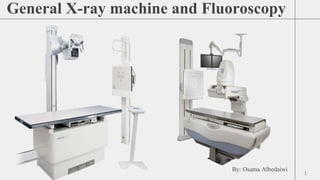
General x-ray machine and fluoroscopy
- 2. CONTENTS 1. What are medical x-rays? 2. History of X-ray. 3. What are some common uses of the procedure? 4. Types of X-Rays. 5. Advantages and disadvantages. 6. How do X-rays work ? 7. Components of X-ray machine. 8. What is Fluoroscopy ? 9. History of fluoroscopy 10. Components of Fluoroscope 11. uses of Fluoroscopy 12. Benefits and Risks of Fluoroscopy Procedures 13. Radiation protection methods 2
- 3. What are medical x-rays? X-rays are a form of electromagnetic radiation, similar to visible light. Unlike light, however, x-rays have higher energy and can pass through most objects, including the body. Medical x-rays are used to generate images of tissues and structures inside the body. If x-rays travelling through the body also pass through an x-ray detector on the other side of the patient, an image will be formed that represents the “shadows” formed by the objects inside the body.
- 4. X-ray ● Non invasive medical test used to produce images of the inside of the body to help diagnose medical conditions. ● X-rays are a form of electromagnetic radiation that is sent through the body. ● Structures that are dense, such as bone, will block most of the X-ray particles and appear white. Metal and contrast media, a special dye used to highlight areas of the body, will appear white. ● Structures containing air will appear black and muscle, fat, and fluid will appear gray. ● Produces two- dimensional images. Examines bones, teeth, lungs, breasts, heart, blood vessels, and the digestive tract. ● Uses ionizing radiation which can increase risk of developing cancer. Particularly effective in bone Not good for analyzing tissue ● Colors: Bone = white Air = black Soft Tissue = gray
- 5. ● Wilhelm Roentgen, Professor of Physics in Wurzburg, Bavaria, discovered X-rays in 1895—accidentally—while testing whether cathode rays could pass through glass. His cathode tube was covered in heavy black paper, so he was surprised when an incandescent green light nevertheless escaped and projected onto a nearby fluorescent screen. Through experimentation, he found that the mysterious light would pass through most substances but leave shadows of solid objects. Because he did not know what the rays were, he called them ‘X,’ meaning ‘unknown,’ rays. ● Roentgen quickly found that X-rays would pass through human tissue too, rendering the bones and tissue beneath visible. News of his discovery spread worldwide, and within a year, doctors in Europe and the United States were using X-rays to locate gun shots, bone fractures, kidney stones and swallowed objects. Honors for his work poured in-- including the first Nobel Prize in physics in 1901. ● Clinical use of the X-ray flourished, with little regard for potential side effects from radiation exposure. There were a few early suspicions from scientists including Thomas Edison, Nikola Tesla, and William J. Morton, each of whom reported injuries they believed resulted from experiments with X-rays. But overall, early use of X-rays was widespread and unrestrained, even to the degree that during the 1930’s and 1940’s, shoe stores offered free X-rays so that customers could see the bones in their feet.
- 6. ● A bone x-ray is used to: - Determine whether a bone has been fractured if a joint is dislocated. - Ensure that a fracture has been properly aligned and stabilized for healing following treatment. - Determine whether there is a build up of fluid in the joint or around a bone.
- 7. Types of X-Rays Several types of X-rays take pictures of different areas inside your body. Some X-rays use contrast material (also known as dye) to make the images clearer. Some of the most common types of X-rays include: ● Abdominal X-ray ● Bone X-ray ● Chest X-ray ● Dental X-ray ● Fluoroscopy ● CT scan (computed tomography) ● Mammogram
- 8. Advantages and Disadvantages Advantages Non invasive test Painless Small radiation dose Can be used with most ill patients Quick medical check up Portable and accessible Disadvantages Exposure to radiation Low contrast and 2D (overlapping structure) Contrast materials sometimes used might produce an allergic reaction Limited information Overused
- 9. ● X-rays are a form of radiation like light or radio waves. X-rays pass through most objects, including the body. Once it is carefully aimed at the part of the body being examined, an x-ray machine produces a small burst of radiation that passes through the body, recording an image on photographic film or a special image recording plate.
- 10. Components of X-ray machine X-ray has three main components 1- Operating Console 2- High Frequency Generator 3- X-ray Tube: Internal and External Other Parts include • Collimator • Grid • Bucky
- 11. What is Fluoroscopy ? ● Fluoroscopy is a common technique used by clinical physicians to obtain real time images of moving body parts and internal structures of a patient ● The fluoroscopy machine ● Takes a continuing stream of x-ray images ● Approximately 25- 30 images per second ● Images are viewed on a monitor
- 12. History of fluoroscopy ● few months later (1896), discovered that calcium tungstate screens produced brighter images. Credited with designing and producing the first commercially available fluoroscope ● Neuilly, France, 1917. Using a fluoroscope, a field doctor examines a wounded soldier for deep-seated bullets. The X-ray tube is visible below the table.
- 13. Components of Fluoroscope X-ray Generator Selection of (kVp) and (mA) X-ray Tube Converts electrical energy provided by the generator into an x-ray beam. Image Intensifier Converts incident x rays into a minified visible light image Focusing lenses To converge Video Camera To record the images CCD Capture real-time image &display Ac current
- 14. Uses of Fluoroscopy ● Used in a variety of procedures ● Examples include: Orthopedic Surgery Catheter Insertion Barium X-Rays Blood Flow Studies Injections into the knees Locating foreign bodies Percutaneous Vertebroplasty Injections into joints or spine
- 15. Benefits and Risks of Fluoroscopy Procedures Benefits •Allows healthcare providers to see movement and function (like in a movie) that cannot be seen in other fixed imaging studies (like a photograph). Guides sometimes life-saving surgical treatments. Risks •Radiation doses are usually higher than in common imaging like x-rays. This means these procedures are slightly more likely to increase the possibility you may get cancer later in life. Some fluoroscopy procedures are longer and use more radiation than others. These could cause skin reddening and hair loss. Contrast dye, if used, can produce an allergic reaction in some people.
- 16. Radiation protection methods 1. Radiation protection, is defined by the International Atomic Energy Agency (IAEA) as "The protection of people from harmful effects of exposure to ionizing radiation, and the means for achieving this". The IAEA also states "The accepted understanding of the term radiation protection is restricted to protection of people.
- 17. 1) Increase the distance between source and personnel. 2) Use of protective barriers. 3) Reduction of exposure factors and unnecessary radiography. 4) Use of radiation monitoring devices. 5) X-ray beam filtration and proper shielding of tube head. 6) Age and sex of involved personnel
- 18. Use of Protective Barriers These barriers are against scatter radiation not against primary beam. Barrier used are; a) Aprons Should have a minimum of 0.25mm lead covering - Material used is lead rubber covered with cloth or plastic impregnated with metallic lead. Aprons should not be folded as protective material tend to separate, should keep flat. Room should also be well coated with lead along with free from any leakage. ▶ The wall of room should be at least 22cm thick and should be of concrete into which iron may be introduced. ▶ Warning signs must be placed near X ray room regarding potential hazards. b) Gloves and Goggles: The lead equivalent of gloves should not be less than 0.33mm. Gloves should be checked by radiography for cracks which can easily be missed on visual inspection. Lead goggles should be used during fluoroscopic examination c) X ray room and equipment: Room should be away from public places Equipment should be checked for possible leakage.
- 19. Reduction of exposure factors and unnecessary radiography Correct exposure factors must be used in ● First attempt. Repeated exposures definitely enhance exposure level. ● Many time owners demand radiography when it is not justified, unnecessary exposure should be avoided. ● X-ray beam filtration and proper shielding of tube head. a) Filtration: Aluminum filters of at least 2.5mm thickness should be used to absorb the soft X rays to reduce the amount of scatter radiation. By this exposure dose can be reduced up to three to four times. b) X-ray tube head: It should be well shielded from all sides except the exit window.
- 20. ALARA ● The guiding principle of radiation safety is “ALARA”. ALARA stands for “as low as reasonably achievable”. ● This principle means that even if it is a small dose, if receiving that dose has no direct benefit, you should try to avoid it. 20