The_nose_and_paranasal_sinuses.ppt
•Download as PPT, PDF•
0 likes•4 views

Has a free tip and attached to forehead by the bridge. External orifices (nares) bounded laterally by the ala & medially by nasal septum. Framework above made up of: nasal bones, frontal process of maxilla, nasal part of frontal bone. Framework below : by plates of hyaline cartilage; upper and lower nasal cartilages, and septal cartilage

Recommended
Recommended
More Related Content
Similar to The_nose_and_paranasal_sinuses.ppt
Similar to The_nose_and_paranasal_sinuses.ppt (20)
More from Dr Ndayisaba Corneille
More from Dr Ndayisaba Corneille (20)
Recently uploaded
Recently uploaded (20)
The_nose_and_paranasal_sinuses.ppt
- 1. Dr. NDAYISABA CORNEILLE CEO of CHG MBChB,DCM,BCSIT,CCNA THE NOSE AND PARANASAL SINUS Supported BY
- 2. The nose
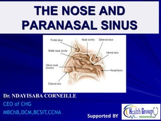
- 3. EXTERNAL NOSE Has a free tip and attached to forehead by the bridge. External orifices (nares) bounded laterally by the ala & medially by nasal septum. Framework above made up of: nasal bones, frontal process of maxilla, nasal part of frontal bone. Framework below : by plates of hyaline cartilage; upper and lower nasal cartilages, and septal cartilage. DR NDAYISABA CORNEILLE 3
- 4. NASAL CAVITY Extends from nares to choanae (posterior nasal apertures) Floor- hard palate Roof- sphenoid and ethmoid bones Medial wall-nasal septum (bony- vomer & perpendicular plate, cartilage anteriorly. Lateral wall-3 projections i.e; superior, middle,& inferior nasal conchae/turbinates. DR NDAYISABA CORNEILLE 4
- 6. Nasal cavity
- 7. NASAL MEATUS Spheno -ethmoid recess- small area above superior concha. Receives opening of sphenoidal air sinus. Superior meatus -below superior concha. Receives opening of posterior ethmoidal air cells. DR NDAYISABA CORNEILLE 7
- 8. Cont… Middle meatus -below middle concha. -Lateral wall has bulla ethmoidalis (bulge of middle ethmoid air cell) which opens there. -Curved cleft(hiatus semilunaris) below bulla leads to funnel shaped channel (infundibulum) anteriorly. -Hiatus semilunaris receives openings of maxillary, frontal & anterior ethmoid sinuses Inferior meatus- below inferior concha . - Receives opening of nasal lacrimal duct. DR NDAYISABA CORNEILLE 8
- 10. Nasal conchae and meatus DR NDAYISABA CORNEILLE 10
- 11. DR NDAYISABA CORNEILLE 11
- 12. OLFACTORY MUCUS MEMBRANE Lines upper surface of superior concha ,spheno -ethmoid recess & corresponding areas of nasal septum. Contains olfactory nerves which receive olfactory stimuli. Olfactory nerves pass through cribriform plate foramina and fuse in cranial cavity to form the olfactory bulb. DR NDAYISABA CORNEILLE 12
- 13. Respiratory mucus membrane. Lines lower part of nasal cavity. Warms ,moistens & cleans inspired air. Plexus of veins in sub mucosa warm air. Mucus comes from glands and goblet cells. Moist and sticky surface removes dust. Contaminated mucus moved backward by ciliated epithelium and swallowed. DR NDAYISABA CORNEILLE 13
- 14. Cont… DR NDAYISABA CORNEILLE 14
- 15. Nerve supply: Olfactory nerves Nerves of general sensation- from ophthalmic and maxillary divisions of trigeminal nerve. Anterior nasal cavity- anterior ethmoidal nerve. Posterior part- nasal, nasalpalatine,& palatine branches of pterygopalatine ganglion. DR NDAYISABA CORNEILLE 15
- 16. DR NDAYISABA CORNEILLE 16
- 17. DR NDAYISABA CORNEILLE 17
- 18. Innervation and blood supply of nasal septum DR NDAYISABA CORNEILLE 18
- 19. Blood supply Sphenopalatine artery- branch of maxillary artery. Anterior and posterior ethmoidal arteries – branches of the ophthalmic artery. Superior labial branch of facial artery. These branches anastomose in the vestibule (kiesselbach’s/little’s area). This is a very common site for Epistaxis. VEINS: form plexus in submucosa and drained by veins accompanying arteries. DR NDAYISABA CORNEILLE 19
- 20. DR NDAYISABA CORNEILLE 20
- 21. Lymphatic drainage Vestibule lymphatics end in submandibular node. Rest of nasal cavity drains to upper deep cervical nodes. DR NDAYISABA CORNEILLE 21
- 23. PARANASAL SINUSES Air-filled extensions of the respiratory part of the nasal cavity into 4 cranial bones: Frontal, Ethmoid, Sphenoid, and Maxilla. The sinuses continue to invade the surrounding bones, and Marked extensions are common in the crania of older individuals. Lined with mucoperiosteum. DR NDAYISABA CORNEILLE 23
- 24. DR NDAYISABA CORNEILLE 24
- 25. DR NDAYISABA CORNEILLE 25
- 26. FUNCTION Mucus produced by the mucous membrane is moved into the nose by ciliary action of the columnar cells. Drainage of the mucus is also achieved by the siphon action created during the blowing of the nose. Sinuses act as resonators to the voice; also reduce the weight of the skull. When the apertures of the sinuses are blocked or they become filled with fluid, the quality of the voice is markedly changed. DR NDAYISABA CORNEILLE 26
- 27. SINUSITIS Infection may spread from the nasal cavities, producing inflammation and swelling of the mucosa of the sinuses (sinusitis) and local pain. Sometimes several sinuses are inflamed (pansinusitis), and the swelling of the mucosa may block one or more openings of the sinuses into the nasal cavities. DR NDAYISABA CORNEILLE 27
- 28. FRONTAL SINUSES Lie between the outer and the inner tables of the frontal bone, posterior to the superciliary arches and the root of the nose. The two frontal sinuses are contained within the frontal bone. They are separated from each other by a bony septum. Each sinus is roughly triangular. DR NDAYISABA CORNEILLE 28
- 29. Cont… Usually detectable in children by 7 years of age. Each sinus drains through a frontonasal duct into the ethmoidal infundibulum, which opens into the semilunar hiatus of the middle nasal meatus. The frontal sinuses are innervated by branches of the supraorbital nerves (CN V1). DR NDAYISABA CORNEILLE 29
- 30. DR NDAYISABA CORNEILLE 30
- 31. VARIATION OF THE FRONTAL SINUSES The right and left frontal sinuses are rarely of equal size, and the septum between them is not usually situated entirely in the median plane. The frontal sinuses vary in size from approximately 5 mm to large spaces extending laterally into the greater wings of the sphenoid. Often a frontal sinus has two parts: a vertical part in the squamous part of the frontal bone and a horizontal part in the orbital part of the frontal bone. One or both parts may be large or small. When the supraorbital part is large, its roof forms the floor of the anterior cranial fossa and its floor forms the roof of the orbit. DR NDAYISABA CORNEILLE 31
- 32. ETHMOIDAL SINUS The ethmoidal cells (sinuses) are small invaginations of the mucous membrane of the middle and superior nasal meatus into the ethmoid bone between the nasal cavity and the orbit. The ethmoidal cells usually are not visible in plain radiographs before 2 years of age but are recognizable in CT scans. The anterior ethmoidal cells drain directly or indirectly into the middle nasal meatus through the ethmoidal infundibulum. DR NDAYISABA CORNEILLE 32
- 33. Cont… The middle ethmoidal cells open directly into the middle meatus and are sometimes called the ethmoidal bulla, a swelling on the superior border of the semilunar hiatus. The posterior ethmoidal cells open directly into the superior meatus. The ethmoidal cells are supplied by the anterior and posterior ethmoidal branches of the nasociliary nerves (CN V1). DR NDAYISABA CORNEILLE 33
- 34. INFECTION OF ETHMOIDAL SINUSES If nasal drainage is blocked, infections of the ethmoidal cells may break through the fragile medial wall of the orbit. Severe infections from this source may cause blindness because some posterior ethmoidal cells lie close to the optic canal, which gives passage to the optic nerve and ophthalmic artery. Spread of infection from these cells could also affect the dural nerve sheath of the optic nerve, causing optic neuritis. DR NDAYISABA CORNEILLE 34
- 35. SPHENOID SINUSES The sphenoidal sinuses are located in the body of the sphenoid and may extend into the wings of this bone. There is extensive pneumatization (formation of air cells or sinuses), rendering the body of the sphenoid fragile. Only thin plates of bone separate the sinuses from several important structures: the optic nerves and optic chiasm, the pituitary gland, the internal carotid arteries, and the cavernous sinuses. DR NDAYISABA CORNEILLE 35
- 36. Cont… The sphenoidal sinuses are derived from a posterior ethmoidal cell that begins to invade the sphenoid at approximately 2 years of age. In some people, several posterior ethmoidal cells invade the sphenoid, giving rise to multiple sphenoidal sinuses that open separately into the sphenoethmoidal recess. The posterior ethmoidal arteries and posterior ethmoidal nerve supply the sphenoidal sinuses. DR NDAYISABA CORNEILLE 36
- 37. DR NDAYISABA CORNEILLE 37
- 38. MAXILLARY SINUS The maxillary sinuses are the largest of the paranasal sinuses. They occupy the bodies of the maxillae and communicate with the middle nasal meatus. The apex of the maxillary sinus extends toward and often into the zygomatic bone. The base of the maxillary sinus forms the inferior part of the lateral wall of the nasal cavity. The roof of the maxillary sinus is formed by the floor of the orbit. DR NDAYISABA CORNEILLE 38
- 39. MAXILLARY SINUS Each maxillary sinus drains by one or more openings, the maxillary ostium (ostia), into the middle nasal meatus of the nasal cavity by way of the semilunar hiatus. The arterial supply of the maxillary sinus is mainly from superior alveolar branches of the maxillary artery; however, branches of the descending and greater palatine arteries supply the floor of the sinus. Innervation of the maxillary sinus is from the anterior, middle, and posterior superior alveolar nerves, which are branches of the maxillary nerve. DR NDAYISABA CORNEILLE 39
- 41. DR NDAYISABA CORNEILLE 41
- 42. END DR NDAYISABA CORNEILLE THANKS FOR LISTENING By DR NDAYISABA CORNEILLE MBChB,DCM,BCSIT,CCNA Contact us: amentalhealths@gmail.com/ ndayicoll@gmail.com whatsaps :+256772497591 /+250788958241 42