THE_MENINGES,CEREBRAL_SINAL_FLUID.pptx
•Download as PPTX, PDF•
0 likes•48 views

1. The document discusses the meninges, cerebral spinal fluid, and dural venous sinuses. It describes the three meningeal layers - dura mater, arachnoid mater, and pia mater. 2. It then provides details on the various dural venous sinuses, including their locations, tributaries, and drainage. Key sinuses discussed include the superior sagittal sinus, straight sinus, transverse sinus, sigmoid sinus, and cavernous sinus. 3. The document also covers cerebral spinal fluid, including its composition and functions. The choroid plexus is described as actively secreting CSF in the ventricles.

Recommended
More Related Content
Similar to THE_MENINGES,CEREBRAL_SINAL_FLUID.pptx
Similar to THE_MENINGES,CEREBRAL_SINAL_FLUID.pptx (20)
More from Dr Ndayisaba Corneille
More from Dr Ndayisaba Corneille (20)
Recently uploaded
Recently uploaded (20)
THE_MENINGES,CEREBRAL_SINAL_FLUID.pptx
- 1. Dr. NDAYISABA CORNEILLE CEO of CHG MBChB,DCM,BCSIT,CCNA THE MENINGES,CEREBRAL SINAL FLUID. Supported BY
- 2. MENINGES • Encloses the brain and spinal cord • Include; • the dura mater, • arachnoid mater, • pia mater. DR NDAYISABA CORNEILLE 2
- 3. The dura mater • Consists of the -outer periosteal layer: attached to the inner periosteum of the skull and continuous on the outside through the foramen magnum -inner meningeal layer: in contact arachnoid mater and continuous with the spinal dura through the foramen magnum. DR NDAYISABA CORNEILLE 3
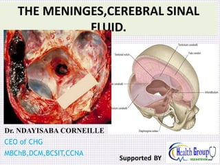
- 4. Cont’n • The layers of dura mater separate to form dural partitions and coatings of the intracranial venous structures.i.e. • falx cerebri: projects downward btn two cerebral hemispheres, anteriorly attached to the ethmoid bone and posteriorly blends with tentorium cerebelli. • Tentorium cerebelli: separates the cerebelli from the cerebral hemispheres posterioly.Attached posterioly to occipital bone and laterally to the temporal bone.ant & lat forms a tentorium notch. DR NDAYISABA CORNEILLE 4
- 6. Cont’n • Falx cerebelli: projects btn two cerebellar ,attached to the internal occipital crest posteriorly and superiorly to the tentorium cerebelli • Diaphragm sellae: this covers the hypophysial fossa of the sellae turcica Arterial supply-anterior meningeal artery -middle and accesory meningeal artery (most important because its susceptible to injury ) -posterior meningeal artery DR NDAYISABA CORNEILLE 6
- 7. Cont’n • Venous supply Meningeal veins, lie lateral to the arteries, middle meningeal vein drains into the sphenoparietal sinus. DR NDAYISABA CORNEILLE 7
- 9. Cont’n • Innervation: trigeminal (v1,v2 ,&v3)vagus and the first three cervical nerves. • Anterior/falx cerebri & tentorium cerebelli: meningeal branches from ethmoidal nvs(ophthalmic nerve,v1) • Middle:maxillary nerve v2,and mandibular nv v3 laterally • Posterior:first to third cervical nvs. DR NDAYISABA CORNEILLE 9
- 10. Arachnoid mater • Is a thin avascular & impermiable layer that lies btn the dura and the pia • Separated from dura and pia by a subdural space and subarachnoid space respectively. • Projects into venous sinuses to form arachnoid villi which when agregated form the arachnoid granulations.(sites for csf diffuson into the blood stream) DR NDAYISABA CORNEILLE 10
- 11. Pia mater • It’s a vascular mebrane that closely covers the brain ,the gyri deep to the sulci. fuses with the nv epineurium. • Clinical Importance: intracranial heamorrhage • Extradural heamorrhage: arterial or venous injury,the middle meningeal artery is mostly injured. • Subdural heamorrhage: damage to the superior cerebral veins. • Subarachnoid heamorrhage: rapture of congenital anuerysms of the circleof willis. • meningitis DR NDAYISABA CORNEILLE 11
- 12. DR NDAYISABA CORNEILLE 12
- 13. DR NDAYISABA CORNEILLE 13
- 14. DR NDAYISABA CORNEILLE 14
- 15. • 15th of all primary tumours • Slow growing and benign • Arise from leptomeninges and cells of arachnoid granules MENINGIOMA DR NDAYISABA CORNEILLE 15
- 16. DR NDAYISABA CORNEILLE 16
- 17. CSF DR NDAYISABA CORNEILLE 17
- 18. Cerebral Spinal Fluid • Found in the ventricles and subarachnoid spaces of the brain and spinal fluid. • Its clear,colourless with same inorganic salt content as blood.(150ml) DR NDAYISABA CORNEILLE 18
- 19. Cont’n Functions of the Cerebrospinal Fluid • Cushions and protects the central nervous system from trauma • Provides mechanical buoyancy and support for the brain • Serves as a reservoir and assists in the regulation of the contents of the skull • Nourishes the central nervous system • Removes metabolites from the central nervous system • Serves as a pathway for pineal secretions to reach the pituitary gland DR NDAYISABA CORNEILLE 19
- 20. Cont’n The Physical Characteristics and Composition of the CSF • Appearance Clear and colorless • Volume c. 150 mL • Rate of production 0.5 mL/minute • Pressure (spinal tap with patient in lateral recumbent position) 60–150 mm of water Composition • Protein 15–45 mg/100 mL • Glucose 50–85 mg/100 mL • Chloride 720–750 mg/100 mL • Number of cells 0–3 lymphocytes/cumm DR NDAYISABA CORNEILLE 20
- 21. Cont’n • Formation: the choroid plexus actively secrets csf in the ventricles and neuronal metabolites from csf into blood DR NDAYISABA CORNEILLE 21
- 22. a DR NDAYISABA CORNEILLE 22
- 23. Sinuses of duramater • Superior sagittal sinus • Inferior sagittal sinus • Straight sinus • Confluence of sinus DR NDAYISABA CORNEILLE 23
- 24. • Transverse sinus • Sigmoid sinus • Superior petrosal sinuses • inferior petrosal sinuses • Cavernous sinus DR NDAYISABA CORNEILLE 24
- 25. Cavernous sinus Sup. ophthalmic v. Inf. ophthalmic v. (not shown) Central v. of retina (inside optic n.) Sphenoparietal sinus Superficial middle cerebral v. Drainage Tributaries Sup. petrosal sinus Inf. petrosal sinus Intercavernous sinus DR NDAYISABA CORNEILLE 25
- 26. Sup. petrosal sinus Inf. petrosal sinus Sigmoid sinus To int. jugular v. DR NDAYISABA CORNEILLE 26
- 27. a DR NDAYISABA CORNEILLE 27
- 28. • All lie between inner and outer layers of dura except the inferior sagittal and straight sinuses • Receive blood from brain and bone • Lack muscular coat • Lined by endothelium (thin) • Valveless, hence blood can floor in either direction Characterstics of dural venous sinuses DR NDAYISABA CORNEILLE 28
- 29. Image of dural sinuses DR NDAYISABA CORNEILLE 29
- 30. • Lies between 2 layers of falx cerebri along its attached margin • Commences at foramen caecum, widens towards internal occipital protuberance • 3-4 lakes of blood project laterally from it Superior sagittal sinus DR NDAYISABA CORNEILLE 30
- 31. • Receives blood from upper and posterior parts of both medial and lateral surfaces of both hemispheres but not frontal pole • Turns at internal occipital protuberance, mostly to the right to become the transverse sinus DR NDAYISABA CORNEILLE 31
- 32. • Begins just posterior to crista galli • Lies between free margin of falxi cerebri layers • Drains inferior parts of medial surfaces of both hemispheres • flows into straight sinus at attachment/junction of falx cebri and tentorium cerebelli Inferior sagittal sinus DR NDAYISABA CORNEILLE 32
- 33. • Lies between folds of fibrous dura at junction of falx cerebri and tentorium cerebelli • Receives inferior sagittal sinus and great cerebral vein of Galen, veins from adjoining occipital lobes , upper surface of cerebellum • Ends in internal occipital protubarence by turning into the transverse sinus usually on the left Straight sinus DR NDAYISABA CORNEILLE 33
- 34. • Commences at internal occipital protuberence between layers of attached margin of tentorium cerebelli • Courses horizontally forwards • Curves downwards at the junction of petrous and mastoid parts of temporal bone as sigmoid sinus • Right usually larger than left because it receives sagittal sinus Transverse sinus DR NDAYISABA CORNEILLE 34
- 35. • Transverse sinuses communicate at their commencement at internal occipital protuberance • Receive tributaries from nearby surfaces of cerebral and cerebellar hemispheres and sagittal sinus Cont/… DR NDAYISABA CORNEILLE 35
- 36. • Commences at termination of transverse sinus • Curves downwards then forwards to posterior margin of jugular foramen , passes through and expands into superior jugular bulb • Internal jugular vein emerges from it Sagittal sinus DR NDAYISABA CORNEILLE 36
- 37. • Is connected to exterior in its upper part by mastoid emissary vein which joins posterior auricular vien • In the lower part by a vein that passes through posterior condylar foramen to suboccipital plexus of veins • Around margins of foramen magnum a pair of sinuses communicate with internal vertebral plexus Sigmoid sinus DR NDAYISABA CORNEILLE 37
- 38. • Runs inferiorly from commencement of transverse sinus through foramen magnum, skirts margin of foramen and drains into the sigmoid sinus • Receives tributaries from cerebellum and medulla • Drains choroid plexus of fourth ventricle Occipital sinus DR NDAYISABA CORNEILLE 38
- 39. • Lies alongside body of sphenoid bone in middle cranial fossa • Receives blood from orbit, vault bones and cerebral hemisphere • Drains by superior and inferior petrosal sinuses to transverse sinus and IJV respectively • Connected to pterygoid plexus by emissary veins Carvenous sinus DR NDAYISABA CORNEILLE 39
- 40. Cont/… • Blood flows in either direction depending on local venous pressures. • It has no valves and so do veins connected to it. • It is a plexus of veins and not a trabeculated venous space as its name suggests • Size 2cm (L) x 1cm (W) DR NDAYISABA CORNEILLE 40
- 41. Image of cavernous sinus - dorsal view DR NDAYISABA CORNEILLE 41
- 42. • Lies in the space between periosteum of body of sphenoid and a fold of the inner layer of dura which forms roof, lateral walls and superior part of medial wall • Medially, roof continuous with diaphragma sellae • Extends from apex of orbit anteriorly to back/apex of petrous part of temporal bone posteriorly Boundaries / location DR NDAYISABA CORNEILLE 42
- 43. • Lateral-medial surface of temporal bone and uncus of temporal lobe • Floor – Greater wing of sphenoid Cont/… DR NDAYISABA CORNEILLE 43
- 44. Relations • Anterior; apex of orbit • Lateral; anterior part of trigeminal ganglion, temporal lobe of cerebral hemisphere • Posteroinferior part of lateral wall; trigeminal (Meckel’s) cave • Posterior; apex of petrous temporal bone • Medial; body of sphenoid bone • Superior; internal carotid aa, uncus of temporal lobe • Inferior; greater wing of sphenoid DR NDAYISABA CORNEILLE 44
- 45. Contents Structures lying within the cavity: • Internal carotid aa • Abducent nn Structures embedded within the lateral wall: • CN III • CN IV • CN V- ophthalmic and maxillary divisions DR NDAYISABA CORNEILLE 45
- 46. Cont/… • Internal carotid artery and abducent nerve run through the sinus • Oculomotor and trochlear nerves and ophthalmic and maxillary divisions of trigeminal nerve lie in the lateral wall of the sinus DR NDAYISABA CORNEILLE 46
- 47. Qn a. What are the tributaries to the cavernous sinus? b. What veins directly drain the cavernous sinus? DR NDAYISABA CORNEILLE 47
- 48. Veins of the cavernous sinus • Characterised by bidirectional floor • Front; -Superior ophthalmic vein -Inferior ophthalmic vein • Roof; -Superficial middle cerebral vein -Shenoparietal sinus • Back; -superior petrosal sinus -inferior petrosal sinus (larger and empties the bulk of blood from cavernous sinus) DR NDAYISABA CORNEILLE 48
- 49. Cont/… • Floor; - emissary veins which communicate with the pterygoid venous plexus • Medial; - intercavernous sinus DR NDAYISABA CORNEILLE 49
- 50. Cavernous sinus and connections DR NDAYISABA CORNEILLE 50
- 51. Sup. ophthalmic v. Inf. ophthalmic v. Cavernous sinus Angular v. Facial v. DR NDAYISABA CORNEILLE 51
- 52. Sup. labial v. Facial v. Angular v. Sup. ophthalmic v. DR NDAYISABA CORNEILLE 52
- 53. Cavernous sinus Optic chiasm Hypophysis Sphenoid sinuses within body of sphenoid bone Similarities with other sinuses Within dura Differences from other sinuses Lined by endothelium Lacks muscular coat Lacks valves III IV V1 V2 VI Int. carotid a. (w/sympathetic plexus) Contains trabeculae DR NDAYISABA CORNEILLE 53
- 54. The flowing of the blood in dural sinus Sup. sagittal sinus Inf. sagittal sinus Straight sinus Confluence of sinus Transverse sinus Cavernous sinus Sup. petrosal sinus Inf. petrosal sinus Internal jugular vein Sigmoid sinus DR NDAYISABA CORNEILLE 54
- 55. Danger triangle of the face DR NDAYISABA CORNEILLE 55
- 56. CASE HISTORY Patient develops a boil on his upper lip after cutting himself shaving on a hunting trip He presents to his physician with a high fever and severe headaches Patient does not improve with penicillin injections and is admitted to a hospital EXAMINATION Rigidity of neck muscles Upper lip swollen and oozing pus Cheek, side of nose and eyelids swollen Exophthalmos Edema of optic nerve at papilla Inability to move eye Paresthesia of forehead, side of nose and upper cheek Blood culture positive for Staphylococcus aureus DIAGNOSIS ??? DR NDAYISABA CORNEILLE 56
- 57. • Carvenous sinus thrombosis from danger area of the face, can spread to dural sinuses and eyes • Veins exit thru- inferior petrosal sinus • Emissary veins • Pterygoid plexus • Inferior ophthalmic • Deep facial vein Qn. In the above case, ocular symptoms were initially unilateral, but later became bilateral. Provide an explanation DR NDAYISABA CORNEILLE 57
- 58. Sup. labial v. Facial v. Angular v. Sup. ophthalmic v. DR NDAYISABA CORNEILLE 58
- 59. • Only anatomic location in the body where an artery travels completely through venous structure • Rupture of ICA within the sinus- ateriovenous fistula ie carotid –carvenous fistula • Pituitary gland lies between two paired carvenous sinuses – pituitary adenoma will compress the sinus—ophthalmoplegia, ophtalmic sensory loss Clinical significance DR NDAYISABA CORNEILLE 59
- 60. END DR NDAYISABA CORNEILLE THANKS FOR LISTENING By DR NDAYISABA CORNEILLE MBChB,DCM,BCSIT,CCNA Contact us: amentalhealths@gmail.com/ ndayicoll@gmail.com whatsaps :+256772497591 /+250788958241 60
Editor's Notes
- 3. a) What are the tributaries to the cavernous sinus? The cavernous sinus receives blood from the superior and inferior ophthalmic veins, central vein of the retina, superficial middle cerebral vein and the sphenoparietal sinus. b) What veins directly drain the cavernous sinus? The superior and inferior petrosal sinuses drain the cavernous sinus directly.