Parotid_Region.ppt
•Download as PPT, PDF•
0 likes•65 views

The region on the lateral surface of the face that comprises the parotid gland & the structures immediately related to it Largest of the salivary glands Located subcutaneously, below and in front of the external auditory meatus Occupies the deep hollow behind the ramus of the mandible Wedge-shaped when viewed externally, with the base above & the apex behind the angle of the mandible

Recommended
More Related Content
Similar to Parotid_Region.ppt
Similar to Parotid_Region.ppt (20)
More from Dr Ndayisaba Corneille
More from Dr Ndayisaba Corneille (20)
Recently uploaded
Recently uploaded (20)
Parotid_Region.ppt
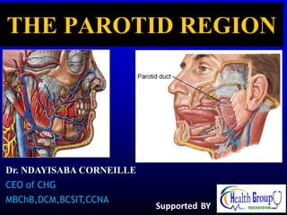
- 1. Dr. NDAYISABA CORNEILLE CEO of CHG MBChB,DCM,BCSIT,CCNA THE PAROTID REGION Supported BY
- 2. The Parotid Region • The region on the lateral surface of the face that comprises the parotid gland & the structures immediately related to it Dr Corneille
- 3. Parotid Gland • Largest of the salivary glands • Located subcutaneously, below and in front of the external auditory meatus • Occupies the deep hollow behind the ramus of the mandible • Wedge-shaped when viewed externally, with the base above & the apex behind the angle of the mandible Dr Corneille
- 4. • Wedge-shaped in horizontal section with the base in the lateral position and apex against the pharyngeal wall. • It exhibits 3 surfaces: Lateral Anteromedial Posteromedial
- 5. Lobes • The facial nerve courses horizontally through the gland and divides it into: Superficial lobe Deep lobe Superficial lobe Deep lobe Facial nerve Dr Corneille
- 6. Processes Glenoid process, that extends upward behind the temporo- mandibular joint, in front of external auditory meatus Facial process, that extends anteriorly onto the masseter muscle Accessory process (part), small part of facial process lying along the parotid duct Pterygoid process, that extends forward from the deeper part, lies between the medial pterygoid muscle & the ramus of mandible Carotid process, that lies posterior to the external carotid artery The gland is an irregular lobulated mass, sends ‘processes’ in various directions. These include: Dr Corneille
- 7. Capsules • The parotid gland is enclosed in two capsules: An inner connective tissue capsule An outer dense fibrous capsule derived from the investing layer of the deep cervical fascia • The deep cervical fascia extends upward, reaches the inferior border of parotid gland, splits into the superficial & the deep layer, to enclose the gland • Above the gland, the: Superficial layer gets attached to the zygomatic arch Deep layer gets attached to the tympanic plate of temporal bone A portion of fascia extending from the styloid process to the angle of mandible is called stylomandibular ligament. It separates the parotid gland from the submandibular gland
- 8. Relations • Superficial (lateral): • Skin & superficial fascia • Great auricular nerve • Parotid lymph nodes • Superior: • External auditory meatus • Temporomandibular joint • Its glenoid process is related to the auriculo- temporal nerve Dr Corneille
- 9. • Anteromedial: • Stylomandibular ligament • Medial pterygoid • Posterior border of the ramus of mandible • Massater • Terminal branches of the facial nerve • Temporo- mandibular joint
- 10. • Posteromedial: • Carotid sheath with its contents • Styloid process & attached muscles • Facial nerve • Posterior belly of digastric muscle • Mastoid process • Sternocleidomastoid
- 11. The Parotid Bed • The structures intimately related to the deep surface of the parotid gland (anteromedial & posteromedial relations) Dr Corneille
- 12. Structures Coursing Within the Parotid Gland Auriculotemporal nerve External carotid artery Retromandibular vein Facial nerve A few lymph nodes are scattered in the substance of the gland Deep Superficial Dr Corneille
- 13. Parotid (Stensen’s) Duct • About 2 inches long • Emerges from the facial process of the gland • Passes forward over the lateral surface of the masseter muscle about a fingerbreadth below the zygomatic arch accompanied by the: transverse facial vessels & upper zygomatic branches of facial nerve above lower zygomatic branches of facial nerve below Dr Corneille
- 14. • Turns around the anterior border of masseter muscle • Pierces the: • Buccal pad of fat • Buccopharyngeal fascia • Buccinator muscle & • Buccal mucosa • Opens into the vestibule of mouth on a small papilla, opposite the second upper molar tooth Parotid duct Buccinator Masseter
- 15. • The oblique passage of the duct in the buccinator muscle acts as a valve-like mechanism & prevents inflation of the duct during blowing Parotid Duct • The duct can be rolled over the clenched masseter muscle • The duct is represented by the middle 1/3 of a line extending from the tragus of the auricle to a point midway between the ala of nose & upper lip
- 16. Venous drainage: Into the retro-mandibular vein Arterial supply: External carotid artery & its terminal branches Retromandibular v. External carotid a. Maxillary a. Superficial temporal a.
- 17. Lymph Drainage: Into the parotid & then into the deep cervical lymph nodes Parotid n. Deep cervical n.
- 18. Nerve Supply • Sensory : Auriculotemporal n. • Autonomic: • Sympathetic through plexus around the arteries (T1→SCG → plexus around ECA) • Parasympthetic through otic ganglion (CN9 → tympanic n. → tympanic plexus → lesser petrosal n. → otic ganglion → auriculotemporal n.) Dr Corneille
- 19. Clinical Anatomy • Parotid duct being a superficial structure, is prone to get damaged in injuries, or during surgical procedures on the face • Parotid neoplasms (malignant) are very invasive and quickly involve the facial nerve causing facial palsy • Inflammation of parotid gland results in painful swelling because of a tight capsule enclosing the gland. The swollen glenoid process exaggerates this pain on chewing
- 20. • Frey’s syndrome: a disorder characterized by recurrent episodes of localized facial flushing and/or sweating in the area over the parotid gland in response to gustatory stimuli • This is due to aberrant nerve regeneration after injury (a communication develops between the auriculo-temporal & greater auricular nerves such that parasympathetic fibers migrate into the cutaneous sympathetic nerves that supply the sweat glands)
- 21. END Dr Corneille THANKS FOR LISTENING By DR NDAYISABA CORNEILLE MBChB,DCM,BCSIT,CCNA Contact us: amentalhealths@gmail.com/ ndayicoll@gmail.com whatsaps :+256772497591 /+250788958241