Azygos ,Thoracic duct and Porto-Systemic anastomosis.pptx
•Download as PPTX, PDF•
0 likes•141 views

The azygos vein connects the inferior vena cava and the superior vena cava The thoracic duct is the largest lymph vessel that ultimately drains lymph from all parts of the body into the blood circulation We shall look at them one at a time

Recommended
More Related Content
What's hot
What's hot (20)
Similar to Azygos ,Thoracic duct and Porto-Systemic anastomosis.pptx
Similar to Azygos ,Thoracic duct and Porto-Systemic anastomosis.pptx (20)
More from Dr Ndayisaba Corneille
More from Dr Ndayisaba Corneille (20)
Recently uploaded
Recently uploaded (20)
Azygos ,Thoracic duct and Porto-Systemic anastomosis.pptx
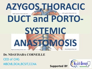
- 1. Dr. NDAYISABA CORNEILLE CEO of CHG MBChB,DCM,BCSIT,CCNA Supported BY AZYGOS,THORACIC DUCT and PORTO- SYSTEMIC ANASTOMOSIS
- 2. Introduction • The azygos vein connects the inferior vena cava and the superior vena cava • The thoracic duct is the largest lymph vessel that ultimately drains lymph from all parts of the body into the blood circulation • We shall look at them one at a time Dr Ndayisaba Corneille
- 3. The azygos veins • Components: – the main azygos vein, – the inferior hemiazygos vein (hemiazygos), – the superior hemiazygos vein (accessory hemiazygos) • Areas of drainage: – the posterior parts of the intercostal spaces, – the posterior abdominal wall, – the pericardium, – the diaphragm, – the bronchi, – the esophagus Dr Ndayisaba Corneille
- 4. Azygos veins (1) Dr Ndayisaba Corneille
- 5. Azygos veins (2) Dr Ndayisaba Corneille
- 6. Azygos vein • Origin: – The origin of this vein is variable – It is often formed by the union of the right ascending lumber vein and the right subcostal vein • Course: – It ascends through the aortic opening in the diaphragm (T12) in the right side of the aorta to the level of the fifth thoracic vertebra Dr Ndayisaba Corneille
- 7. Cont/… – Here, it arches forward above the root of the right lung to empty into the posterior surface of the superior vena cava • Branches/tributaries: – The 8 lower intercostal veins – The right superior intercostal vein – The superior and inferior hemiazygos veins – Mediastinal veins Dr Ndayisaba Corneille
- 8. Inferior hemiazygos vein • Origin: – Often formed by the union of the left ascending lumber vein and the left subcostal vein. • Course: – It ascends through the left crus of the diaphragm – At about the level of the 8th thoracic vertebra, it turns to the right and joins the azygos vein Dr Ndayisaba Corneille
- 9. Cont/… • Branches/tributaries: – Some lower left intercostal veins – Mediastinal veins Superior hemiazygos vein • This vein is formed by the union of the 4th to 8th intercostal veins • It joins the azygos vein at the level of the 7th thoracic vertebra Dr Ndayisaba Corneille
- 10. The azygos system (1) Dr Ndayisaba Corneille
- 12. The azygos system (2) Dr Ndayisaba Corneille
- 13. Clinical significance of the azygos veins • In obstruction of the superior and inferior venae cavae, the azygos veins provide an alternative pathway for the return of venous blood to the right atrium of the heart. • This is possible since these veins and their tributaries connect the superior and inferior venae cavae Dr Ndayisaba Corneille
- 14. THORACIC DUCT
- 15. Thoracic duct • Also called the left lymphatic duct • Origin: – Begins below in the abdomen as a dilated sac, the cysterna chyli • Course: – It ascends through the aortic opening in the diaphragm (T12) on the right side of the descending aorta Dr Ndayisaba Corneille
- 16. Cont/… – It gradually crosses the median plane behind the esophagus and reaches the left border of the esophagus at the level of the lower border of T4 (sternal angle) – It then runs upwards along the left edge of the esophagus to enter the root of the neck – Here, it bends laterally behind the carotid sheath and infront of the vertebral vessels Dr Ndayisaba Corneille
- 17. Cont/… – It turns downwards infront of the left phrenic nerve and crosses the subclavian artery to enter the beginning of the left brachiocephalic vein (confluence of the left internal jugular and left subclavian veins) – At the root of the neck, the thoracic duct receives the left jugular, subclavian, and bronchomediastinal lymph trunks – They may alternatively drain directly into the adjacent large veins Dr Ndayisaba Corneille
- 18. Course of the thoracic duct Dr Ndayisaba Corneille
- 19. Areas of drainage • The thoracic duct conveys to the blood all lymph from: – The lower limbs – Pelvic cavity – Abdominal cavity – Left side of the thorax – Left side of the head and neck – Left arm Dr Ndayisaba Corneille
- 20. Right lymphatic duct • The right jugular, subclavian, and bronchomediastinal trunks, which drain the right side of the head and neck, the right upper limb, and the right side of the thorax, respectively, may join to form the right lymphatic duct • This common duct, if present, is about ½ inch (1.3cm) long and opens into the beginning of the right brachiocephalic vein (confluence of right internal jugular and right subclavian veins) Dr Ndayisaba Corneille
- 21. Cont/… • Alternatively, the trunks may open independently into the great veins at the root of the neck Dr Ndayisaba Corneille
- 22. Azygos, thoracic duct, & their areas of drainage Dr Ndayisaba Corneille
- 24. Thoracic duct on the cadaver Dr Ndayisaba Corneille
- 25. Orders Of Lymphatic Vessels • Lymph capillaries - smallest lymph vessels, first to receive lymph • Lymphatic collecting vessels - collect from lymph capillaries • Lymph nodes - scattered along collecting vessels • Lymph trunks - collect lymph from collecting vessels • Lymph ducts - empty into veins of the neck
- 26. Lymphatic Collecting Vessels • Accompany blood vessels • Composed of the same three tunics as blood vessels • Contain more valves than veins do – helps direct the flow of blood • Lymph propelled by: – contraction of skeletal muscles – pulse pressure of nearby arteries – Tunica media of the lymph vessels Dr Ndayisaba Corneille
- 27. Lymph Nodes • Cleanse the lymph of pathogens • Human body contains around 500 • Lymph nodes are organized in clusters
- 28. Microscopic Anatomy of a Lymph Node • Fibrous capsule – surrounds lymph nodes • Trabeculae – connective tissue strands • Lymph vessels – Afferent lymphatic vessels – Efferent lymphatic vessels
- 29. Lymph Trunks • Lymphatic collecting vessels converge • Five major lymph trunks – Lumbar trunks • Receives lymph from lower limbs – Intestinal trunk • Receives chyle from digestive organs – Bronchomediastinal trunks • Collects lymph from thoracic viscera – Subclavian trunks • Receive lymph from upper limbs and thoracic wall – Jugular trunks • Drain lymph from the head and neck
- 30. Lymph Ducts • Cisterna chyli - located at the union of lumbar and intestinal trunks • Thoracic duct - ascends along vertebral bodies – Empties into venous circulation – Junction of left internal jugular and left subclavian veins – Drains three quarters of the body • Right lymphatic duct - empties into right internal jugular and subclavian veins
- 31. The Immune System • Recognizes specific foreign molecules • Destroys pathogens effectively • Key cells – lymphocytes • Also includes lymphoid tissue and lymphoid organs Dr Ndayisaba Corneille
- 33. INTRODUCTION • Porto-systemic anastomosis is also known as portocaval anatomoses • This occurs between the veins of the portal circulation and those of the systemic circulation. • It serves as a collateral communication between the portal and systemic venous system. • The importance of this anastomosis is to provide alternative routes of circulation when there is blockage in the liver or portal vein. Dr Ndayisaba Corneille
- 35. Porto-systemic anastomosis i) The superior rectal vein drains into the portal circulation and the inferior rectal vein drains into the systemic circulation (inferior vena cava) • The anastomosis between the superior and inferior rectal vein is known as porto-systemic anastomosis • Blockage of the portal vein in liver disease will cause portal hypertension, hence portal blood will move retrograde and enlarge the anastomoses between the superior rectal and inferior rectal veins, thereby causing varicosity (hemorrhoids or piles) Dr Ndayisaba Corneille
- 37. Cont/… ii). Esophageal branches of the left gastric vein (portal circulation) anastomose with esophageal branches of the azygous vein (systemic circulation) • In portal hypertension as a result of liver disease, blood will move retrograde and expand these anastomoses to form varicosities (esophageal varicosities). • These varicosities usually rupture and result into vomiting of blood (hematemesis) Dr Ndayisaba Corneille
- 39. Cont/… iii) The right colic vein, the middle colic vein and the left colic vein (portal circulation) anastomose with the renal vein, suprarenal vein, paravertebral vein and testicular or ovarian veins which are found on the posterior abdominal wall (systemic circulation) • This is yet another portal-systemic anastomosis Dr Ndayisaba Corneille
- 41. Cont/… iv) The paraumblical veins, tributaries of the portal vein (portal circulation) anastomose with superficial epigastric vein on the anterior abdominal wall (systemic circulation) • In portal hypertension as a result of liver disease, blood will move retrograde towards the anterior abdominal wall through the anastomosis • Stagnation or slowing down of blood flow may result into ascites (accumulation of fluid within the peritoneum). • Veins on the anterior abdominal wall will be logged with blood and cause a condition known as caput medusae. • This condition can be used to diagnose liver disease Dr Ndayisaba Corneille
- 44. CLINICAL SIGNIFICANCE A portosystemic shunt is the diversion of portal blood into a systemic vein without the blood passing through the liver. This occurs naturally in developing fetus This may be intrahepatic or extrahepatic, congenital or acquired. Extrahepatic occurs in congenital atresia of the portal vein Intrahepatic results from a connection between the portal venous system within the liver and either the hepatic veins or inferior vena cava Signs/Symptoms: tremors, epileptic seizures, weight loss, bladder stones, vomitting Dr Ndayisaba Corneille
- 45. Portal Hypertension • This is increase in blood pressure in the veins of the venous system Cause: blockage in the veins of the liver due to pathological condition such as cirrhosis and the inability of the blood to flow through. Signs/symptoms: varicose veins on the abdominal wall called caput medusa, oesophageal varices, enlargement of the spleen, accumulation of fluid in the peritoneal cavity and bleeding in the GIT. Dr Ndayisaba Corneille
- 46. END Dr Ndayisaba Corneille THANKS FOR LISTENING By DR NDAYISABA CORNEILLE MBChB,DCM,BCSIT,CCNA Contact us: amentalhealths@gmail.com/ ndayicoll@gmail.com whatsaps :+256772497591 /+250788958241