Anatomy of Suprarenal (Adrenal) Glands.pptx
•Download as PPTX, PDF•
1 like•952 views

At the end of the presentation ,we should be able to describe the: Location, shape and relations of the right and left adrenal glands. Blood supply, lymphatic drainage and nerve supply of right and left adrenal glands Parts of adrenal glands and function of each part. Development of adrenal gland and common anomalies.

Recommended
More Related Content
What's hot
What's hot (20)
Similar to Anatomy of Suprarenal (Adrenal) Glands.pptx
Similar to Anatomy of Suprarenal (Adrenal) Glands.pptx (20)
More from Dr Ndayisaba Corneille
More from Dr Ndayisaba Corneille (20)
Recently uploaded
Recently uploaded (20)
Anatomy of Suprarenal (Adrenal) Glands.pptx
- 1. ANATOMY OF SUPRARENAL (ADRENAL) GLANDS Dr. NDAYISABA CORNEILLE CEO of CHG MBChB,DCM,BCSIT,CCNA Supported BY
- 2. Objectives • At the end of the presentation ,we should be able to describe the: • Location, shape and relations of the right and left adrenal glands. • Blood supply, lymphatic drainage and nerve supply of right and left adrenal glands • Parts of adrenal glands and function of each part. • Development of adrenal gland and common anomalies. Dr Ndayisaba Corneille
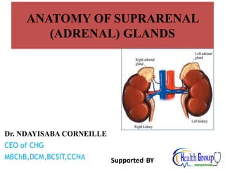
- 3. Suprarenal Glands • The suprarenal (adrenal) gland is a component of the hypothalamic- pituitary-suprarenal axis that is responsible for coordinating stress response and metabolism. • They are yellowish retroperitoneal organs that lie on the upper poles of the kidneys, • just above the level of the last thoracic vertebra (T12). • They are surrounded by renal fascia (but are separated from the kidneys by the perirenal fat). • Each gland has an outer yellow cortex and an inner dark brown medulla. Dr Ndayisaba Corneille
- 4. •Suprarenal glands surrounded by connective tissue containing considerable perinephric fat • Suprarenal glands are enclosed by renal fascia by which they are attached to the crura of the diaphragm • Each gland has a hilum, where the veins and lymphatic vessels exit the gland; whereas arteries and nerve enter the glands at multiple sites • They are separated from the kidneys by a thin septum (part of the renal fascia) •The medial borders of the suprarenal glands are 4-5 cm apart. In this area, from right to left, are the IVC, right of the diaphragm, celiac ganglion, celiac trunk, SMA and the left of the diaphragm. Suprarenal Glands Dr Ndayisaba Corneille
- 5. The right suprarenal gland Is pyramid shaped. Caps the upper pole of the right kidney. • Relations: • Anterior: right lobe of the liver and inferior vena cava. • Posterior: diaphragm. Dr Ndayisaba Corneille
- 6. The left suprarenal gland Is crescentic in shape Extends along the medial border of the left kidney from the upper pole to the hilus. • Relations: • Anterior: pancreas, lesser sac, and stomach • Posterior: diaphragm. Dr Ndayisaba Corneille
- 7. PARTS OF SUPRA RENAL GLANDS •Each suprarenal gland has two parts : ~ Suprarenal cortex ~ Suprarenal medulla Dr Ndayisaba Corneille
- 8. SUPRARENAL CORTEX • Derives from mesoderm Three zones in suprarenal cortex Outer zone / zona glomerulosa Mineralocorticoid hormone middle zone / zona fasciculata Glucocorticoid hormone Inner zone / Zona reticularis Androgen hormone Dr Ndayisaba Corneille
- 9. • Aldosterone is the major mineralocorticoid. • It regulates homeostasis of two mineral ions, namely sodium ions (Na) and potassium ions (K), and helps adjust blood pressure and blood volume. • Aldosterone also promotes excretion of H+ in the urine; this removal of acids from the body can help prevent acidosis Mineralocorticoid Dr Ndayisaba Corneille
- 10. •include cortisol (hydrocortisone), corticosterone, and cortisone •Function : Protein breakdown, glucose formation, lipolysis, resistance to stress, anti-inflammatory effects, depression of immune responses Glucocorticoid Dr Ndayisaba Corneille
- 11. •The major androgen secreted by the adrenal gland is dehydroepiandrosterone (DHEA) •Function : promote libido (sex drive) and are converted into estrogens (feminizing sex steroids) by other body tissues; after menopause, when ovarian secretion of estrogens ceases all female estrogens come from conversion of adrenal androgens; stimulate growth of axillary and pubic hair in boys and girls and contribute to the prepubertal growth spurt. Androgen Dr Ndayisaba Corneille
- 12. SUPRARENAL MEDULLA • Derive from neural crest cells associated with the sympathetic nervous system • The cromaffin cells secrete catecholamines / epinephrine and norepinephrine (NE), also called adrenaline and noradrenaline, respectively. • Unlike the hormones of the adrenal cortex, the hormones of the adrenal medulla are not essential for life since they only intensify sympathetic responses in other parts of the body. Dr Ndayisaba Corneille
- 13. Action of catecholamine : 1. Increased heart rate 2. Increased force of cardiac muscle contraction 3. Elevated blood pressure 4. Increased breathing rate 5. Decreased activity in the digestive system Dr Ndayisaba Corneille
- 14. Arteries: The arteries supplying each gland are three in number: superior, middle, and inferior suprarenal arteries arise from; inferior phrenic artery, abdominal aorta, and renal artery, respectively. • Veins: A single vein emerges from the hilum of each gland and drains • into the inferior vena cava on the right and • into the left renal vein on the left. Dr Ndayisaba Corneille
- 15. Nerve Supply: Preganglionic sympathetic fibers derived from the splanchnic nerves supply the glands. Most of the nerves end in the medulla of the gland. Lymph Drainage: The lymph drains into the lateral aortic nodes. Dr Ndayisaba Corneille
- 16. Functions The cortex of the suprarenal glands secretes hormones that include: Mineral corticoids, which are concerned with the control of fluid and electrolyte balance Glucocorticoids, which are concerned with the control of the metabolism of carbohydrates, fats, and proteins Small amounts of sex hormones, which probably play a role in the prepubertal development of the sex organs. The medulla of the suprarenal glands secretes the catecholamines: epinephrine and norepinephrine Dr Ndayisaba Corneille
- 17. Development of the Adrenal Glands The two parts of the adrenal gland i.e. the cortex and the medulla develop from two different origins. Cortex • is mesodermal in origin; • develops from the celomic epithelium of the posterior abdominal wall. Medulla • is ectodermal in origin; • develops from the neural crest cells. Dr Ndayisaba Corneille
- 18. The cortex • During the 6th week of development, • by aggregation of the mesenchymal cells • between dorsal mesentery and developing gonads. • This fetal cortex is derived from the mesothelium lining the posterior abdominal wall. The medulla • It forms a mass medial to the fetal cortex • derived from the adjacent sympathetic ganglion; • from neural crest cells. Dr Ndayisaba Corneille
- 19. Permanent cortex •A second wave of mesenchymal cells arise from the mesothelium, enclose the fetal cortex. •forms a thinner definitive (permanent) cortex. Dr Ndayisaba Corneille
- 20. • Differentiation of the characteristic suprarenal cortical zones begins during the late fetal period. • Zona glomerulosa & • zona fasciculata are present at birth, but • zona reticularis is not recognizable until the end of third year. Dr Ndayisaba Corneille
- 21. • The suprarenal gland of the fetus is 10-20 times larger than the adult glands relative to the body weight, and are large compared with the kidneys. This is because of the extensive size of the fetal cortex. The medulla remains relatively small until after birth. • The suprarenal glands rapidly become smaller during the first 2-3 weeks after birth, due to the rapid regression of the fetal cortex. • Its involution is largely completed in the first year of life. • During the process of involution, the cortex is friable and susceptible to trauma at birth leading to severe hemorrhage. • Congenital adrenal hyperplasia (CAH): • An abnormal increase in the cortical cells results in excessive androgen production; during the fetal period. • In females, it may lead to musculization of external genitalia and enlargement of clitoris. • In males, it may remain undetected in early infancy. • Later in childhood, in both sexes, androgen excess may lead to rapid growth and accelerated skeletal maturation. Dr Ndayisaba Corneille
- 22. END THANKS FOR LISTENING By DR NDAYISABA CORNEILLE MBChB,DCM,BCSIT,CCNA Contact us: amentalhealths@gmail.com/ ndayicoll@gmail.com whatsaps :+256772497591 /+250788958241 Dr Ndayisaba Corneille