Differential Diagnosis of Pulmonary Cysts
•Download as PPTX, PDF•
1 like•768 views

For quick reference you can learn this ppt in short period of time. 4 main d/d is discussed with beautiful CT-Scan findings.

Recommended
More Related Content
What's hot
What's hot (20)
Similar to Differential Diagnosis of Pulmonary Cysts
Similar to Differential Diagnosis of Pulmonary Cysts (20)
More from Dr.Bijay Yadav
More from Dr.Bijay Yadav (20)
Recently uploaded
Recently uploaded (20)
Differential Diagnosis of Pulmonary Cysts
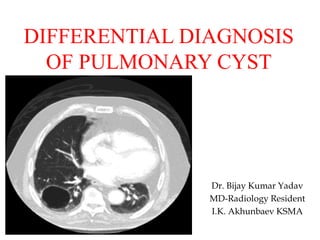
- 1. DIFFERENTIAL DIAGNOSIS OF PULMONARY CYST Dr. Bijay Kumar Yadav MD-Radiology Resident I.K. Akhunbaev KSMA
- 2. Pulmonary Cyst: • Pulmonary cysts are round, thin-walled, low attenuation spaces/lucencies in the lung. • Lung cysts usually contain air but occasionally also contain fluid or solid material. • Pulmonary cysts can be congenital or acquired.
- 3. Differential Diagnosis Of pulmonary Cyst 1. Bullous Emphysema 2. Cystic Bronchiectasis 3. Histiocytosis (Pulmonary Langerhans cell Histiocytosis) 4. Pneumatoceles
- 4. 1. Bullous Emphysema • BULLOUS emphysema is usually associated with strictures of the bronchi. These narrowed bronchi permit the inspired air to enter the alveoli, but on expiration the outlet closes and back-pressure is created, • A mechanism which in time causes dilatation, atrophy, & rupture of the alveoli. • Finally, large emphysematous bullae evolve from the coalescing alveoli.
- 5. Emphysematous Changes More On The Right Side With Apical Bullae
- 6. Axial CT Bullous Emphysematous
- 8. Coronal Lung Window Bullous Emphysema
- 10. Bullae Cyst
- 11. 2. Cystic Bronchiectasis • Cystic bronchiectasis is one of the less common morphological forms of bronchiectasis. • It may be present on its own or may occur in combination with other forms of bronchiectasis. • Bronchiectasis is defined as an irreversible abnormal dilatation of the bronchial tree. It has a variety of underlying causes, with a common aetiology of chronic inflammation. • According to macroscopic morphology, Bronchiectasis is of three types. a. Cylindrical bronchiectasis : b. Varicose bronchiectasis : c. Cystic bronchiectasis :
- 12. Radiographic features: • It is characterized by saccular dilatation of bronchi that extends to the pleural surfaces. When aggregated these may give a "bunch of grapes" like appearance.
- 14. Cystic Bronchiectasis Causing Pneumothorax
- 15. Multiple Diffuse Cystic Dilatation Of The Bronchioles
- 16. 3. Histiocytosis Pulmonary Langerhans cell histiocytosis (PLCH): • May be seen as part of widespread involvement in patients with disseminated Langerhans cell histiocytosis or more frequently as a distinct entity in young adult age group 20-40 years smokers. • In adults, PLCH is most common & the lung is the most common and often only organ involved. Radiographic Findings: • Thin-section computed tomography (CT) performed during early- stage disease shows ill-defined 1–10-mm centrilobular nodules that may be cavitary. • As disease progresses, cystic lesions predominate over nodules. • Cysts may be thin or thick walled and often have irregular & bizarre shape.
- 17. PLCH in a 44-year-old woman. (a) Axial thin-section CT image shows poorly defined nodules predominantly in the upper lobes (arrowheads), typical of the early cellular stage of PLCH. Some nodules have central lucency, indicating developing cysts (arrows). (b) Axial thin-section CT image shows spared lung bases. (a) (b)
- 18. PLCH in a 31-year-old man. Axial (a) and coronal reformatted (b) thin-section CT images show irregular cysts of variable thickness and size (arrow) in the upper lobes and sparing of the lung bases. (a) (b)
- 19. PLCH in a 28-year-old man. (a) Axial thin-section CT image shows bizarrely shaped cysts (arrow) in the upper lobes. (b) Two-year follow-up axial thin-section CT image obtained after chemotherapy for systemic LCH shows smaller cysts (arrow).
- 20. PLCH in a 25-year-old man. Axial thin-section CT image shows irregular cysts (arrow) of variable size in the upper lobes and pneumothorax on the left side (★).
- 21. 4. Pneumatoceles • Pneumatoceles are intrapulmonary gas-filled cystic spaces that can have a variety of sizes and appearances. • They may contain gas-fluid levels and are usually the result of ventilator-induced lung injury in neonates or post-infectious. • Although pneumatoceles are seen in all age groups, they are most frequently encountered in infant. • The majority of pneumatoceles occur as a result of pneumonia also due to blunt trauma.
- 22. Radiographic features: CT Scan • Smooth inner margins • Little if any fluid content • The wall, if visible, is thin and regular • Tend to persist despite an absence of symptoms.
- 23. Pneumatoceles developing in 13-year-old boy with staphylococcal pneumonia. A) CT scan shows thick-walled pneumatoceles (white arrows) in the early phase of pneumonia in the right upper lobe. B) in another patient, follow-up CT scan obtained 20 months after initial infection shows numerous residual pneumatoceles (white arrows) in the right lower lobe.