Brad Bernardini, MD: Sub-Pectoralis Biceps Tenodesis Using the TenoLok™ Tenodesis Anchor
•
0 likes•125 views

A new addition to CONMED’s Shoulder Restoration System,™ the versatile new TenoLok™ Dual Expanding Tenodesis anchor is used for subpectoral biceps tenodesis repair. The TenoLok™ is designed to provide strong tendon-to-bone fixation, reduced tendon damage and tendon wrap as well as a fast, efficient technique.

Recommended
Recommended
More Related Content
What's hot
What's hot (20)
Similar to Brad Bernardini, MD: Sub-Pectoralis Biceps Tenodesis Using the TenoLok™ Tenodesis Anchor
Similar to Brad Bernardini, MD: Sub-Pectoralis Biceps Tenodesis Using the TenoLok™ Tenodesis Anchor (20)
Recently uploaded
Recently uploaded (20)
Brad Bernardini, MD: Sub-Pectoralis Biceps Tenodesis Using the TenoLok™ Tenodesis Anchor
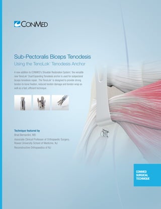
- 1. Technique featured by Brad Bernardini, MD Associate Clinical Professor of Orthopaedic Surgery, Rowan University School of Medicine, NJ Reconstructive Orthopaedics of NJ Sub-Pectoralis Biceps Tenodesis Using the TenoLok™ Tenodesis Anchor A new addition to CONMED’s Shoulder Restoration System,™ the versatile new TenoLok™ Dual Expanding Tenodesis anchor is used for subpectoral biceps tenodesis repair. The TenoLok™ is designed to provide strong tendon-to-bone fixation, reduced tendon damage and tendon wrap as well as a fast, efficient technique. CONMED SURGICAL TECHNIQUE
- 2. 2 | Sub-Pectoralis Biceps Tenodesis Using the TenoLok™ Tenodesis Anchor Sub-Pectoralis Biceps Tenodesis Using the TenoLok™ Tenodesis Anchor Pathology of the proximal long head biceps tendon is common and often requires surgical management in isolation or in combination with other pathologic conditions of the shoulder. Up to 50% of patients with shoulder pain are found to have a diagnosable pathology involving the long head of the biceps proximally. Surgical indications include refractory biceps tendonitis, instability, interstitial and surface tears, and biceps anchor pathology, i.e. SLAP tears. Surgical treatment options include attempts at primary repair, biceps sling reconstruction, tenotomy, and tenodesis. Despite the options, biceps tenodesis has gained recent favor given its ability to resolve pain, restore a proper length-tension relationship, avoid cosmetic deformity, and improve functional strength. Currently there exists a number of accepted techniques, with arthroscopic and open options for long head biceps tenodesis. Both categories have been shown to provide satisfactory results, however sub-pec biceps tenodesis has the additional benefit of addressing pathologic conditions that exist in the bicipital groove distal to the intra-articular location. Additionally, it may result in decreased post-operative stiffness and intra-operative swelling. Regardless of the preferred technique or location, a critical factor in providing a successful surgical outcome is to restore the proper biceps length-tension relationship with solid fixation that allows early rehabilitation. Introduction by Dr. Brad Bernardini
- 3. Shoulder Restoration System™ | 3 Partner of Reconstructive Orthopedics of NJ Director of Sports Medicine forVirtua Sports Medicine, Virtua Health &Wellness Centers, TotalTurf Experience,and Endeavor Sports Performance – NJ Associate Clinical Professor of Orthopaedic Surgery, Rowan University School of Medicine – NJ Brad Bernardini, MD is a partner of Reconstructive Orthopedics of NJ and is an Associate Clinical Professor of Orthopaedic Surgery for Rowan University School of Medicine and Orthopaedic Surgery Residency Program. Dr. Bernardini is currently the Medical Director of Sports Medicine forVirtua Sports Medicine, theVirtua Health & Wellness Centers, the Total Turf Experience, and Endeavor Sports Performance in Southern NJ. Dr. Bernardini completed his residency at the University of Connecticut Health Sciences Center in Farmington, CT., and was fellowship trained in Sports Medicine at the Taos Orthopedic Institute & Research Foundation in Taos, NM. He is Board Certified in Orthopedic Surgery and Subspecialty Certified in Orthopedic Sports Medicine. He has been distinguished as an Associate Masters Instructor of Arthroscopic Knee and Shoulder Surgery by the Arthroscopy Association of North America, and is a published author in the American Journal of Sports Medicine. Dr. Bernardini remains an active member of the United States Ski & Snowboard Team Physician Pool Program and is affiliated with numerous regional athletic teams. He is a former Division IAA Academic All-American Football Player and Patriot League 60 meter dash Champion at Bucknell University. Currently, Dr. Bernardini maintains his passion for athletics as a competitive triathlete and 4X Ironman finisher. Brad Bernardini, MD Dr. Brad Bernardini is a paid consultant for CONMED Corporation.
- 4. 4 | Sub-Pectoralis Biceps Tenodesis Using the TenoLok™ Tenodesis Anchor MINI-OPEN APPROACH Sub-Pectoralis Biceps Tenodesis Using the TenoLok™ Tenodesis Anchor An anatomically precise LH Biceps Tenodesis placed in the Sub-Pec location through a mini-open approach has been shown to provide solid fixation, restore proper length-tension relationship, and prevent cosmetic deformity. When long head biceps pathology is present, non-operative management should be optimized, but if tenodesis is deemed necessary, Sub-Pec Tenodesis utilizing the TenoLok™ implant is an excellent surgical option. The dual-expansion and push-in style of the TenoLok™ Tenodesis anchor provides tendon preservation without the issues of tendon wrap or tissue damage as commonly seen with interference screws. With proper technique, the Sub-Pec tenodesis is a time efficient procedure with reproducible results. NON-DEPLOYED ANCHOR
- 5. Shoulder Restoration System™ | 5 CONMED SURGICAL TECHNIQUE CONMED SURGICAL TECHNIQUE 1 3 2 Place a drill tip guide pin centered within the bicipital groove 20mm proximal to the inferior border of the pec major tendon insertion. An 8mm Badger drill bit is placed over the guide pin, and is then drilled 20mm or unicortically to create a socket. NOTE: For the 6.0mm TenoLok, drill a 7.5mm to 8.0mm hole. For the 5.0mm TenoLok, drill a 6.5mm to 7.0mm hole. 4 POSITIONING AND DRILLING A 3cm incision is made, centered over the palpable inferior border of the pec major muscle belly. Dissect through the skin and fascia to the pec major tendon insertion on the humerus. With proximal retraction of the pec major, the long head of the biceps tendon will be located just medial to the pec major insertion. Arthroscopically, release the long head of the biceps at its insertion point of the superior labrum. Ensure that there are no adhesions to prevent tendon retraction. BADGER™ DRILL BIT
- 6. 6 | Sub-Pectoralis Biceps Tenodesis Using the TenoLok™ Tenodesis Anchor The biceps tendon will be measured and marked 20mm proximal from the musculo-tendinous junction or the created socket. Pass the biceps tendon through the Hi-Fi® suture loop at the leading edge of the TenoLok implant, and secure it at this 20mm location on the tendon. Tension and cleat the suture to secure the tendon to the implant. 5 6 Sub-Pectoralis Biceps Tenodesis Using the TenoLok™ Tenodesis Anchor IMPLANT INSERTION TENOLOK™ TENODESIS ANCHOR
- 7. Shoulder Restoration System™ | 7 CONMED SURGICAL TECHNIQUE Orient the biceps tendon so that it exits the bone inferiorly, toward its distal insertion point. Insert the implant while maintaining same angle of approach used during the drilling step. Deliver the implant and tendon construct into the socket with light mallet technique until the laser line is flush with the humeral cortex. NOTE: DO NOT over insert, as anchor could possibly fall in humeral canal. While holding the delivery handle firmly, turn the knob clockwise to deploy the TenoLok. NOTE: Hold delivery handle steady as you turn the black knob. Do not advance the implant into the socket during the deployment step. ANCHOR DEPLOYMENT 7 An audible pop will sound to motion the user that the implant has fully deployed and the driver has disengaged from the implant. Loosen the cleated sutures and remove the driver. NOTE: Upon deployment, the 6.0mm TenoLok will expand to 10mm subcortically and 7.5mm within the cortex. Once the driver is removed, excess tendon is cut, and multiple half-hitches are tied. Cut the remaining suture. Final construct shows the biceps tendon implanted into the humerus and on both sides of the dual- expanding TenoLok Tenodesis Anchor for a fast, easy, and reproducible technique. 9 10 8
- 8. TENOLOK TIPS AND PEARLS POST-OPERATIVE CARE 1. Socket Preparation • Upsize drill hole: Drill hole diameter based on tendon and anchor size. • Adequately remove all soft tissue around drill hole. • Socket depth: - Subpectoral: 20mm - Arthroscopic suprapectoral or high in the groove: 20-25mm 2. Angle of Delivery: Ensure anchor delivery matches angle of drilled bone socket. 3. Insertion: Insert so that laser line is flush with cortex – do not over-insert! 4. Deployment: Listen for audible“POP”; uncleat sutures prior to removing inserter. * Large tendons may require technique modification to aid in anchor insertion into drill hole. For tendons 8mm and larger, an alternative technique option is to leave a 1cm tail through the anchor. TenoLok Anchor Size Tendon Size 4mm 5mm 6mm 7mm 8mm 5.0mm TenoLok 6.5mm 7mm Use 6.0mm anchor for tendons 6mm + 6.0mm TenoLok 7.5mm 7.5mm 7.5mm 8mm 8mm* 8 | Sub-Pectoralis Biceps Tenodesis Using the TenoLok™ Tenodesis Anchor With isolated Sub-Pec biceps tenodesis using the TenoLok™ implant, patients are placed in a simple sling with immediate active and passive range of motion exercises of the elbow. We initiate formal physical therapy on post-op day #5 where range of motion is started through a full arc of elbow and shoulder motion.The sling is discharged at 4 weeks post-op and motion remains the focus of therapy. At 4 weeks, scapular stabilizer and rotator cuff strengthening is allowed. However, we do not allow any additional weight application to the distal extremity for the first 8 weeks. • 8 -10 weeks post-op, graduated strength training exercises involving the biceps are initiated. • Full Strength training is allowed after 12 weeks post-op. If concomitant procedures are performed, such as a rotator cuff repair, those procedures should determine if any further limitations are required during the post-operative course.
- 9. FEATURES AND BENEFITS Shoulder Restoration System™ | 9 CONMED SURGICAL TECHNIQUE Designed to MinimizeTendon Disruption Tendon Wrap Currently a surgeon’s primary options for sub-cortical tenodesis fixation are threaded interference screw devices.These can potentially cause the tendon to wrap around the anchor as it screws in or even slice and damage the tendon. To avoid this, the unique dual expanding feature of TenoLok™ anchor provides an all-inside repair that’s designed to minimize tendon disruption. Simple, ReproducibleTechnique TenoLok™ anchors feature a fast technique that’s designed to help reduce procedure times. Straightforward delivery and fixation combined with no whip-stitching creates a technique that eliminates steps and is easy to learn and replicate. The sliding suture also allows surgeons to gather and tension the tendon prior to fixation for additional ease. SMARTER TENODESIS STARTS HERE Sub-Pectoralis Biceps Tenodesis Using the TenoLok™ Tenodesis Anchor
- 10. 10 | Sub-Pectoralis Biceps Tenodesis Using the TenoLok™ Tenodesis Anchor ORDERING INFORMATION To order any of our TenoLok™ products including Badger® Drill Bits, Guide Pins, Tendon Sizing Rings and additional instrumentation please call CONMED Customer Service at: (US) 800-237-0619 or, (Global) 727-392-6464. ADDITIONAL INSTRUMENTATION TENOLOK™ TENODESIS ANCHOR 6.0mm TenoLok Tenodesis Anchor with one #2 Hi-Fi® Suture . . . . . . . . . . . . . . . . . . . . . . . . . . . . . . . . . . T60S 5.0mm TenoLok Tenodesis Anchor with one #2 Hi-Fi® Suture . . . . . . . . . . . . . . . . . . . . . . . . . . . . . . . . . . T50S 6.0mm TenoLok Tenodesis Anchor . . . . . . . . . . . . . . . . . . . . . . . . . . . T60A GUIDE PIN AND DRILL BIT OPTIONS 9 High Strength Guide Pin, 2.4mm diameter . . . . . . . . . . . . . . . 9742D 6.5 mm Badger® Drill Bit . . . . . . . . . . . . . . . . . . . . . . . . . . . . . . . . . . C8593 7.0 mm Badger® Drill Bit . . . . . . . . . . . . . . . . . . . . . . . . . . . . . . . . . . C8582 7.5 mm Badger® Drill Bit . . . . . . . . . . . . . . . . . . . . . . . . . . . . . . . . . . . 8593 8.0 mm Badger® Drill Bit . . . . . . . . . . . . . . . . . . . . . . . . . . . . . . . . . C8599 TENOLOK™ TENODESIS KITS 5.0mm TenoLok Tenodesis Anchor with one #2 Hi-Fi® Suture, Guide Pin, and 6.5mm Drill Bit. . . . . . . . . . . . . . . . . . . . . . . . . . . . T50S65 5.0mm TenoLok Tenodesis Anchor with one #2 Hi-Fi® Suture, Guide Pin, and 7.0mm Drill Bit. . . . . . . . . . . . . . . . . . . . . . . . . . . . T50S70 6.0mm TenoLok Tenodesis Anchor with one #2 Hi-Fi® Suture, Guide Pin, and 7.5mm Drill Bit. . . . . . . . . . . . . . . . . . . . . . . . . . . . T60S75 6.0mm TenoLok Tenodesis Anchor with one #2 Hi-Fi® Suture, Guide Pin, and 8.0mm Drill Bit. . . . . . . . . . . . . . . . . . . . . . . . . . . . T60S80 6.0mm TenoLok Tenodesis Anchor, Guide Pin, and 7.5mm Drill Bit. . . . . . . . . . . . . . . . . . . . . . . . . . . T60A75 6.0mm TenoLok Tenodesis Anchor, Guide Pin, and 8.0mm Drill Bit. . . . . . . . . . . . . . . . . . . . . . . . . . . T60A80 TENDON SIZER Biceps tendon sizing ring, 5.0 mm -8.0 mm. . . . . . . . . . . . . . . . . GFT-SM HALL™ POWER SET 1-Trigger Modular Drill. . . . . . . . . . . . . . . . . . . . . . . . . . . . . . . . PRO7100B Pin Driver, Range 1.8 – 4.0mm. . . . . . . . . . . . . . . . . . . . . . . . . . . PRO6140 ¼”(6.35mm) Jacobs Chuck. . . . . . . . . . . . . . . . . . . . . . . . . . . . . . PRO2041 Small Lithium Battery. . . . . . . . . . . . . . . . . . . . . . . . . . . . . . . . . . L3000SM ½ Size Sterilization Container. . . . . . . . . . . . . . . . . . . . . . . . . . . . . TR12R ½ Size Drill Set Inner Tray. . . . . . . . . . . . . . . . . . . . . . . . . . . . . . PRO7001T
- 11. Shoulder Restoration System™ | 11 CONMED SURGICAL TECHNIQUE Advancing the Future of Minimally Invasive and Orthopaedic Surgery. Together.
- 12. Shoulder Restoration System™ 525 French Road Utica, New York 13502 Local 727-392-6464 Toll Free 800-237-0169 CONMED.com customer_service@conmed.com CONMED SURGICAL TECHNIQUE ©2016 CONMED Corporation M2016001 8/16